FIGURE 1. Illustrations of the three dragons investigated here. 1.1. Belon’s dragon, from Belon (1588). 1.2. Belon’s dragon, from Gessner (1589). 1.3. Belon’s dragon, from Aldrovandi (1640). 1.4. Aldrovandi’s dragon, from the Tavole di animale (Biblioteca Universitaria di Bologna, 2013). 1.5. Aldrovandi’s dragon, from Aldrovandi (1640). 1.6. Cardinal Barberini’s dragon, from Faber (1651). 1.7. Cardinal Barberini’s dragon, from Kircher (1664).
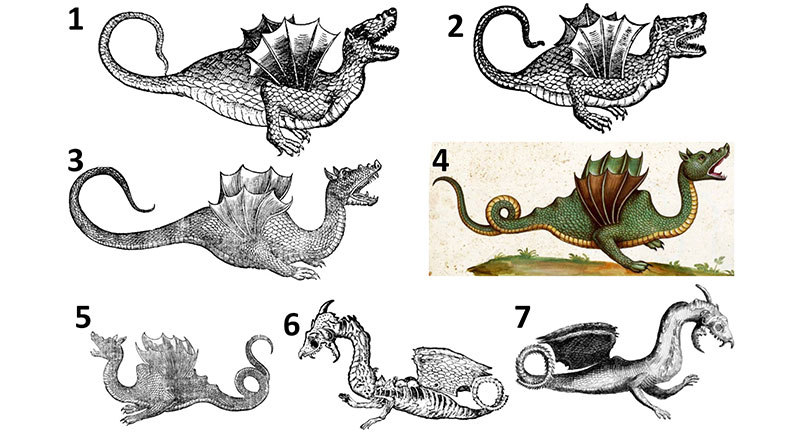
FIGURE 2. Skeletons of pterosaurs. 2.1. AMNH castof Pterodactylus antiquus. 2.2. Pteranodon longiceps, AMNH (American Museum of Natural History, New York City, New York) 6158. 2.3. AMNH cast of Rhamphorhynchus muensteri. 2.4. AMNH cast of Campylognathoides zitteli. 2.5. Vertebrae of the long-tailed pterosaur Rhamphorhychus muensteri in right lateral view, modified from Wellnhofer (1975); left: series of caudal vertebrae, showing the network of elongate dorsal and ventral processes that stiffens the tail; right: a single caudal vertebra and hemal arch, showing the elongate dorsal and ventral processes that contribute to the network.
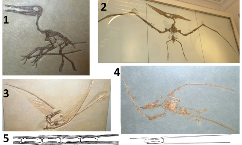
FIGURE 3. Comparison of the skin of the three dragons to that of snakes and lizards. 3.1. Belon’s dragon, scales of torso (above) and tail (below, with original cross-hatching removed from one scale row for clarity), from Belon (1588). 3.2. Aldrovandi’s dragon, skin of neck (above) and tail (below, rotated clockwise relative to its position in Aldrovandi’s original illustration), from Aldrovandi (1640). 3.3. Torso skin of Cardinal Barberini’s dragon, from Kircher (1664). 3.4. Agkistrodon piscivorus (cottonmouth), live specimen at Reptile Lagoon (Hamer, South Carolina), showing the diagonal scale rows that are present both on the torso and the tail in snakes; note that the scale rows are diagonal on both torso and tail in Belon’s and Aldrovandi’s dragons. 3.5. Dendroaspis viridis (western green mamba), live specimen at Riverbanks Zoo (Columbia, South Carolina), showing diagonal torso and tail scale rows, and showing the transversely elongated ventral scutes that are unique to snakes; note that the three dragons also have ventral scutes of this type. 3.6. Torso of Gerrhosaurus validus (giant plated lizard), live specimen at United States National Zoo (Washington, D.C.), showing that the scale rows of the torso are vertical, as in many lizards; note that the scale rows are also vertical on the torso of Cardinal Barberini’s dragon. 3.7. Tail of Cyclura lewisi (blue iguana), live specimen at United States National Zoo, showing that the scale rows are vertical, as in the tails of most lizards; note that the scale rows are not vertical on the tails of Belon’s and Aldrovandi’s dragons. 3.8. Juvenile Python sebae (African rock python) after a large meal, drawn from a photo by P.S., showing that in a snake the torso skin distends without tearing when a large object is in the digestive tract; note the similarity with the bulbous torsos of Belon’s and Aldrovandi’s dragons in Figure 1.
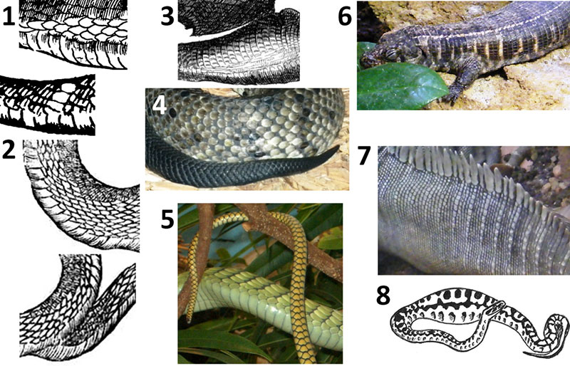
FIGURE 4. Heads of Belon’s and Aldrovandi’s dragons compared with those of African fruit bats and with those of snakes. 4.1. Belon’s dragon. 4.2. Aldrovandi’s dragon. 4.3. left to right: Rousettus aegyptiacus (Egyptian fruit bat), USNM (United States National Museum, Washington, D.C.) 114691; Eidolon helvum (straw-colored fruit bat) , USNM 378787. 4.4. Snakes (live specimens at Reptile Lagoon, Hamer, South Carolina), showing that the eye is closer to the tip of the snout than in Belon’s and Aldrovandi’s dragons; left to right: Python molurus (Asian rock python), Cerastes cerastes (Sahara horned viper), Naja haje (Egyptian cobra).
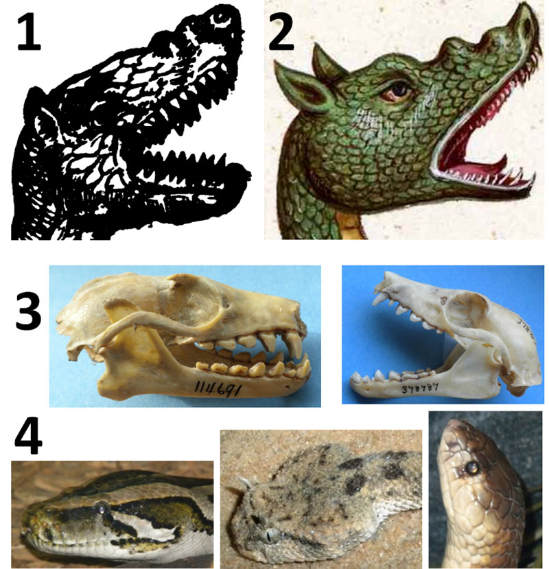
FIGURE 5. Comparison of the wings of Belon’s and Aldrovandi’s dragons to pterosaur wings, bat wings, and the pectoral fins of flying gurnards and a sea robin. 5.1. Underside of right wing of Belon’s dragon. 5.2. Underside of left wing of Aldrovandi’s dragon. 5.3. Dactylopterus volitans (flying gurnard), AMNH 44472 in ventral view, with right pectoral fin partially spread. 5.4. Partially-spread pectoral fin of D. volitans , AMNH 52457. 5.5. Partially-spread pectoral fin of D. volitans , AMNH 44472; note that as in the two dragons’ wings, the fin is narrow at the base, widens distally, and has multiple internal struts that proceed from the shoulder distally. 5.6. Model of D. volitans with pectoral fins fully spread; note the resemblance to the two dragon’s wings, both in the scalloped margin of the fins and the width of the soft tissue between the internal struts. 5.7. Illustration of D. volitans from Bloch (1790), showing the life colors of the fish; note that the colors and spotted pattern fade after death, as shown in 5.3-5, explaining the lack of spots in Belon’s and Aldrovandi’s illustrations. 5.8. Model of Prionotus evolans (striped sea robin), showing that despite similar morphology in the pectoral fin, the spread of soft tissue between the internal struts is much less than in the two dragons and D. volitans. 5.9. Antrozois pallidus (pallid bat), NCSM (North Carolina Museum of Natural Sciences, Raleigh, North Carolina) 5314, showing that in bats the wing has only four internal struts, two of which are closely appressed so that there appear to be only three; note that the wings of the two dragons have more internal struts than this. 5.10. AMNH cast of Rhamphorhynchus muensteri with soft-tissue impressions, showing that the wing of a pterosaur is vastly different in shape from that of the two dragons and has only one internal strut.
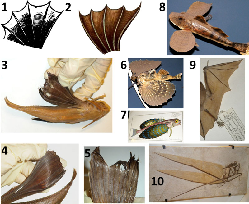
FIGURE 6. Skull of Cardinal Barberini’s dragon and those of weasels (Mustela). All photos but 6.6. and 6.8 are to scale (rulers are marked in mm). Note that in Mustela the upper molar is hidden by the much larger third premolar, and the second lower molar is tiny, making both easy to miss. 6.1. Cardinal Barberini’s dragon, from Faber (1651). 6.2. Mustela nivalis (common weasel), USNM 115207, lateral view; note the resemblance to the dragon’s skull. 6.3. M. nivalis, USNM 115207, palatal view. 6.4. M. nivalis, USNM 115207, anterior view, showing that the incisors match Faber’s description. 6.5. M. nivalis mandible, USNM 115207, occlusal view. 6.6. M. nivalis, USNM 115207, enlargement of dentition. 6.7. M. erminea (stoat), USNM 38493. 6.8. M. erminea, USNM 38493, enlargement of dentition. cp = coronoid process; m = molar; p = premolar.
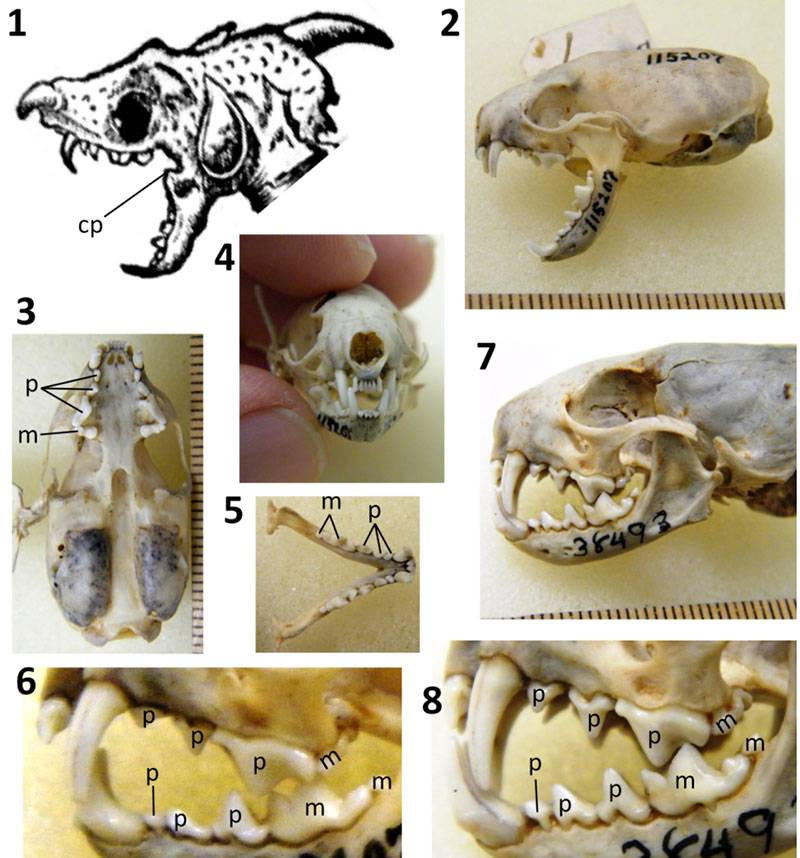
FIGURE 7. Wings of Cardinal Barberini’s dragon compared with patagium of Draco (flying lizards) and frill of Chlamydosaurus kingii (frilled lizard). 7.1. Wings of Cardinal Barberini’s dragon, from Faber (1651). 7.2. Wings of Cardinal Barberini’s dragon, from Kircher (1664). 7.3. Draco haematopogon (personal collection of P.S.), showing spots on patagium. 7.4. Detached right patagium of specimen in 7.3, with yellow arrows showing proximal ends of ribs and black arrows showing distal tips of ribs. 7.5. Same specimen after detachment of left patagium plus part of the body wall. 7.6. Internal view of detached left patagium, showing that no clawlike structures are present. 7.7. Draco maximus, AMNH 29976, ventral view, showing spots on patagium. 7.8-11. Chlamydosaurus kingii, showing projections at edge of frill that resemble the claws on Cardinal Barberini’s dragon, showing the three strong creases in the ventral half of the frill ( 7.8, 7.10-11: AMNH 86512), and showing spots on the frill of one specimen ( 7.9: AMNH 99843).
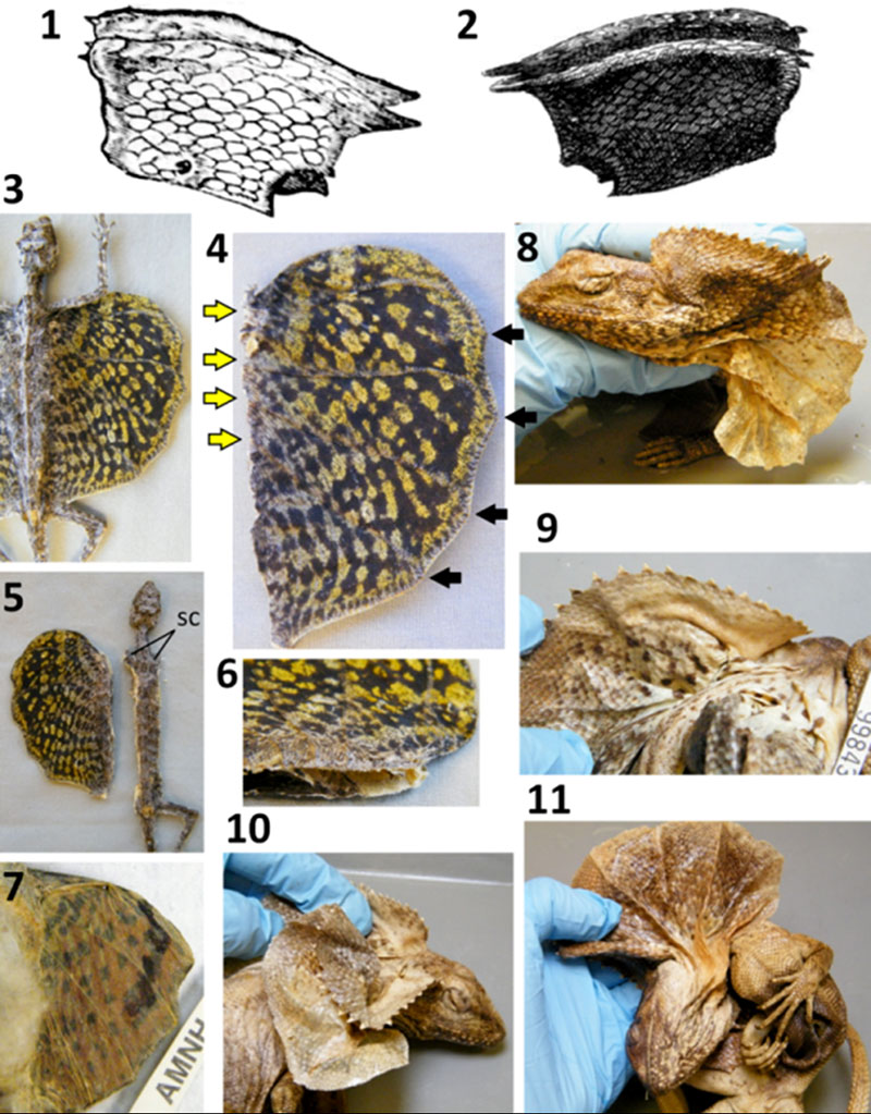
FIGURE 8. Hands of Cardinal Barberini’s dragon compared with those of a common weasel (Mustela nivalis) (USNM 115211) and an ocellated lizard (Lacerta lepida) (MCZ [Museum of Comparative Zoology, Cambridge, Massachusetts] R-29871). Thumbs are circled in yellow. 8.1. Cardinal Barberini’s dragon, from Faber (1651). 8.2. M. nivalis, showing that the size of the hand relative to the head resembles that in Cardinal Barberini’s dragon (see Figure 1.5-6). 8.3. M. nivalis, showing that the thumb is shorter than the other fingers. 8.4. M. nivalis, showing curvature of the finger claws. 8.5. L. lepida, showing that the thumb is shorter than the other fingers.
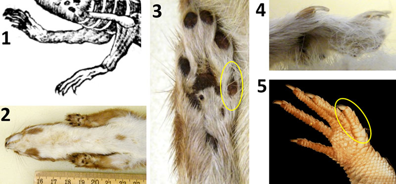
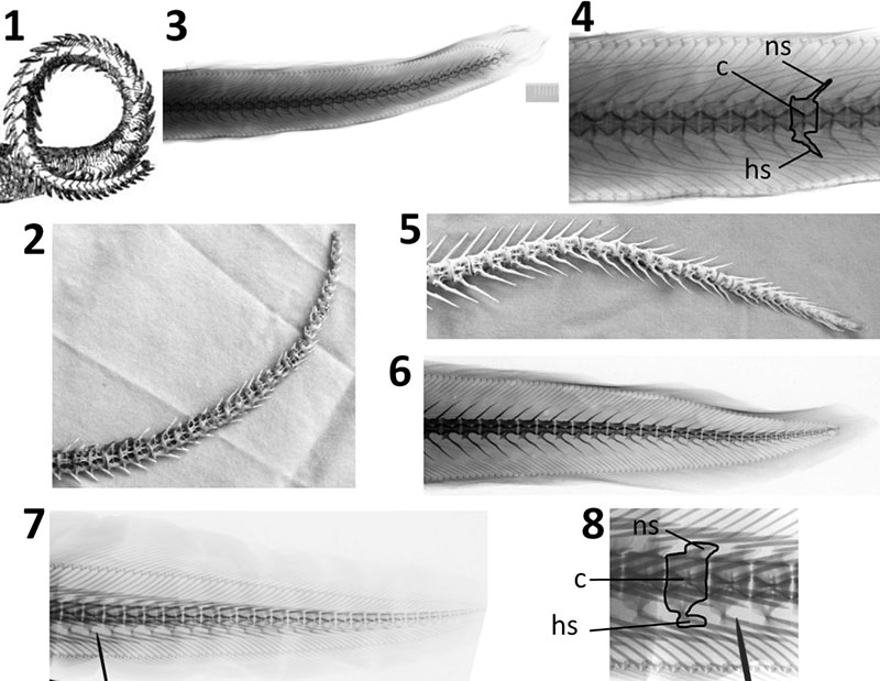
FIGURE 9. Tail of Cardinal Barberini’s dragon compared with the tails of eels. In the photos and x-ray images of eels, including the close-ups, the anteriormost complete vertebra shown is the thirtieth vertebra from the tip of the tail, to facilitate comparison with Cardinal Barberini’s dragon, on which Faber counted 30 tail vertebrae (Appendix 1). 9.1. Cardinal Barberini’s dragon, from Kircher (1664). 9.2. Tail skeleton of Anguilla rostrata (American eel) (NCSM 69208), showing the shortness of the neural and hemal spines in Anguilla, as in Cardinal Barberini’s dragon. 9.3. X-ray image of Anguilla anguilla (European eel) (MCZ 9244), the one Mediterranean species of Anguilla. 9.4. Close-up of 9.3 with one vertebra outlined, showing that the neural and hemal spines are short relative to centrum height. 9.5. Tail skeleton of Conger oceanicus (American conger) (NCSM 69209), showing the great length of the neural and hemal spines. 9.6. Conger conger (European conger) (MCZ 9293), showing that in the one Mediterranean species of Conger, the neural and hemal spines are longer relative to the centrum than they are in Anguilla, and in fact are longer than the height of the centrum. 9.7. Muraena helena (Mediterranean moray) (MCZ 9293). 9.8. Close-up of 9.7, with one vertebra outlined, showing that the shapes of the neural and hemal arches are not simple spikes and therefore do not match Cardinal Barberini’s dragon. c = centrum; hs = hemal spine; ns = neural spine.