FIGURE 1. Maps of the Ryukyu Archipelago (1, 2) and Okinawajima Island (3). The map of Okinawajima shows topography, distribution of the Pleistocene limestone, and study sites. Geological data were obtained from Kizaki (1985).
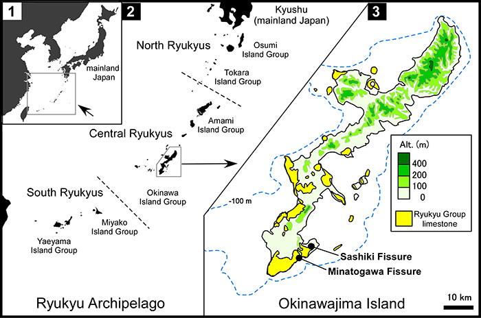
FIGURE 2. Photograph of the Sashiki Fissure (left) and schematic figure showing the structure of the fissure (right). In the right figure, broken lines represent the outline of the fissure behind rock, shaded areas represent studied sediments, and open circles represent the locations of the dating samples.
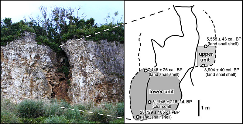
FIGURE 3. Fossils referred to Limnonectes namiyei (1-4) and Babina holsti (5-11). 1-3, left female humerus lacking the proximal and distal parts and the crista ventralis (YMHF-MA 001) in ventral (1), medial (2) and dorsal (3) views; 4, left ilium lacking the anterior part (RUMF-GF-04000) in lateral view; 5-7, right female humerus (one of six registered as RUMF-GF-04003) in ventral (5), medial (6) and dorsal (7) views; 8-10, right male humerus lacking the proximal part (one of two registered as RUMF-GF-04004) in ventral (8), medial (9), and dorsal (10) views; and 11, right ilium lacking the anterior part (one of nine registered as RUMF-GF-04005) in lateral view. Abbreviations: acet, acetabulum; acet.m, acetabular margin; cr.dors, crista dorsalis; cr.lat, crista lateralis; cr.med, crista medialis; cr.par, crista paraventralis; cr.ven, crista ventralis; e.cap, eminentia capitata; ep.rad, epicondylus radialis; ep.ul, epicondylus ulnaris; fo.div, fossula dividens; il.sh, ilial shaft; ol.sc, olecranon scar; p.asc, pars ascendens; stm, spina tuberculi medialis; supr.fo, supracetabular fossa; tub.sup, tuber superior. Scale bars equal 5 mm.
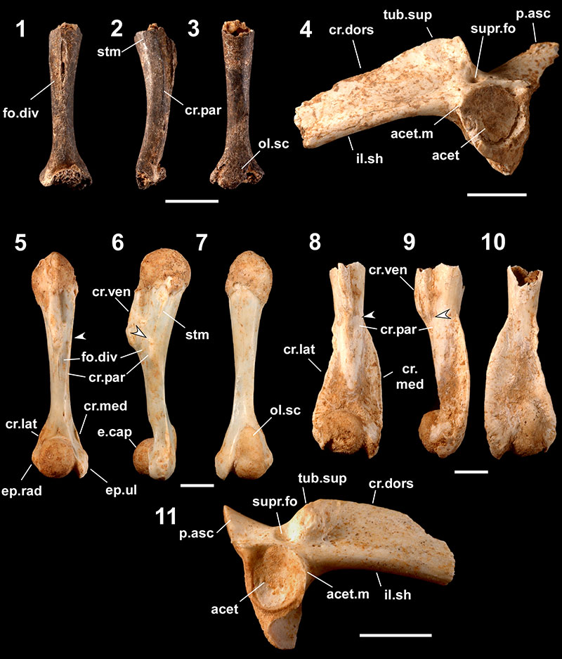
FIGURE 4. Fossils referred to Odorrana ishikawae (1-7) and Odorrana narina (8-14), 1-3, right female humerus (one of 10 registered as RUMF-GF-04009) in ventral (1), medial (2), and dorsal (3) views; 4-6, right male humerus (one of three registered as RUMF-GF-04010) in ventral (4), medial (5), and dorsal (6) views; 7, right ilium lacking the anterior part with part of the ischium (one of five registered as RUMF-GF-04011) in lateral view; 8-10, right female humerus (one of eight registered as RUMF-GF-04014) in ventral (8), medial (9), and dorsal (10) views; 11-13, right male humerus lacking the proximal part of the shaft and the distal part of the epicondylus ulnaris (RUMF-GF-04015) in ventral (11), medial (12), and dorsal (13) views; and 14, pelvic girdle (fused right and left ilia [lacking anterior parts] with the ischium and the pubis: RUMF-GF-04016) in right lateral view. Abbreviations are the same as Figure 3. Arrows indicate the proximal ends of the crista paraventralis. Scale bars equal 5 mm.
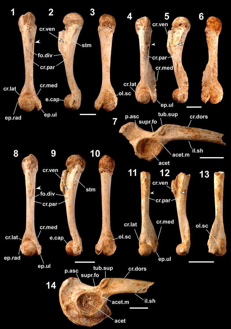
FIGURE 5. Fossils referred to Rana ulma (1-7) and Microhyla okinavensis (8). 1-3, right female humerus (one of 176 registered as RUMF-GF-04019) in ventral (1), medial (2), and dorsal (3) views; 4-6, right male humerus (one of 87 registered as RUMF-GF-04020) in ventral (4), medial (5), and dorsal (6) views; 7, right ilium (one of 93 registered as RUMF-GF-04021) in lateral view; and 8, right ilium (one of two registered as RUMF-GF-04023) in lateral view. Abbreviations are the same as Figure 3. Arrows indicate the proximal ends of the crista paraventralis. Scale bars equal 1 mm.
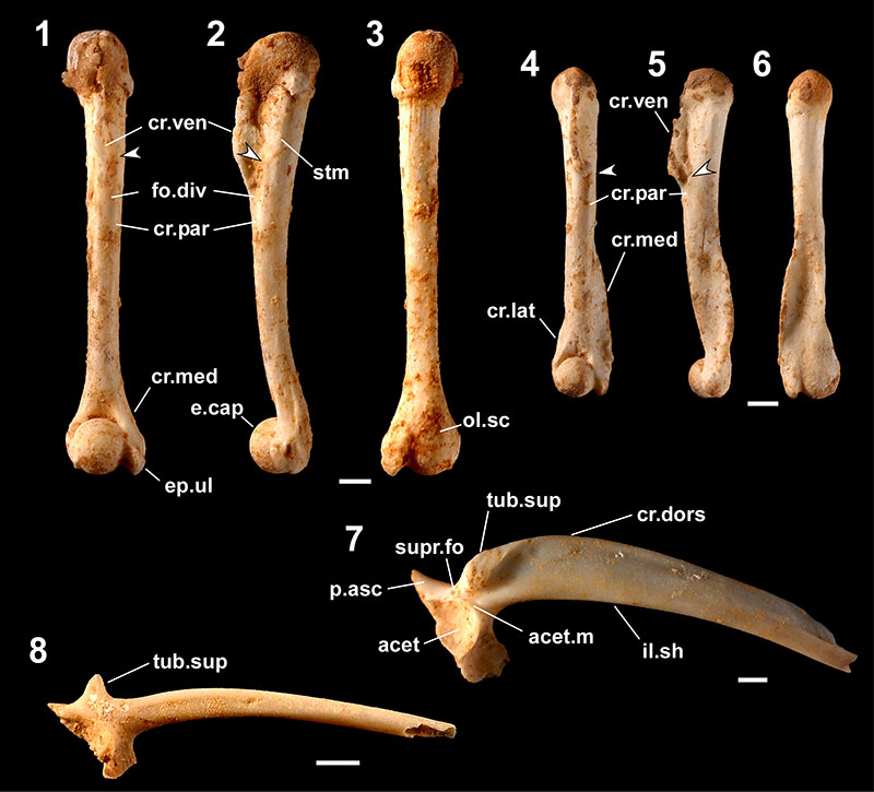
FIGURE 6. Fossils referred to Buergeria japonica (1-4) and Rhacophorus viridis viridis (5-11). 1-3, right female humerus lacking the proximal part (YMHF-MA 010) in ventral (1), medial (2), and dorsal (3) views; 4, left ilium lacking most part of the crista dorsalis (RUMF-GF-04024) in lateral view; 5-7, left female humerus (one of seven registered as RUMF-GF-04025) in ventral (5), medial (6), and dorsal (7) views; 8-10, left male humerus (RUMF-GF-04027) in ventral (8), medial (9), and dorsal (10) views; and 11, pelvic girdle (fused right and left ilia [lacking anterior parts] with the ischium: RUMF-GF-04029) in left lateral view. Abbreviations: pre.acet, preacetabular zone; the others are the same as Figure 3. Arrows indicate the proximal ends of the crista paraventralis. Scale bars equal 1 mm.
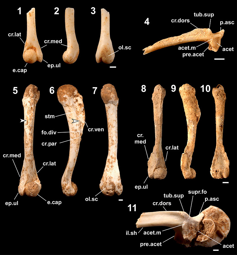
FIGURE 7. Fossils referred to Cynops ensicauda. 1-4, postatlantal precaudal vertebra (one of 108 registered as RUMF-GF-04039 ) in anterior (1), left lateral (2), dorsal (3), and ventral (4) views; 5 and 6, atlas (one of five registered as RUMF-GF-04038) in anterior (5) and left lateral (6) views; 7, parietal-prootic-exoccipital (RUMF-GF-04033) in dorsal view; 8, right maxilla (one of three registered as RUMF-GF-04032) in lateral view; 9, right dentary (one of nine registered as RUMF-GF-04037) in medial view; 10, right rib (one of 25 registered as RUMF-GF-04040) in posterior view; 11, right humerus (one of 85 registered as RUMF-GF-04041) in lateral view; and 12, right femur (one of 92 registered as RUMF-GF-04042) in posterior view. Abbreviations: con, condyle; diap, diapophyses; epi.pr, epipleural processes; neu.sp, neural spine; n.prep, notch for prearticular; parap, parapophyses; pos.pr, posterior process; subd.d, subdental ditch; zygap, zygapophyses. The arrow in 12 indicates the concavity (see text). Scale bars equal 1 mm.
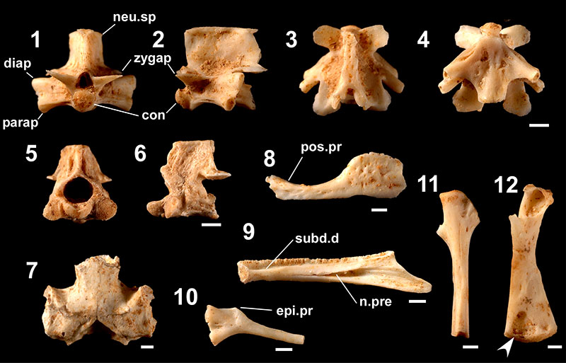
FIGURE 8. Fossils referred to Echinotriton andersoni. 1-4, postatlantal precaudal vertebra (one of 290 registered as RUMF-GF-04052) in anterior (1), left lateral (2), dorsal (3), and ventral (4) views; 5 and 6, atlas (one of 11 registered as RUMF-GF-04051) in anterior (5) and left lateral (6) views; 7, parietal-prootic-exoccipital (RUMF-GF-04047) in dorsal view; 8, right maxilla (one of 30 registered as RUMF-GF-04045) in lateral view; 9, right quadrate (one of five registered as RUMF-GF-04049) in dorsal view; 10, right dentary (one of 70 registered as RUMF-GF-04050) in medial view; 11, right rib (one of 181 registered as RUMF-GF-04053) in posterior view; 12, right humerus (one of 144 registered as RUMF-GF-04054) in lateral view; and 13, right femur (one of 163 registered as RUMF-GF-04055) in posterior view. Abbreviations: tro, trochanter; the others are the same as Figure 7. Scale bars equal 1 mm.
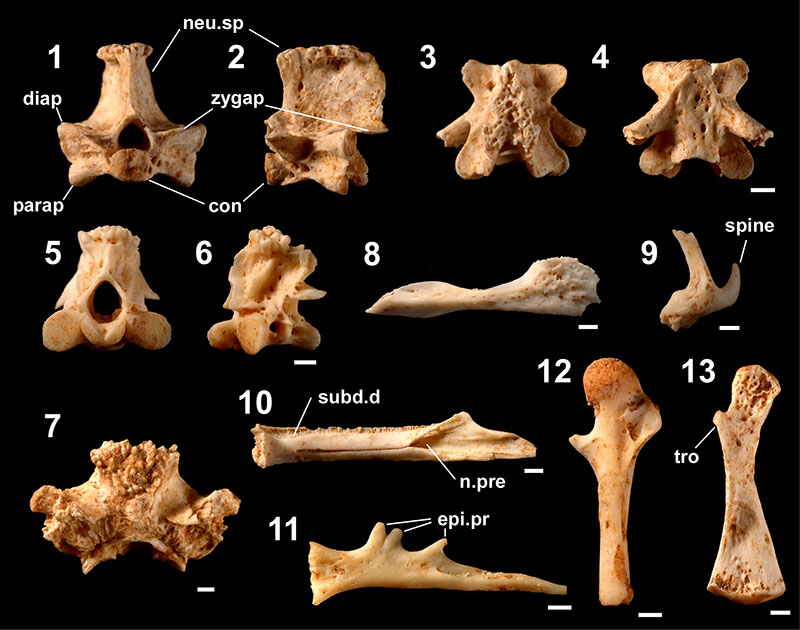
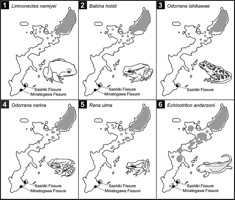
FIGURE 9. Maps showing current distributions (shaded areas) of five forest frogs (Limnonectes namiyei a, b, d, f-h, k, n [ 1], Babina holsti a, b, d, f-k, m [ 2 ], Odorrana ishikawae a, b, d, f-h, j-l, o [ 3 ], O. narina a, b, f-h [ 4 ], and Rana ulma a, b, d, f-k, o [5]) and the newt Echinotriton andersoni a-j (6) on Okinawajima Island and adjacent islets. Superscripts after the species names indicate the data sources—a and b: Chigira (1977, 2003); c: Honda et al. (2012); d: Koba (1957); e: Matayoshi et al. (1977); f-h: Okinawa General Bureau, North Dam Construction Office (1997, 1998, 2002); i and j: Sato (1989, 1993); k: Tanaka (1993); l: Toyama (1988); m: Tominaga et al. (2014); n: Utsunomiya (1996b); and o: Y.N. personal observations at Oku. “?” denotes a population lacking a record or observation in the past 30 years.