FIGURE 1. Chancelloria eros Walcott, 1920, lectotype, USNM 66524. Figured by Walcott (1920) as pls 86:2 and 88:1f. 1. Wet. 2. Dry, coated with ammonium chloride. Detail of 1 (position marked by frame). 3. Dry, coated with ammonium chloride. Detail of 2 (position marked by frame). Top arrow (grey): 4+0 sclerite. Middle arrow (black): 7+1 sclerite, preserved in negative relief (ascending ray towards the viewer). Bottom arrow (white): 7+1 sclerite, preserved in positive relief (ascending ray pointing away from the viewer).
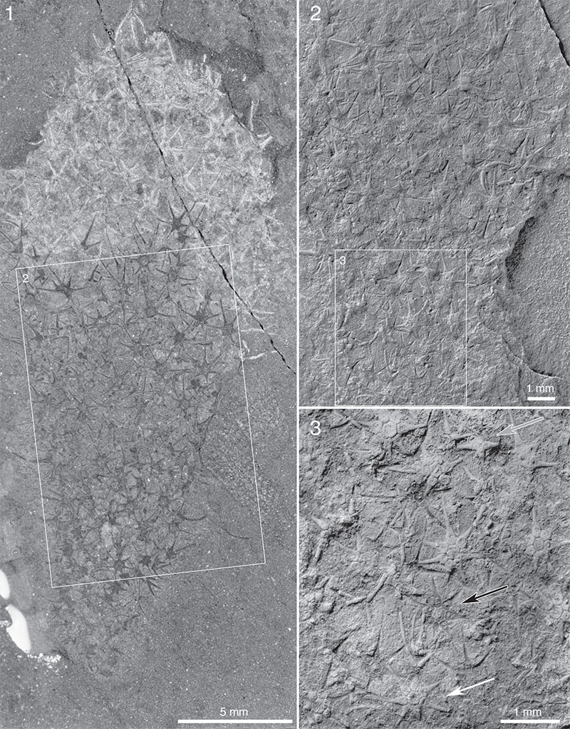
FIGURE 2. Allonnia tintinopsis n.sp., USNM 66526 (specimen figured as Chancelloria eros by Walcott, 1920, pl. 88:1, 1a, and by Briggs et al., 1994, figure 176). 1. Wet. 2, 3. Details of 1 (positions marked by frames), dry.
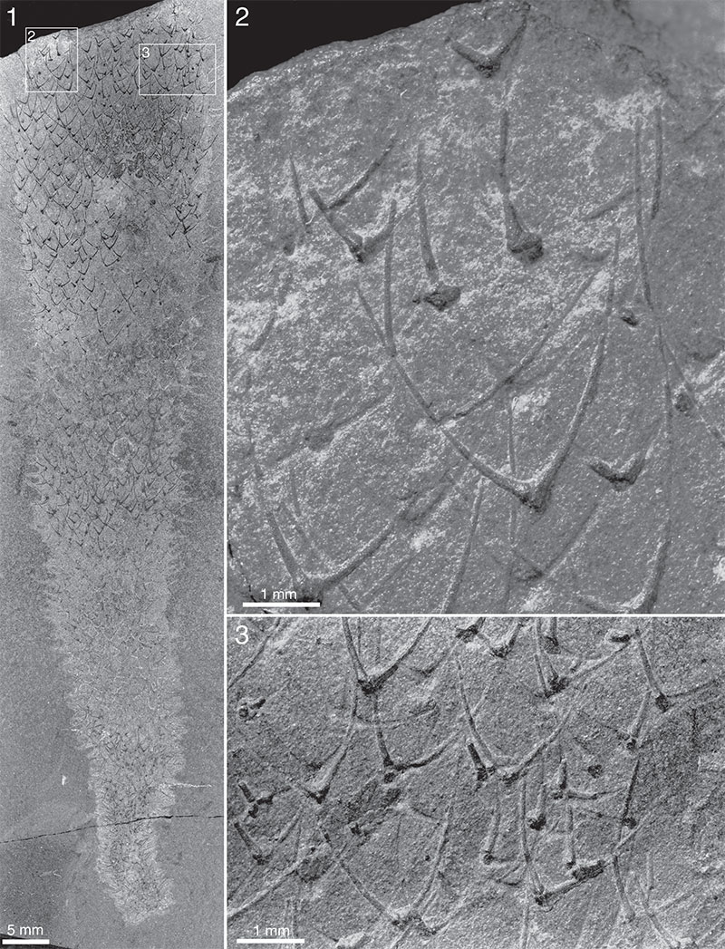
FIGURE 3. Allonnia tintinopsis n.sp., USNM 66528, upper part of body (specimen figured as Chancelloria eros by Walcott, 1920, pl. 88:1d, 1e). 1. Wet. 2. Detail of 1 (position marked by frame), wet. 3. Detail of 2 (position marked by frame), wet. 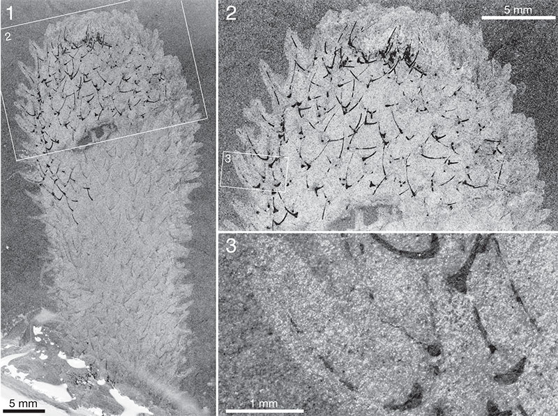
FIGURE 4. Archiasterella coriacea n.sp. USNM 66527 (specimen figured as Chancelloria eros by Walcott,1920, pl. 88:1c, and de Laubenfels, 1955, figure 76). 1. Dry. 2. Detail of 1 (position marked by frame), dry.
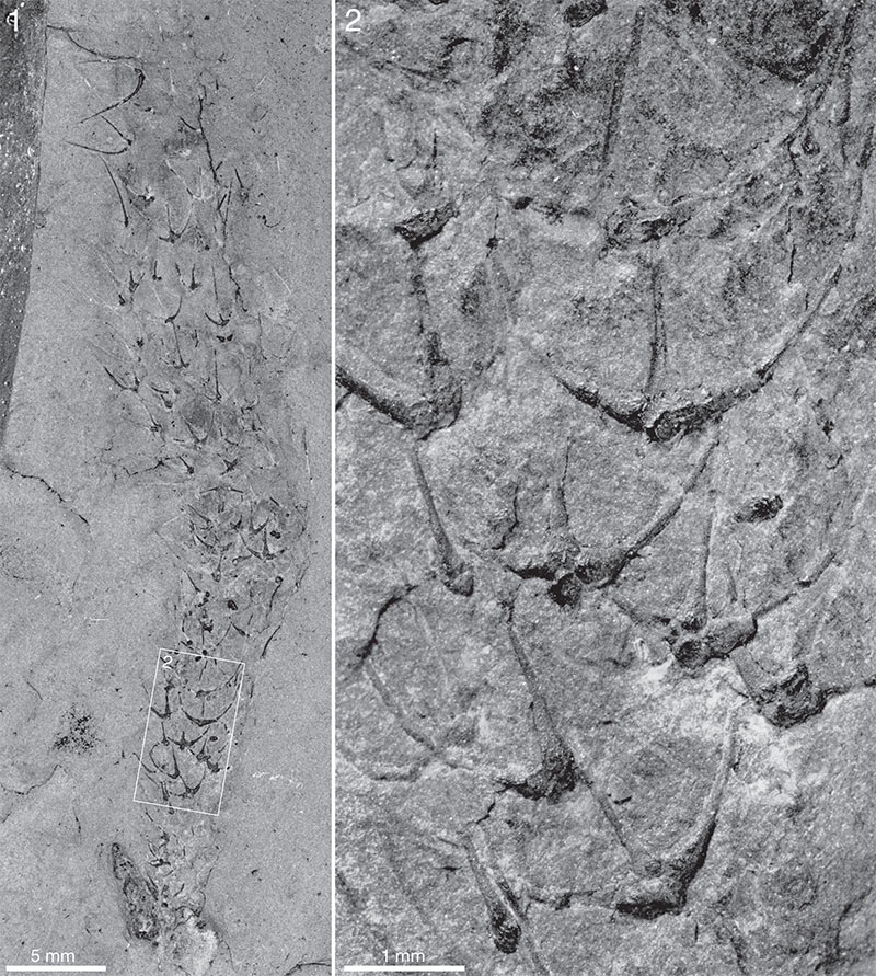
FIGURE 5. Chancelloria eros Walcott, 1920. 1. ROM 62538, ST talus, wet. Arrow points to empty area that may represent an apical orifice. 2. Detail of 1 (position marked by frame), wet. 3. ROM 62535A, BW -300, dry.
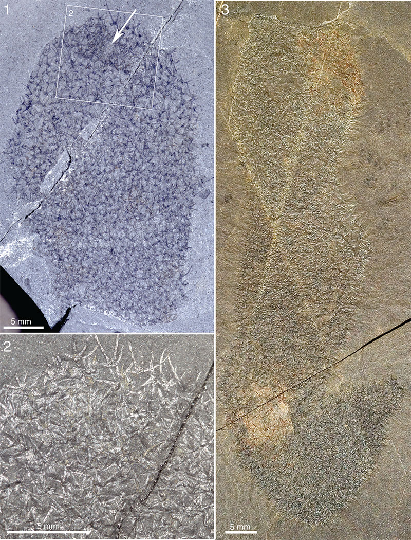
FIGURE 6. Chancelloria eros Walcott, 1920. ROM 62539, BW -320. 1. Dry. 2. Detail of 1 (position marked by frame), wet. 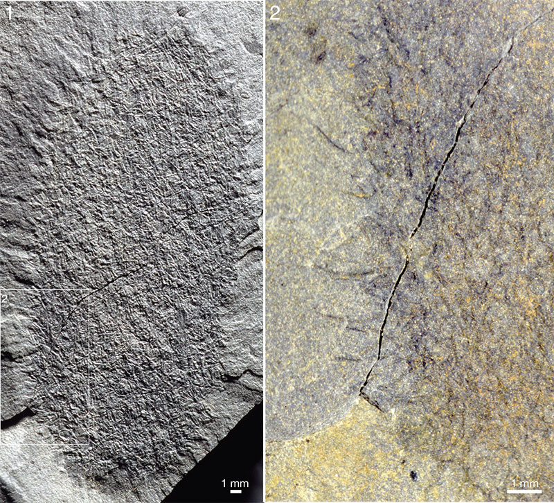
FIGURE 7. Chancelloria eros Walcott, 1920. ROM 62588B, BW -320. 1. Dry. 2. Detail of 1 (position marked by frame), dry. 3. Detail of 1 (position marked by frame), wet.
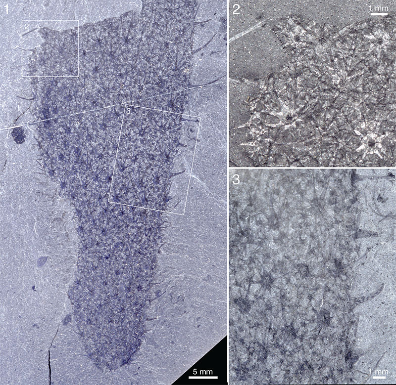
FIGURE 8. Chancelloria eros Walcott, 1920. 1, 2. ROM 49580, ST. 1. Dry. 2. Detail of 1 (position marked by frame), dry. 3. ROM 62590, ST, dry. 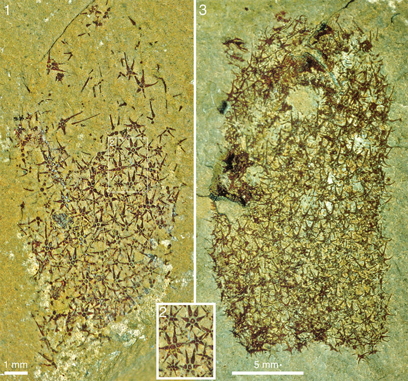
FIGURE 9. Chancelloria eros Walcott, 1920. 1 - 3. ROM 57604, BW -130. Specimens of the lingulate brachiopod Acrothyra gregaria appear to be attached to the spines of the Chancelloria. The brachiopods are concentrated to what is interpreted as the apical part of the chancelloriid body. 1. Wet. 2. Detail of 1 (position marked by frame), dry. 3. Detail of 2 (position marked by frame), dry. 4. ROM 63055, ST, dry. 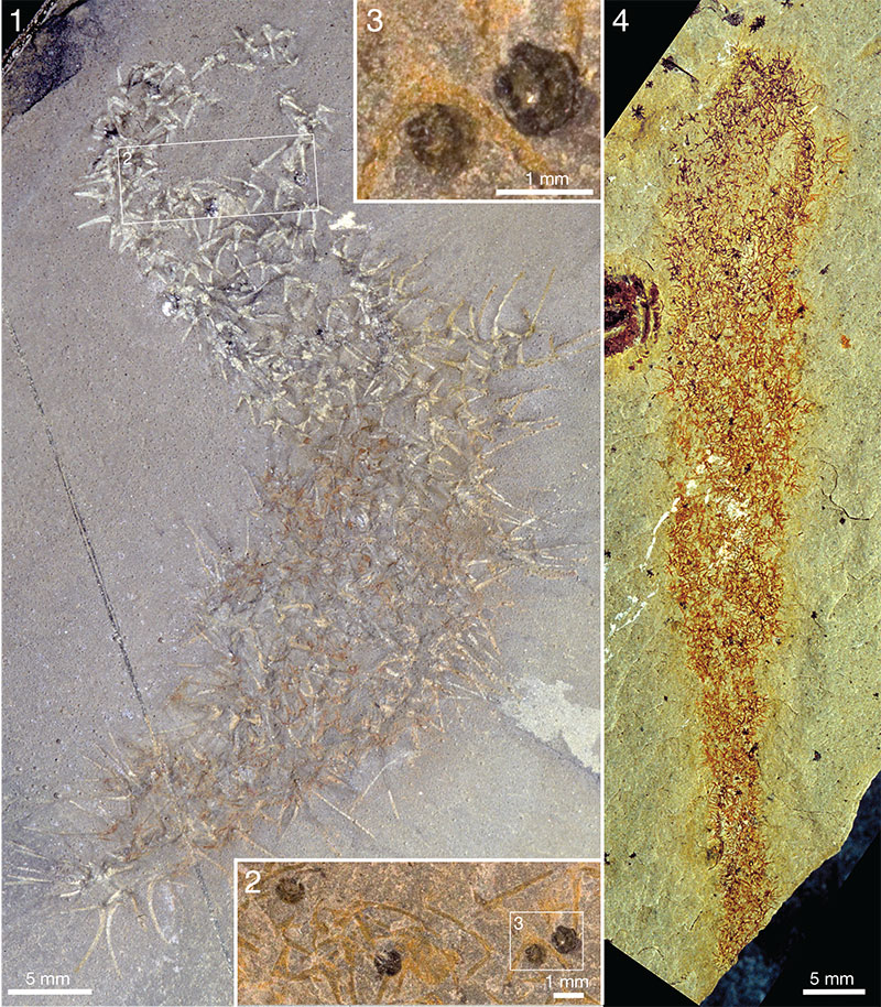
FIGURE 10. Chancelloria eros Walcott, 1920. ROM 62534, RT. 1. Wet. 2. Detail of 1 (position marked by frame), wet. Note the common presence of 3+0 Allonnia -type sclerites (arrows) in a scleritome dominated by 5–7+1 Chancelloria-type sclerites. 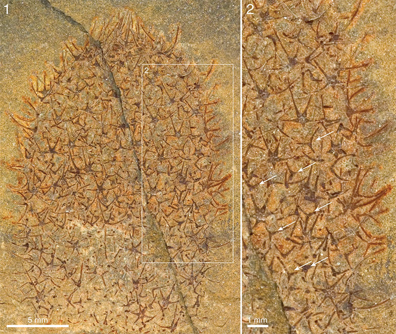
FIGURE 11. Chancelloria eros Walcott, 1920. ROM 49599A, RQ 8.0. Arrows point to examples of 3+0 sclerites. 1. Dry. 2. Detail of 1 (position marked by frame), dry. 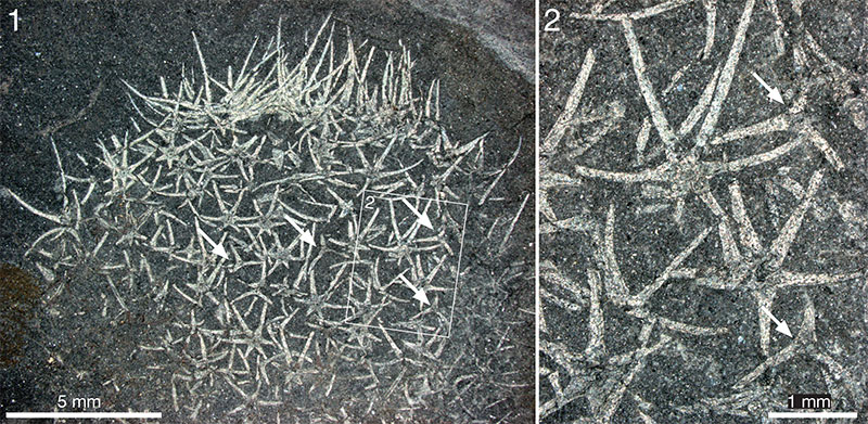
FIGURE 12. Allonnia tintinopsis n.sp. intergrown with or superimposed on Chancelloria eros Walcott, 1920. 1 - 3. ROM 49605(2) (intergrown with a specimen of Vauxia), RQ 9.2. 1. Same as figure 12B in Bengtson, 2000; see that paper for details on settings. 2, 3. Details of 1 (positions marked by frames), dry. 4 - 6. ROM 62537B, RQ 8.4. 4. Wet. 5, 6. Details of 4 (positions marked by frames), dry. Net-like patches are fragments of Micromitra shells. 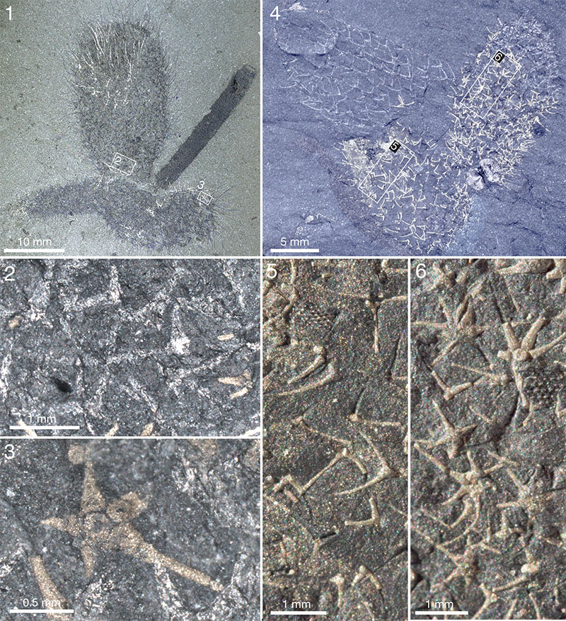
FIGURE 13. Chancelloria eros Walcott, 1920. 1. ROM 49578, ST, dry. 2. ROM 43126, ST, dry.
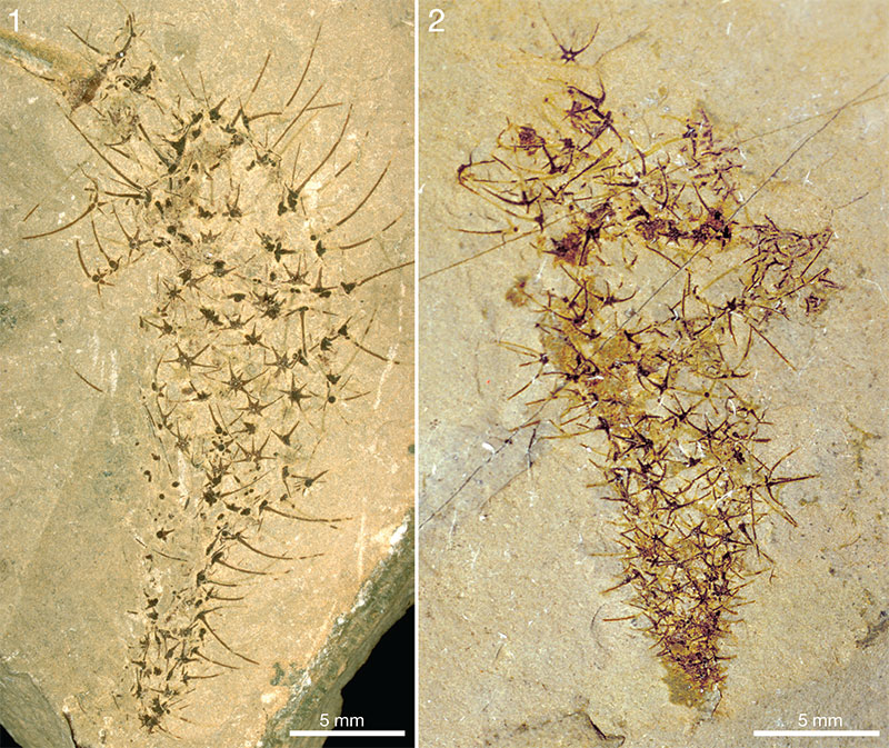
FIGURE 14. Chancelloria eros Walcott, 1920 (1, 2), and C. cf. eros (3-5), ST. 1. ROM 49574, dry. 2. ROM 49572, dry. 3 - 5. ROM 49576, dry. Items 4 and 5 show details of the upper and lower portions, respectively, of 3.
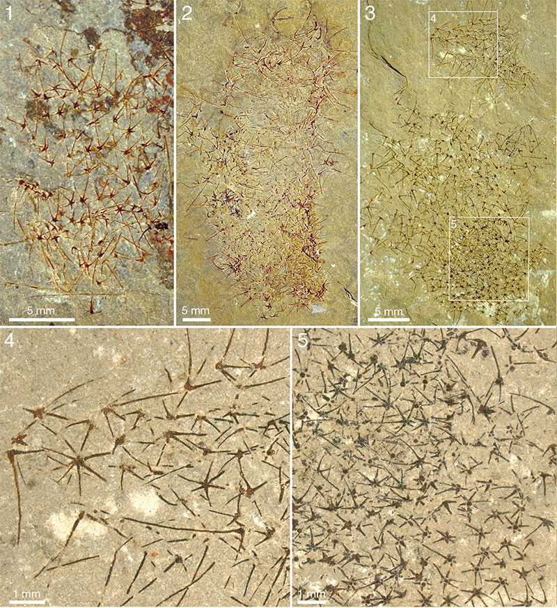
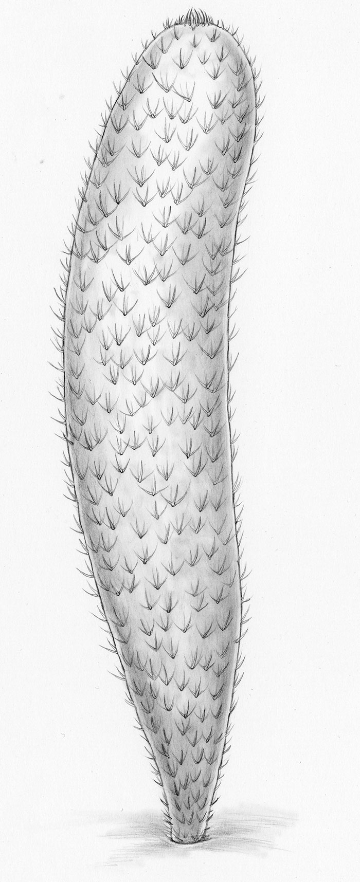
FIGURE 15. Life reconstruction of Chancelloria eros, Walcott, 1920. Artwork Pollyanna von Knorring.
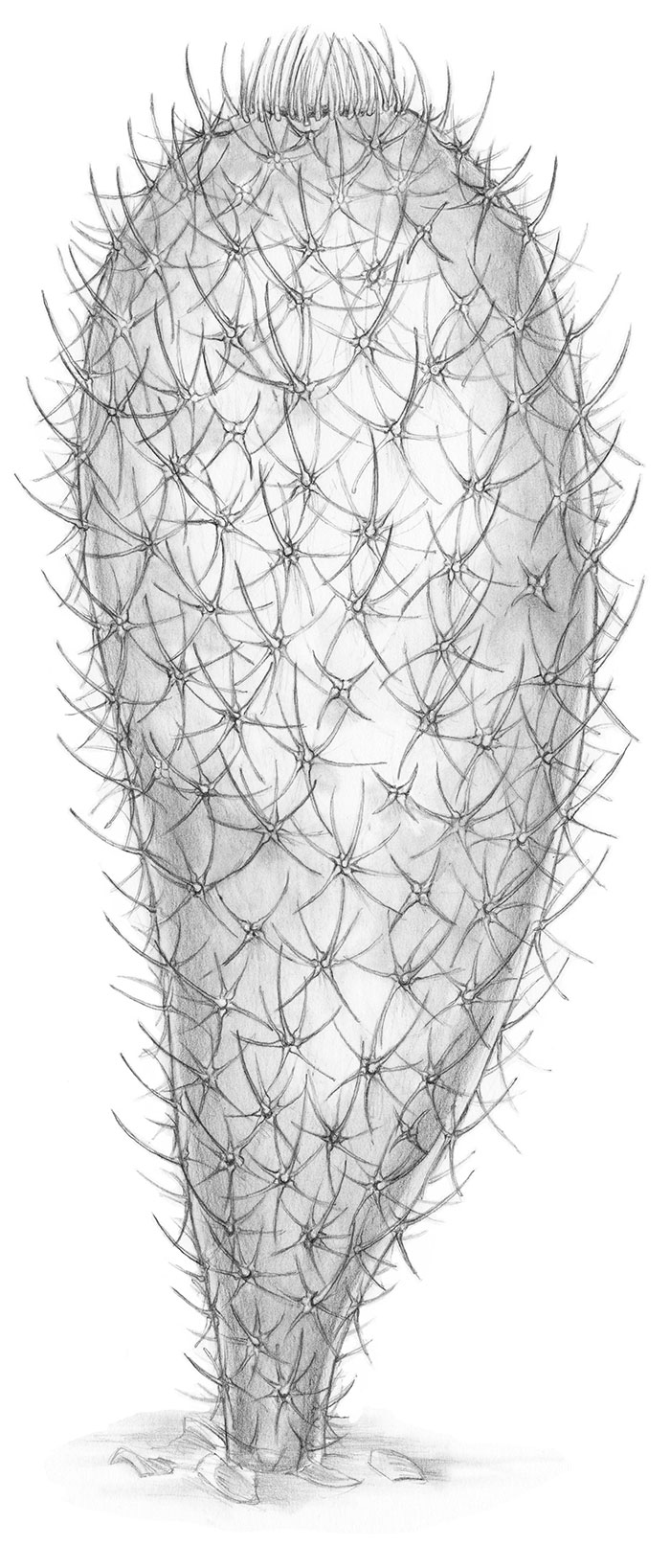
FIGURE 16. Allonnia tintinopsis n.sp., ROM 62527, part (1, 2) and counterpart (3), BW -110. 1. The larger specimen to the right in 1 is the holotype (ROM 62527[1]). 2. Holotype, detail of apical part (position marked in 1), with tuft. 3. Detail of lower part of body of holotype (counterpart; position on part marked in 1), showing morphology and arrangement of sclerites. Small letters at mid right denote lateral (L) and ascending (A) rays of selected sclerites. All pictures taken wet. 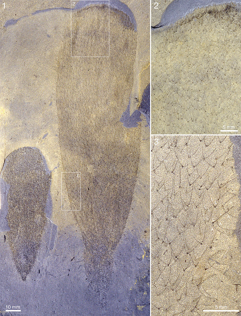
FIGURE 17. Allonnia tintinopsis n.sp. Apical tufts and concentration gradient of pyrite. 1. ROM 62524A (also in Figure 21.1), RQ 8.7, wet. 2. ROM 62516B, RQ 8.8, wet. 3. ROM 62519B, RQ 8.2, wet. 4. ROM 62586, wet. 5. ROM 62517, RQ 8.8, wet. 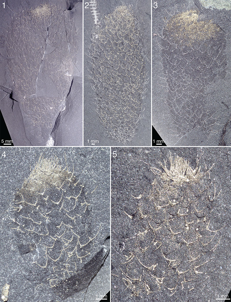
FIGURE 18. Ray length against distance from base in Allonnia tintinopsis n.sp. ROM 49610A, RQ 11.4 (left; white squares) and ROM 49607A, RQ 10.7 (right; black circles). 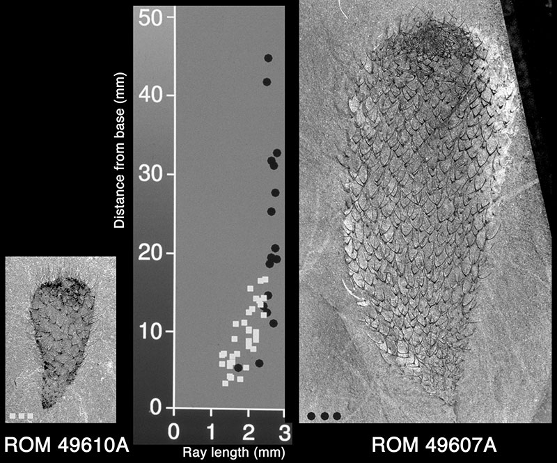
FIGURE 19. Allonnia tintinopsis n.sp. with stalk-like constriction. ROM 49573, ST. 1. Complete specimen, dry. 2. Detail of 1 (position marked by frame), dry, showing lower part of stalk with densely packed sclerites and debris.
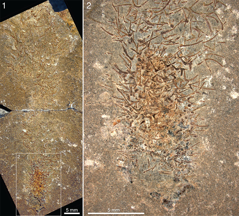
FIGURE 20. Allonnia tintinopsis n.sp. with stalk-like constrictions. 1. ROM 49616B, RQ 9.1, dry. 2. ROM 49596A, RQ 8.4, dry. 3, 4. ROM 49603, RQ 8.9. 3, Dry/pol. 4 . Detail of 3 (position marked by frame), dry/pol.
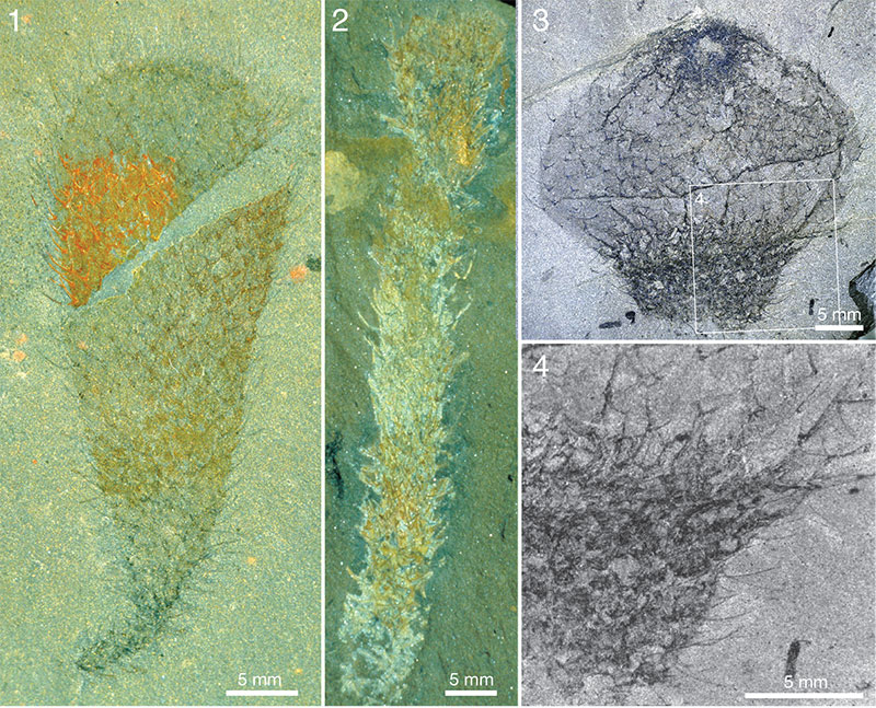
FIGURE 21. Allonnia tintinopsis n.sp. Apical tufts and orifices. 1. ROM 62524A (also in Figure 17.1), RQ 8.7, dry. 2. ROM 49608a, RT, dry/pol. 3. ROM 49608b, RT, dry/pol. 4. ROM 49607A, RQ 10.7, wet/pol. 5. ROM 62525B, RQ 8.4, dry. 6. ROM 62525B, RQ 8.4, dry/pol. 7. ROM 62520B (also in Figure 24.3), RQ 9.0, wet. 8, 9. ROM 62587, RQ 8.4, wet/pol (8), wet (9). 10. ROM 49614B, RQ 9.0, dry/pol. 11, 12. ROM 49620B (counterpart to the upper right specimen in Figure 27.3), RQ 9.0, 11. Dry/pol. 12. Detail of 11 (position marked by frame), dry/pol. Scale bars in all pictures equal 1 mm.
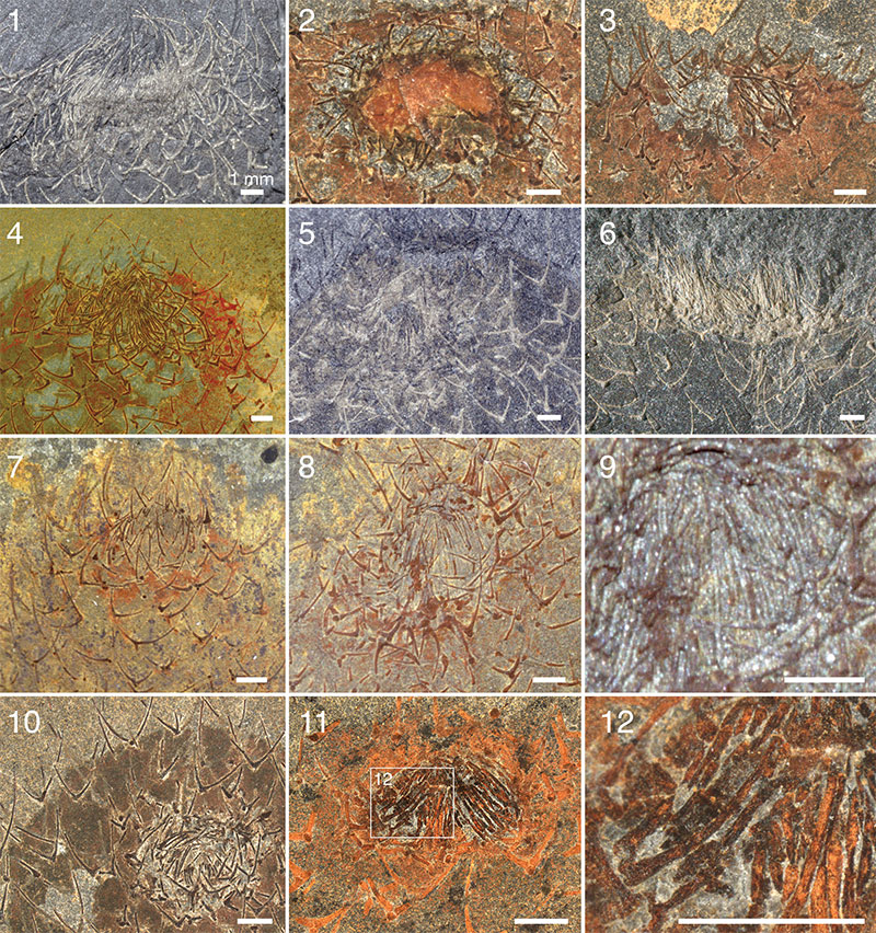
FIGURE 22. Allonnia tintinopsis n.sp. ROM 49612A, RQ 9.8, dry/pol. 1. Apical part of body, with tuft. 2, 3. Details of 1 (positions marked by frames). 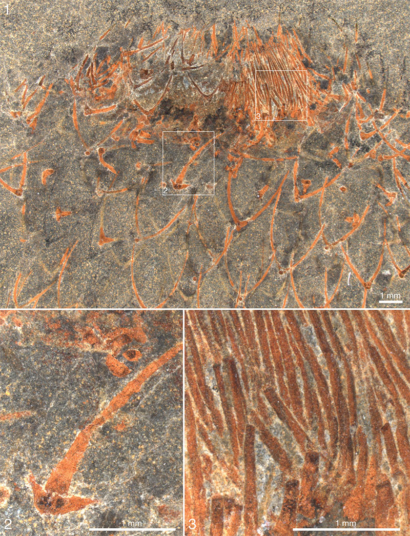
FIGURE 23. Allonnia tintinopsis n.sp. Apical tufts. 1. ROM 49605(1), RQ 9.2, wet/pol. 2, 3. ROM 62591, UE. 2. Wet. 3. Detail of 2 (position marked by frame), wet. 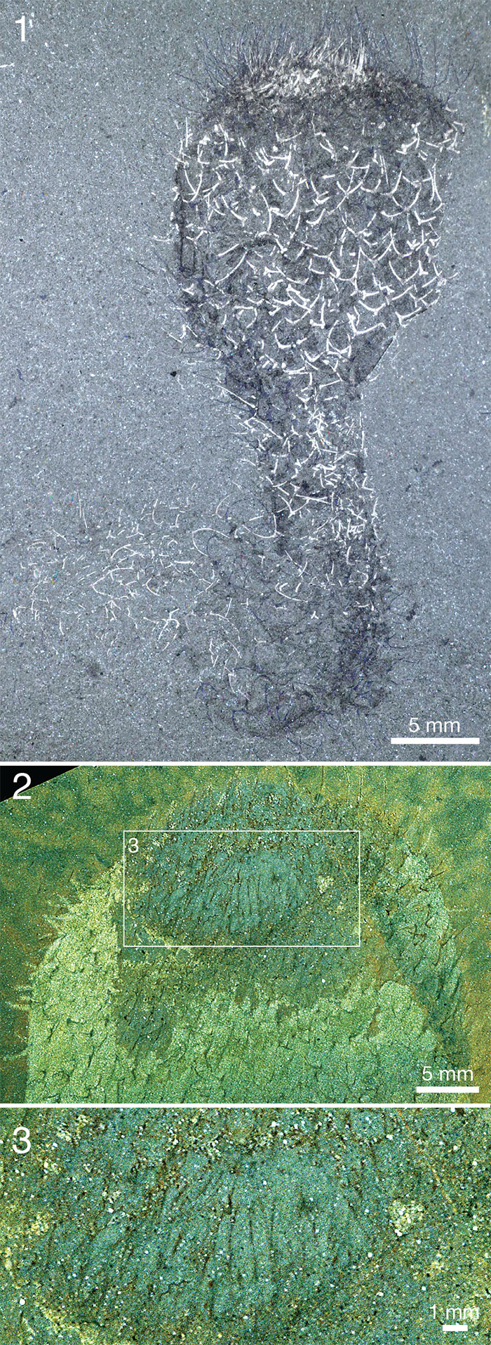
FIGURE 24. Allonnia tintinopsis n.sp. intergrown with Vauxia (1, 2) and having Micromitra attached to the spines (1, 3, 4). 1. ROM 49588B, RQ 9.0, wet/pol. 2. ROM 49606, RQ 8.9, wet. 3. ROM 62520B (also in Figure 21.7), RQ 9.0, wet. 4. ROM 62515B, RQ 8.8, wet.
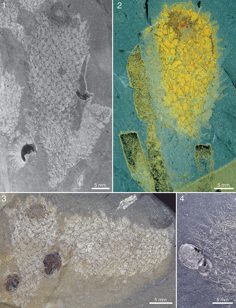
FIGURE 25. Allonnia tintinopsis n.sp. associated with Vauxia. 1. ROM 49587B, RQ 8.3, dry. 2. ROM 49584A, RQ 8.1, wet.
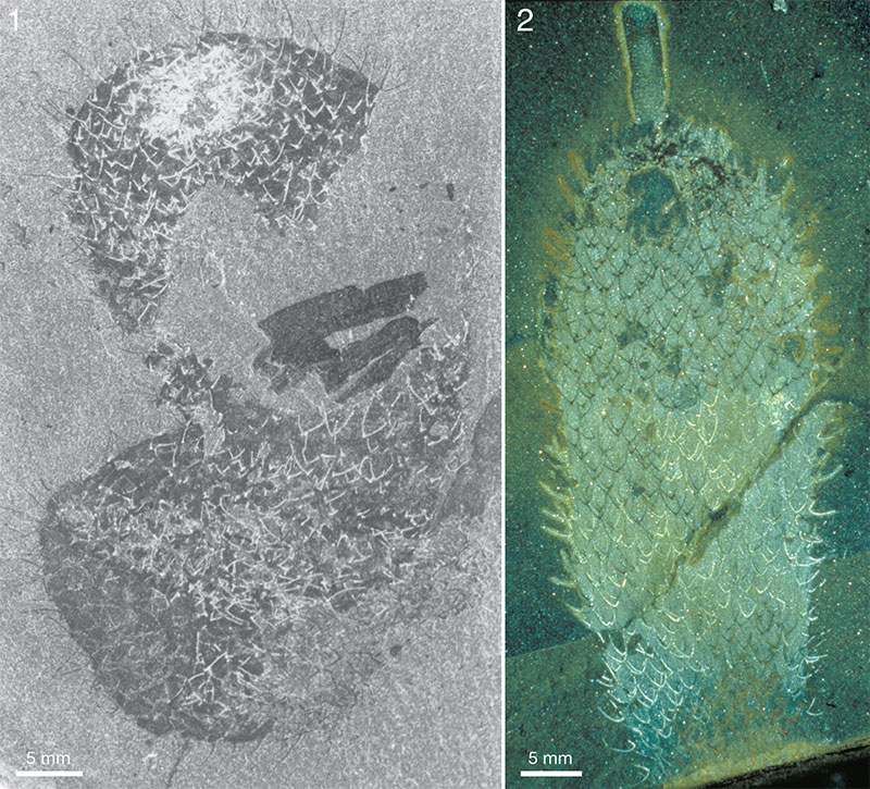
FIGURE 26. Allonnia tintinopsis n.sp. ROM 57574, RQ 8.5. Apical tuft and orifice. 1. Dry. 2 - 4. Details of preceding pictures (positions marked by frames), dry.
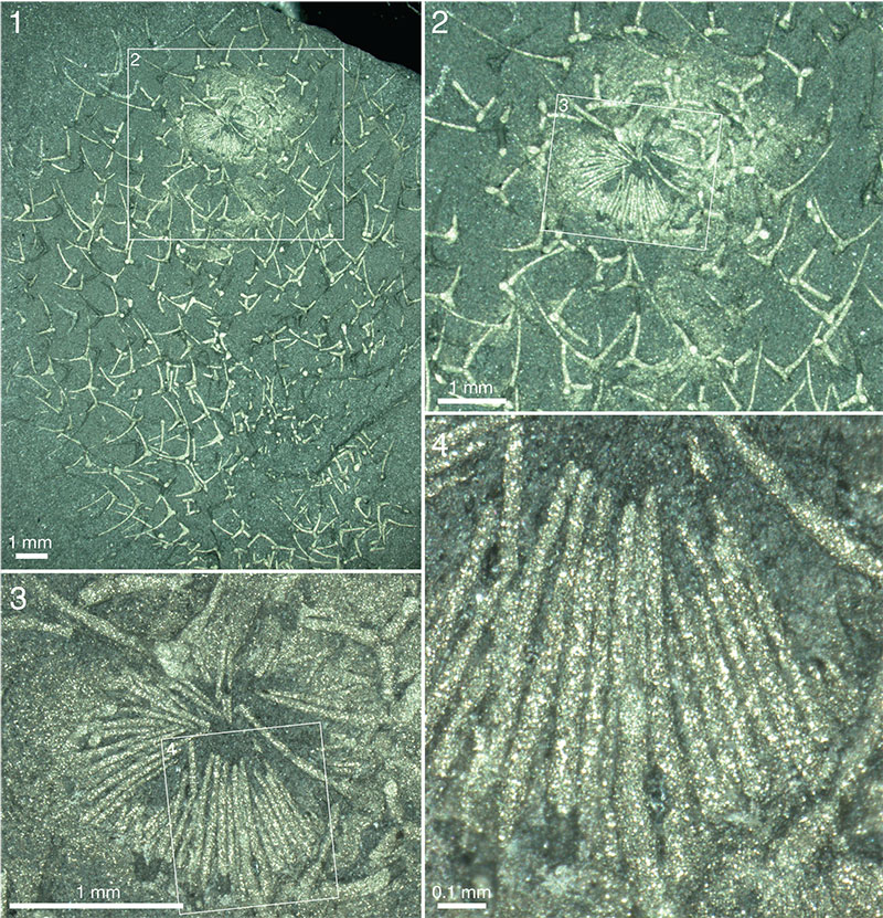
FIGURE 27. Allonnia tintinopsis n.sp., attachment to Vauxia. 1 - 2. ROM 62523A, RQ 8.4. 1. Wet. 2. Detail of 1 (position marked by frame), wet. 3. ROM 49620A (upper right-hand specimen is part to the one in Figure 21.11-12), RQ 9.0, wet. 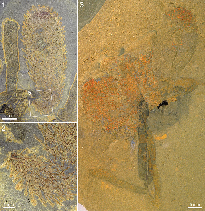
FIGURE 28. Allonnia tintinopsis n.sp., two specimens on the same slab showing probable attachment to hyoliths ( 1 , wet) and Vauxia ( 2, wet). ROM 62518A, RQ 8.2. Inset in 2 (dry) shows blowup of isolated, probably extraneous, Chancelloria -like sclerite in Allonnia scleritome. 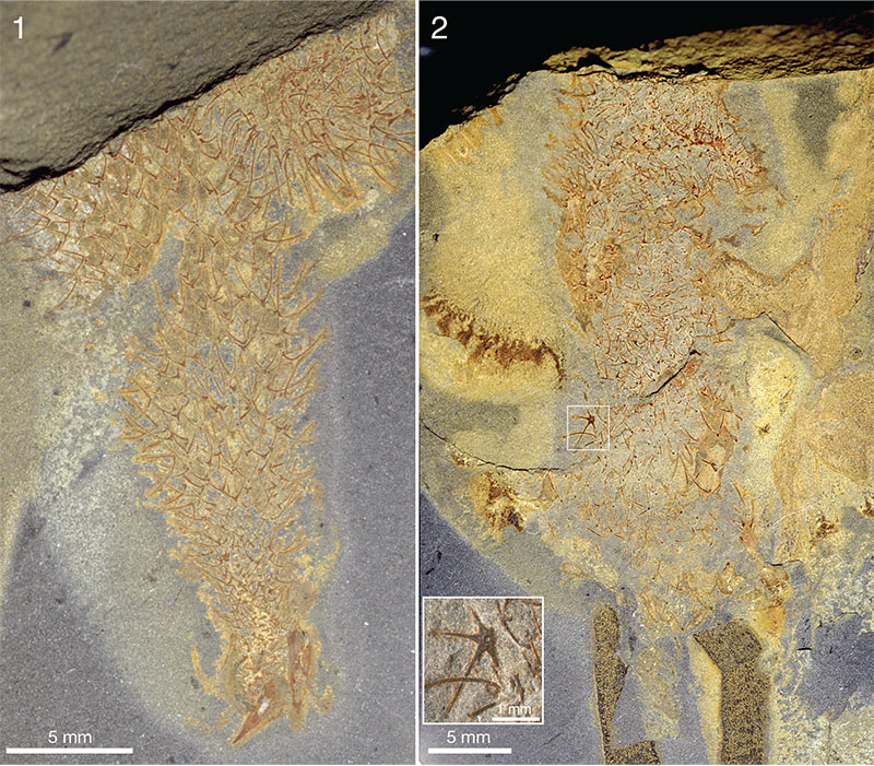
FIGURE 29. Intergrowths of Allonnia tintinopsis n.sp. and Vauxia. Part, ROM 62526A (2) and counterpart, ROM 62526B (1, 3), RQ 8.4. 1. Wet. 2. Detail of part (position marked by frame in 1), wet. 3. Detail of 1 (position marked by frame), wet.
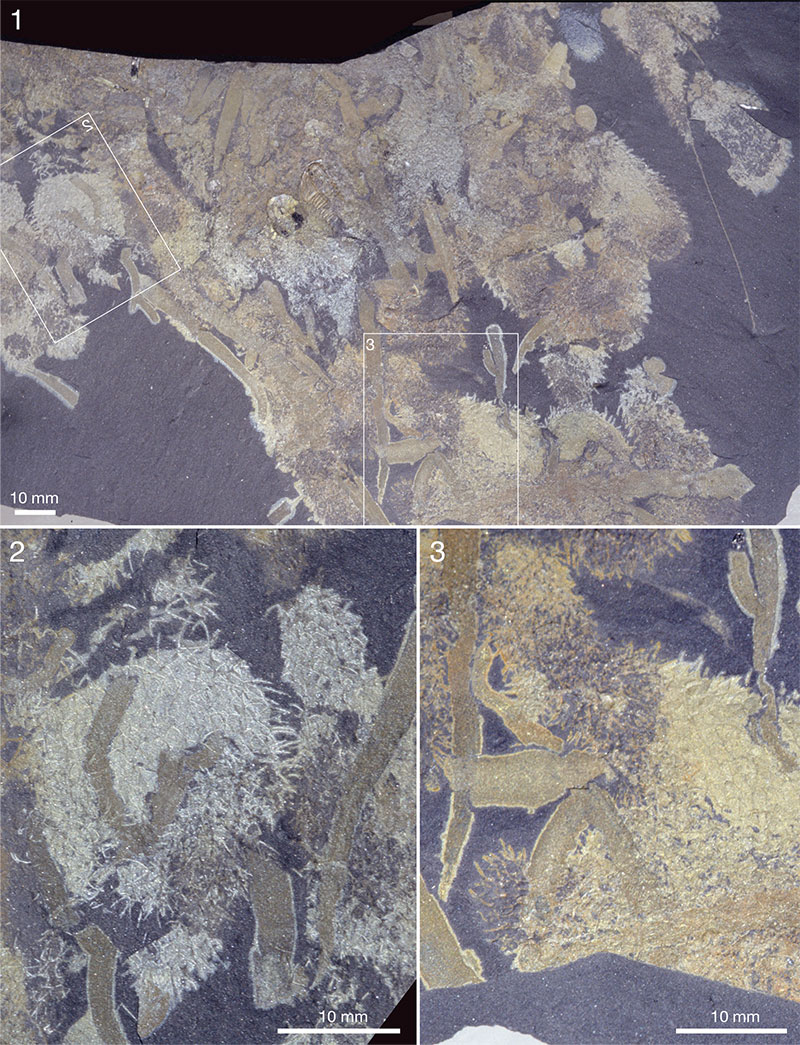
FIGURE 30. Allonnia tintinopsis n.sp, apical orifice with pyrite concentration. 1. ROM 49609A, RQ 9.0, dry. 2. ROM 62536, BW -400, wet/pol. 3. ROM 49582B, RQ 9.0, wet/pol. 4. ROM 49601A, RQ 9.7 (figure 1A2 in Bengtson, 2000; see that paper for details on settings).
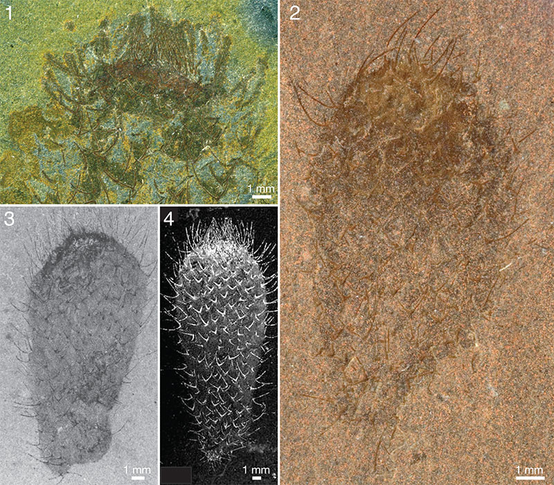
FIGURE 31. Integument structure of Allonnia and Archiasterella. 1. Allonnia tintinopsis n.sp. ROM 62514B, RQ 9.8, dry. 2, 3. Allonnia tintinopsis n.sp., ROM 49600A, UE. 2. Dry. 3. Detail of 2 (position marked by frame), dry. 4. Archiasterella coriacea n.sp., holotype, ROM 62531A, BW -130, dry. 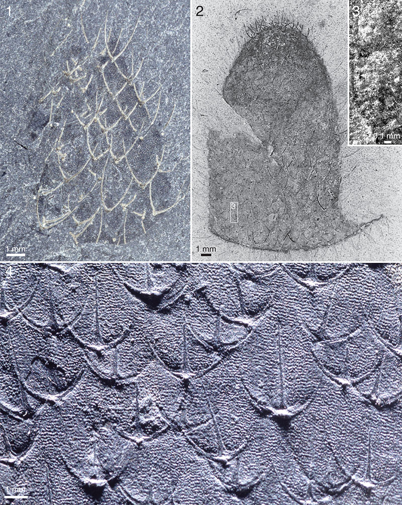
FIGURE 32. Allonnia tintinopsis n.sp. 1. ROM 49615A, WT, dry/pol. 2. ROM 49621, UE, dry. 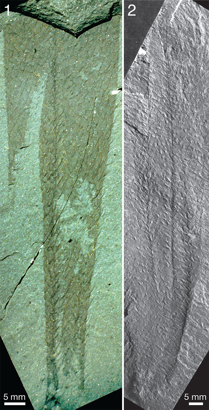
FIGURE 33. Association of Allonnia tintinopsis n.sp., bodies and attachments, on bedding slab. ROM 62585, RQ 8.4. 1. Part; arrow points to specimen represented by counterpart in 3, dry. 2. Tracing of bodies (red; darker tones toward the apex) and root bulbs (grey), based on both part and counterpart. 3. Counterpart of specimen with root bulb marked by arrow in 1, dry.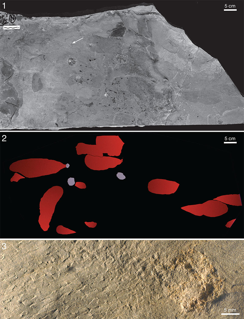
FIGURE 34. Two Allonnia tintinopsis n.sp. associated with a Vauxia. Ottoia to the right. ROM 49589A, RQ 9.0, wet.
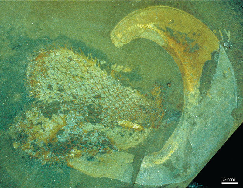
FIGURE 35. Intergrowth of Allonnia tintinopsis n.sp. with Vauxia. ROM 62521B, RQ 8.4. 1. Dry. 2. Detail of 1 (position marked by frame), dry.
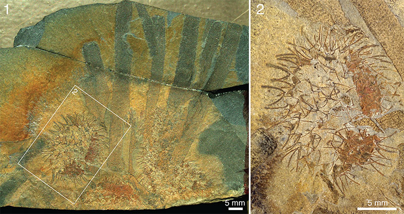
FIGURE 36. Allonnia tintinopsis n.sp. together with Micromitra shells and carapace of Isoxys. ROM 49602B, RQ 8.7, dry.
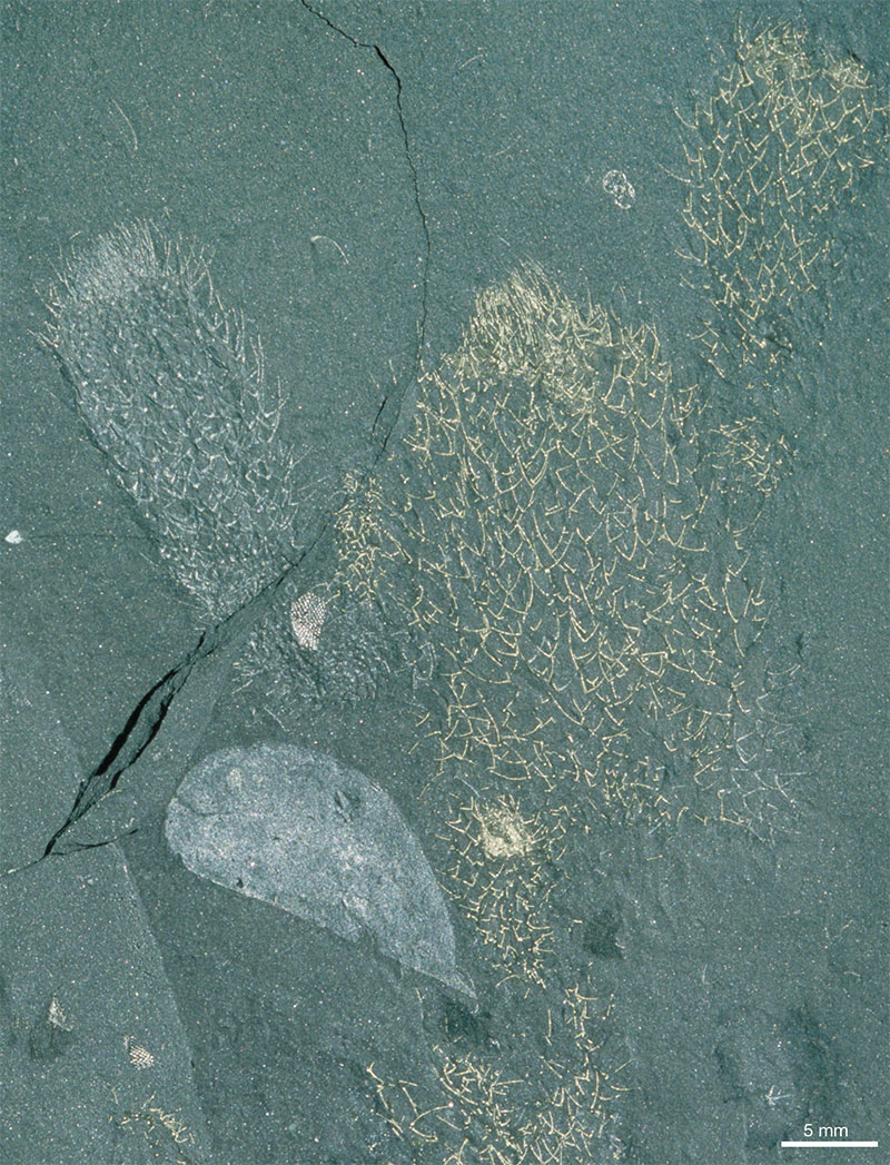
FIGURE 37. Life reconstruction of Allonnia tintinopsis, n.sp., growing on the sponge Vauxia. Artwork Pollyanna von Knorring.
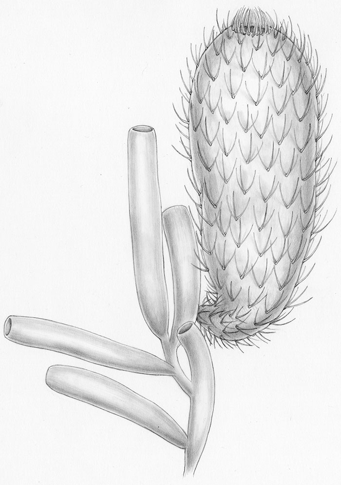
FIGURE 38. Archiasterella coriacea n.sp., holotype, ROM 62531A (same specimen as in Figures 31.4 and 39), BW -130. 1. Complete specimen showing alignment of sclerites, wet. 2. Detail of 1 (position marked by frame) showing the apical tuft, wet. 3. Detail of 1 (position marked by frame) showing the attitude of sclerites relative to the body wall, wet.
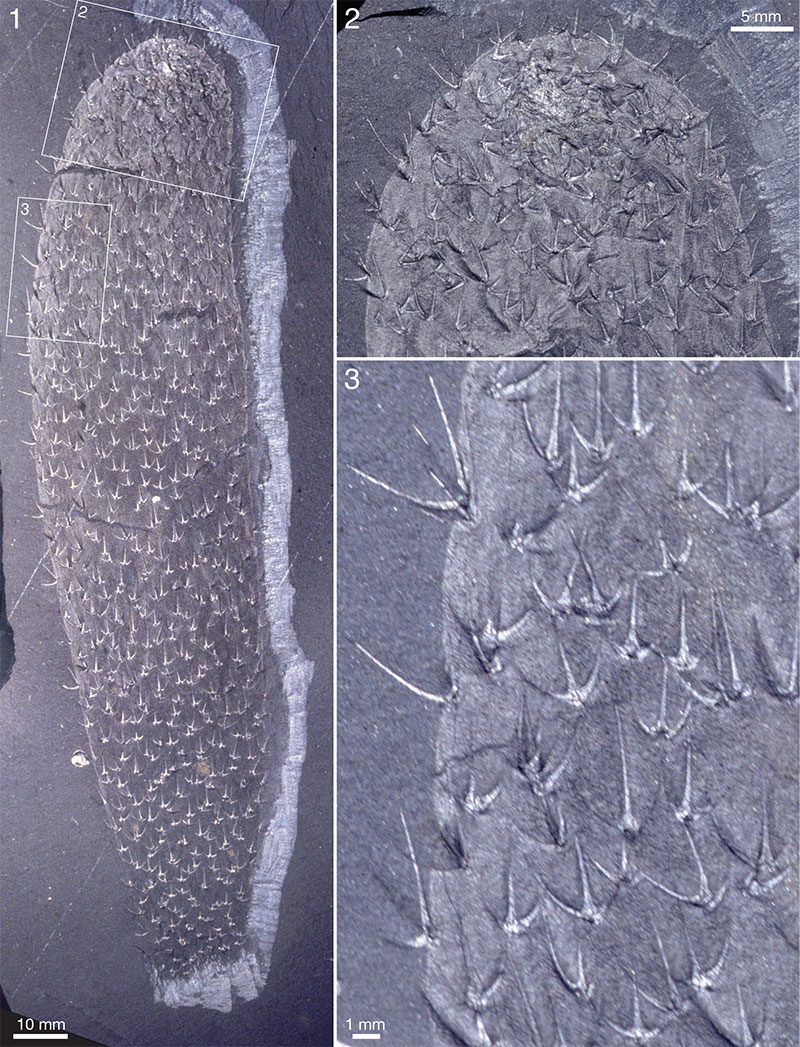
FIGURE 39. Archiasterella coriacea n.sp., holotype, ROM 62531A, BW -130, superposition of sclerites from two sides of body. △ - basal side down; ▼ - basal side up. Cf. Figures 31.4 and 38.
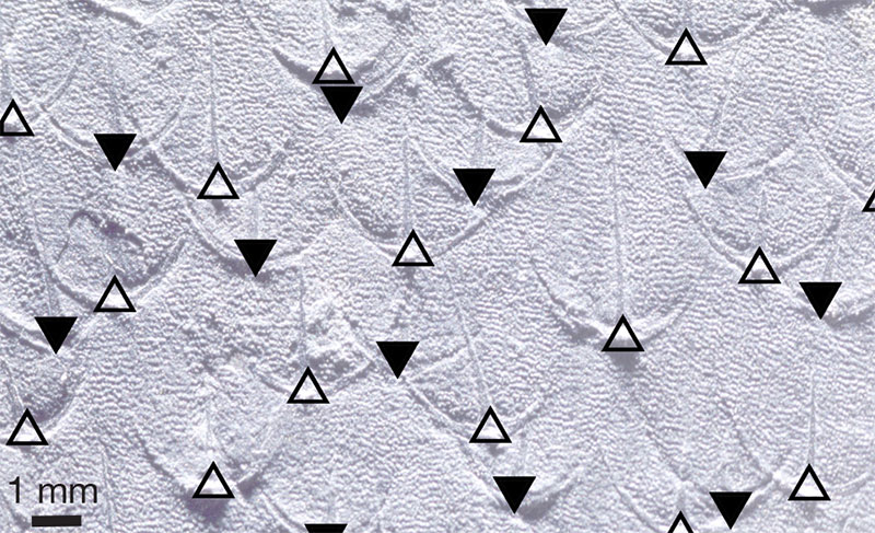
FIGURE 40. Archiasterella coriacea n.sp, ROM 57573, WQ -130. 1. Wet. 2, 3. SEM stereographs (red/blue anaglyphic images) of latex casts taken of counterpart to show structure of integument. Frames show positions of details 2 and 3.
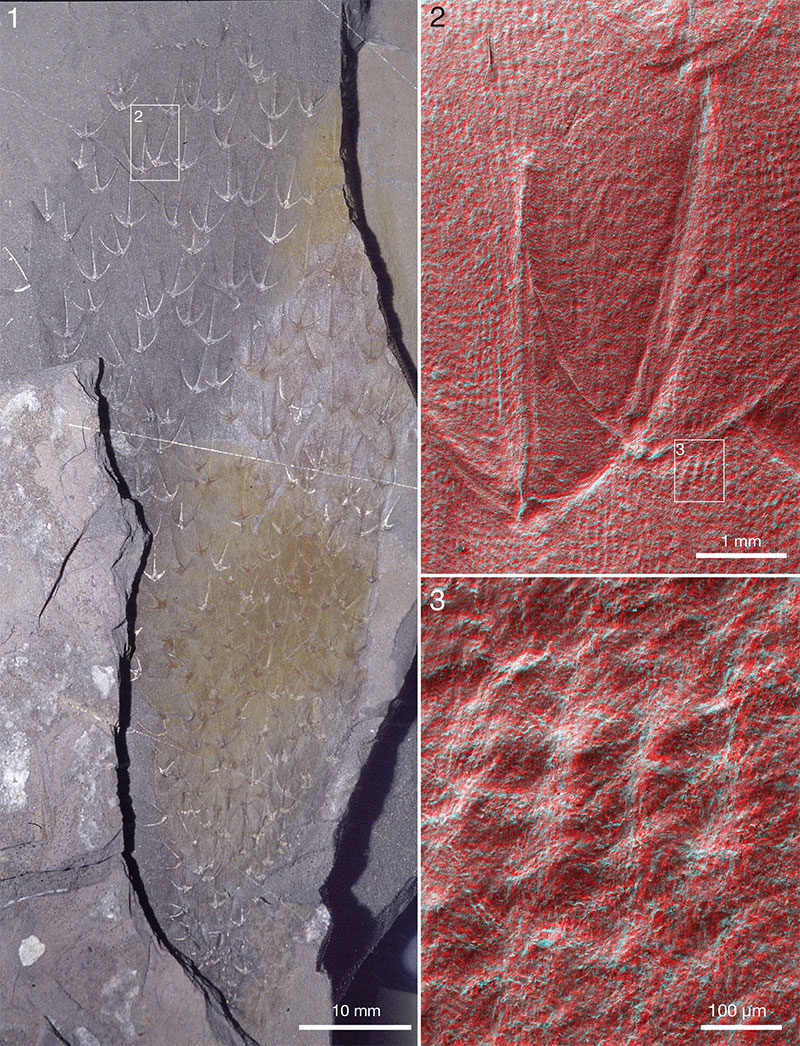
FIGURE 41. Archiasterella coriacea n.sp. 1, 2. ROM 62529B, BW -130. 1. Dry. 2. Detail of 1 (position marked by frame), wet/pol. 3. ROM 62530A, BW -130, wet. 4. ROM 62532, BW -130, apical part, wet.
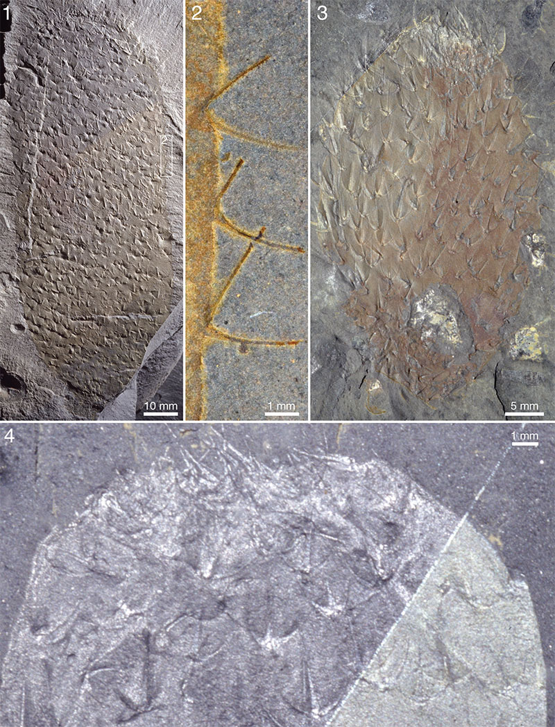
FIGURE 42. Archiasterella coriacea n.sp. 1, 2. ROM 62528A, RQ 8.4. 1. Showing distribution of sclerites, wet. 2. Showing integument, wet/pol. 3. ROM 62533A, BW -130, wet. 4. ROM 49583B, RQ 9.0, wet. Specimen attached to a Vauxia.
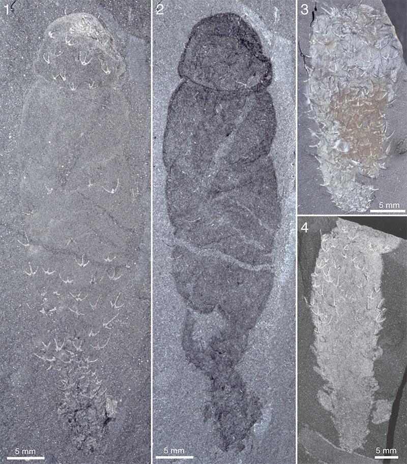
FIGURE 43. Archiasterella coriacea n.sp. ROM 49617B, RQ 13.8. 1. Complete specimen, wet. 2. Detail of upper specimen in 1 (position marked by frame), wet. 3. Detail of 1 (position marked by frame), wet, showing stalk uniting upper and lower specimen.
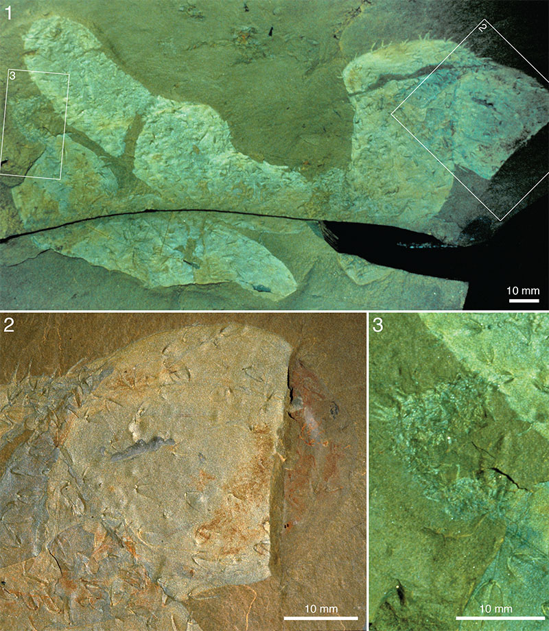
FIGURE 44. Archiasterella coriacea n.sp. 1. ROM 49619A, RQ, wet. 2. ROM 49567A, UE, wet. Arrow points to tuft.
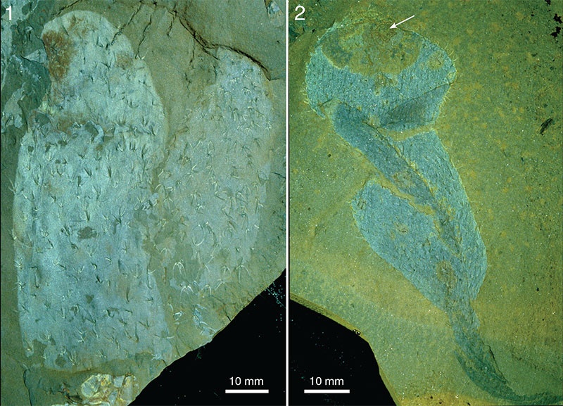
FIGURE 45. Life reconstruction of Archiasterella coriacea, n.sp. Artwork Pollyanna von Knorring.