

APPENDIX 1.
List of characters used in the phylogenetic analysis. Characters indicated by an asterisk are ordered in the analysis.
1.* Number of upper incisors (modified from Springer et al., 1997, Rougier et al., 1998, Wroe et al., 2000, Horovitz and Sánchez-Villagra, 2003 and Black et al., 2012): (0) More than three; (1) Two or Three; (2) One.
2. Size of I 1 relative to I 2 (Black et al., 2012): (0) Equal or similar in length; (1) Large, elongate.
3. Upper canine (modified from Springer et al., 1997, Horovitz and Sánchez-Villagra, 2003 by Black et al., 2012): (0) Present; (1) Absent or vestigal.
4.* Number of upper premolars (Black et al., 2012): (0) Three; (1) Two; (2) One.
5. Posterobuccal cusp on P 3 (Woodburne et al., 1987; Black and Archer, 1997; Myers and Archer, 1997; Black et al., 2012): (0) Absent; (1) Present.
6. Enamel extending down the buccal surface of P 3 and onto the root: (0) Absent; (1) Present.
7.* Size of P 3 relative to M 1 (modified from Black and Archer, 1997 and Black et al., 2012): (0) P 3 length greater than M 1 length; (1) P 3 length less than or equal to M 1 length; (2) P 3 length less than or equal to 80% of M 1 length.
8.* Molar morphology (Black et al., 2012): (0) Tribosphenic; (1) Selenodont; (2) Semi-lophodont/bunodont; (3) Lophodont but stylar cusps still evident on lophs; (4) Fully lophodont.
9. Hypselodont cheek teeth (modified from Murray, 1998; Black et al., 2012): (0) Absent; (1) Present.
10. Enamel noticeably thicker on trailing edge (lingual surface on upper molars and buccal surface on lower molars) relative to leading edge of molars (buccal surface on upper molars and lingual surface on lower molars): (0) Absent; (1) Present.
11.* Presence of enamel tracts that extends down the lingual surface of upper molars and buccal surface of lower molars: (0) Absent; (1) Moderate enamel tracts / changes in enamel height around the tooth; (2) Extensive enamel tracts / changes in enamel height around the tooth.
12. Anteriorly concave lower / anteriorly convex upper molar lophs (Black et al., 2012): (0) Absent; (1) Present.
13. M 1 with decrease in enamel height on lingual surface at constriction or just posterior to this: (0) Absent; (1) Present.
14.* Parastyle development on M 1 (Black and Archer, 1997; Black et al., 2012): (0) Absent/small; (1) Moderately developed expansion of anterobuccal tooth corner; (2) Large, cuspate and pyramidal.
15. Presence of a cusp on posterobuccal side of M 1 (and occasionally M 2) in position of St-E: (0) Absent; (1) Present.
16.* St-C larger and better developed than St-B on M 2 : (0) Absent; (1) Similar in size; (2) Present.
17. Presence of crest connecting paracone to St-B on M 2 : (0) Absent; (1) Present.
18.* Separation of stylar cusps C and D (modified from Myers and Archer, 1997 and Black et al., 2012): (0) Close together; (1) Separated; (2) Separated by a large trough.
19. Postprotocrista (Archer, 1984; Black et al., 2012): (0) Present; (1) Absent.
20. M 2 width of anterior moiety versus tooth length (modified from Springer et al., 1997; Horovitz and Sánchez-Villagra, 2003 by Black et al., 2012): (0) M 2 longer than wide; (1) M 2 width is greater or equal to length.
21. M 4 metaconule (Black et al., 2012): (0) Absent / significantly reduced and retracted towards posterior cingulum; (1) Distinct / cuspate.
22. Posteriorly increasing (upper) molar gradient (modified from Black, 2007 and Black et al., 2012): (0) Absent, M 4 / M 1 is less than or equal to 1; (1) Present, M 4 / M 1 is greater than 1.
23.* Number of lower incisors (modified from Springer et al., 1997, Rougier et al., 1998, Wroe et al., 2000, Luo et al., 2002, Horovitz and Sánchez-Villagra, 2003 and Black et al., 2012): (0) Two or more; (1) One.
24. Inclination angle of I 1 (Marshall et al., 1990; Springer et al., 1997; Horovitz and Sánchez-Villagra, 2003; Black, 2007; Black et al., 2012): (0) High (greater or equal to 30 degrees); (1) Low (less than 30 degrees).
25. Development of paraconid and paracristid on M 1 (modified from Myers and Archer, 1997, Horovitz and Sánchez-Villagra, 2003 by Black, 2007; Black et al., 2012): (0) Paraconid present and paracristid well-developed; (1) Paraconid weak or absent; paracristid present and distinct; (2) Paraconid and paracristid absent.
26. Protostylid on M 1 (Woodburne et al. 1987; Black and Archer 1997; Myers and Archer 1997; Springer et al. 1997; Black 2007): (0) Absent; (1) Present.
27. Cristid obliqua (modified from Black, 2007 and Black et al., 2012): (0) Well-developed, meets or approaches postprotocristid lingual to horizontal tooth midline; (1) Well-developed, meets or approaches postprotocristid buccal to horizontal tooth midline; (2) Weak / absent.
28. Distinct and separate paraconid on M 2-4 : (0) Absent; (1) Present.
29. Paraconid located on lingual side of tooth on M 2-3 (or preprotocristid / paracristid terminates on lingual side of tooth): (0) Present; (1) Absent, located close to midline or buccal side of tooth.
30. Paraconid connected to protoconid via crest on M 2-3 (or well-developed preprotocristid / paracristid): (0) Present; (1) Absent.
31. Presence of a crest connecting entoconid and hypoconid: (0) Absent; (1) Present.
32. Incisive foramen posterior (modified from Dawson, 1983b; Murray, 1998; Tedford, 2002): (0) Absent; (1) Present, the incisive foramina lie posteriorly, just anterior to P 3 , in a deep pocket at the rear of the diastema. The posterior position of the foramina correspondingly displaces the premaxillary-maxillary suture to a position just anterior to the cheek tooth.
33. Lateral edge of palate emarginated from I 1 to P 3 in ventral view (modified from Tedford, 2002): (0) Absent; (1) Present.
34. Palate width opposite first molar (ventral view) (Merrilees, 1967; Scott et al., 1988): (0) Narrow, less than buccolingual width of M 1 ; (1) Wide, equal to or greater than buccolingual width of M 1.
35. Median premaxillary process (Tedford, 2002): (0) Absent; (1) Well-developed or present as a low spine at the midline dorsal surface of the premaxilla.
36.* Position of infraorbital foramen: (0) Close to level of alveolar process; (1) Approximately midway between dorsal surface of skull and alveolar process; (2) Closer to dorsal surface of skull than alveolar process of tooth row.
37.* Posterior palatal vacuities (maxillopalatine vacuity) (modified from Archer, 1984, Reig et al., 1987, Rougier et al., 1998, Wroe et al., 2000, Horovitz and Sánchez-Villagra, 2003 and Black et al., 2012): (0) Extends anteriorly to a point anterior of M 3; (1) Confined within palate, opposite M 3-4; (2) Absent.
38.* Nasal aperture retracted beyond incisor arcade (modified from Black et al., 2012): (0) Absent or just posterior to incisor arcade; (1) Retracted to above diastema (or level of premaxillary-maxillary suture); (2) Retracted to above cheek tooth row.
39. Nasomaxillary suture length relative to nasopremaxillary suture length (dorsal view) (Scott et al., 1988): (0) Nearly same or greater; (1) Far less than nasopremaxillary suture length.
40. Separate left and right parietal ridges along entire length of parietal bone: (0) Absent; (1) Present.
41.* Masseteric process (Wroe et al., 1998; modified version in Black et al., 2012): (0) Absent; (1) Short; (2) Elongate.
42. Dorsoventral position of ventral extent of anterior zygomatic root (relative to the cheek tooth row): (0) Close to level of alveolar process; (1) Distant to alveolar process.
43. Anteroposterior position of ventral extent of anterior zygomatic root (relative to the cheek tooth row): (0) Above M 3 or M 4; (1) Above junction between M 2 and M 3 ; (2) Above M 2; (3) At junction between M 1 and M 2 or anterior to M 2.
44. Infraorbital shelf (Black et al., 2012): (0) Well-developed; (1) Weak.
45. Postglenoid process (Springer et al., 1997; Horovitz and Sánchez-Villagra, 2003; modified version in Black et al., 2012): (0) Present; (1) Absent.
46. Tympanic cavity roof elements (modified from Travoullion et al., 2010 and Black et al., 2012): (0) Alisphenoid (no squamosal contribution); (1) Alisphenoid and squamosal; (2) Squamosal (no alisphenoid contribution).
47. Tympanic floor elements (Black et al., 2012): (0) Alisphenoid; (1) Alisphenoid and squamosal; (2) Squamosal.
48.* Alisphenoid tympanic wing (modified from Springer et al., 1997, Wroe et al., 1998, Horovitz and Sánchez-Villagra, 2003 and Black et al., 2012): (0) Absent; (1) Short; (2) Moderate to elongate, extends under periotic.
49. Epitympanic fenestra developed in postglenoid cavity (Black et al., 2012): (0) Absent; (1) Present.
50. Non-auditory sinuses (Murray, 1986; Black et al., 2012): (0) Absent; (1) Present.
51. Posterior epitympanic fossa (modified from Murray, 1986; modified from Black et al., 2012): (0) Absent / weak; (1) Moderate to deep.
52.* Postglenoid cavity / epitympanic fossa (modified from Murray, 1998, Tedford, 2002, Louys, 2004 and Black et al., 2012): (0) Absent; (1) Present, but shallow; (2) Present and moderately extensive / deep.
53.* Rostral tympanic process of periotic (Rougier et al., 1998; Sánchez -Villagra and Wible, 2002; Horovitz and Sánchez-Villagra, 2003 modified version; Black et al., 2012): (0) Strong; (1) Weak / absent.
54. Posterior parietal width (Black et al., 2012): (0) Broad; (1) Narrow.
55. Interparietal (Louys et al., 2009; Black et al., 2012): (0) Present; (1) Absent.
56. Narrow mastoid strip on occiput (Murray, 1986; Black et al., 2012): (0) Absent; (1) Present.
57. Ventrolaterally flared mastoid process on occiput (Murray 1986; Black et al., 2012): (0) Absent; (1) Present.
58.* Length of articular surface (mandible) relative to length of cheek tooth row (modified from Scott et al., 1988): (0) Short, ratio of 0.4 or less; (1) Medium, ratio of 0.5 to 0.6; (2) Long, ratio of 0.7 or greater.
59.* Forward extent of ectalveolar groove of mandible (Dawson, 1983a): (0) Posterior of M 4; (1) Junction between M 3 and M 4 to mid M 4; (2) Adjacent to M 3.
60.* Depth of masseteric fossa (mandible) (Merrilees, 1967; Scott et al., 1988): (0) Shallow to medium; (1) Deep; (2) Very deep.
61. Angle of the anterior border of the ascending ramus (mandible) (modified from Hope and Wilkinson, 1982; Black, 2007; Black et al., 2012): (0) < 70°; (1) > or = 70°.
62.* Posterior extent of mandibular symphysis (mandible) (Black, 2007; Black et al., 2012): (0) Anterior to P 3; (1) Below P 3; (2) Below M 1; (3) Below M 2-3.
63.* Diastema length between I 1 and P 3 (modified from Black, 2007 and Black et al., 2012): (0) < 30% horizontal ramus length; (1) Approximately 30% horizontal ramus length; (2) > 30% horizontal ramus length.
64. Distance between anterior edge of P 3 and posterior edge of M 4 relative to length of horizontal ramus (mandible) (Black et al., 2012): (0) > 60%; (1) < or = 60%.
65. Flared masseteric eminences (mandible) (Murray, 1998; Black et al., 2012): (0) Absent / weak; (1) Moderately to strongly flared.
66. Masseteric foramen (Black, 2007; Black et al., 2012): (0) Absent; (1) Present.
67. Fused mandibular symphysis: (0) Absent; (1) Present.
68. Cervical vertebrae: combined length of C3-C7 divided by width of C7 (Munson, 1992): (0) > 1.0 (long neck); (1) < 1.0 (short neck).
69. Humerus: max. width across distal end divided by length (Munson, 1992): (0) < or = 0.3; (1) > 0.3.
70. Humerus: deltoid ridge (Munson, 1992): (0) Has no lateral overhang, or a very minor one; (1) Has a large flaring overhang.
71.* Humerus: deltoid tuberosity (modified from Scott and Richardson, 1988): (0) Angle between deltoid and pectoral crests < 30°; (1) Angle between 30° and 40°; (2) Angle > 40°.
72. Ulna: anteroposterior thickness at semilunar notch divided by bone length (modified from Munson, 1992): (0) < or = 0.10; (1) > 0.10.
73. Ulna: length of olecranon process divided by bone length (Munson, 1992): (0) < 0.20; (1) > or = 0.20.
74. Ulna: max. anteroposterior thickness at coronoid process divided by bone length (Munson, 1992): (0) < 0.2; (1) > or = 0.2.
75. Radius: length (Munson, 1992): (0) > length of humerus; (1) < or = length of humerus.
76. Radius: anteroposterior thickness of distal end divided by bone length (Munson, 1992): (0) < or = 0.1; (1) > 0.1.
77. Cuneiform: position relative to scapholunar (Munson, 1992; Weisbecker and Archer, 2008): (0) Directly lateral to scapholunar; (1) More proximal than the scapholunar.
78. Cuneiform: ridge between ulnar and pisiform facets (Weisbecker and Archer, 2008): (0) Middle section sinks below the rim of the cuneiform; (1) High rising throughout, straight.
79. Pisiform: area of ulnar facet (Munson, 1992; Weisbecker and Archer, 2008): (0) < area of cuneiform facet; (1) > or = area of cuneiform facet.
80. Pisiform: width divided by thickness (Munson, 1992): (0) < 0.5; (1) > or = 0.5.
81. Scaphoid: distal surface (Weisbecker and Archer, 2008): (0) Single facet, smooth; (1) Separated into two distinct facets.
82. Scaphoid: distolateral process (Weisbecker and Sánchez-Villagra, 2006): (0) Absent; (1) Present.
83. Unciform: area of MC V facet (Munson, 1992; Weisbecker and Archer, 2008): (0) < MC IV facet; (1) > or = area of MC IV facet.
84. Unciform: hamate process (modified from Munson, 1992, Weisbecker and Sánchez-Villagra, 2006 and Weisbecker and Archer, 2008): (0) Large, hooked and curves medially; (1) Reduced, not as hooked and does not curve as far medially.
85.* MC I: max. width divided by length (modified from Munson, 1992 by Weisbecker and Archer, 2008): (0) < 0.4 ; (1) 0.4 to 0.5; (2) > 0.5.
86. MC II: proximal end (Munson, 1992; Weisbecker and Archer, 2008): (0) Mediolaterally compressed; (1) Expanded and concave.
87. MC III: facet for MC IV (modified from Munson, 1992): (0) Proximal only to ligament pit (no dorsal component); (1) Dorsal to ligament pit (no proximal component); (2) Dorsal and proximal to ligament pit.
88. MC III: facet for MC IV (modified from Munson, 1992 by Weisbecker and Archer, 2008): (0) Facing laterally or only slightly distally slanted; (1) Strongly distally slanted or completely facing distally.
89. MC III: magnum facet (modified from Munson 1992): (0) Grooved or concave; (1) Convex or flat to slightly convex.
90. MC III: MC II facet (Munson, 1992; Weisbecker and Archer, 2008): (0) Separate, distinct, and lateral to the magnum facet; (1) Continuous with the magnum facet.
91.* MC V: max. width of distal end divided by length (modified from Munson, 1992 and Weisbecker and Archer, 2008): (0) < 0.4; (1) 0.4 to 0.5; (2) > 0.5.
92.* Proximal phalanges II to V of manus, IV and V only in pes: mid-shaft width divided by length (modified from Munson, 1992 and Weisbecker and Archer, 2008): (0) < 0.3; (1) 0.3 to 0.4; (2) > 0.4.
93. Proximal phalanges, manus or pes: distal ends (Munson, 1992; Weisbecker and Archer, 2008): (0) Not tapered; (1) Dorsoventrally tapered, so that the articulation for the medial phalanx faces ventrally.
94.* Medial phalanx III of manus or IV of pes: length divided by length of corresponding metapodial (modified from Munson, 1992 and Weisbecker and Archer, 2008): (0) < 0.30; (1) 0.30 to 0.40; (2) > 0.40.
95. Pelvis: dorsoventral height at symphysis divided by width at mid-ischium (Munson, 1992): (0) < 0.8; (1) > or = 0.8.
96. Pelvis: ilium (Munson, 1992): (0) Only slightly dorsoventrally flattened, or mediolaterally flattened; (1) Greatly dorsoventrally flattened.
97.* Pelvis: width of iliac crest divided by length of ilium (Munson, 1992): (0) < 0.30; (1) 0.30 to 0.50; (2) > 0.50.
98. Epipubics: width at mid-bone divided by length X 100 (Munson, 1992): (0) < 10; (1) > or = 10.
99. Femur: width of proximal end divided by length (Munson, 1992): (0) < 0.3; (1) > or = 0.3.
100. Tibia: length (Munson, 1992): (0) > or = length of femur; (1) < length of femur.
101. Fibula: lateral notch on distal end (Munson, 1992): (0) Clearly present; (1) Extremely slight or absent.
102. Fibula: width of proximal end divided by bone length (modified from Munson, 1992): (0) < 0.10; (1) > or = 0.10.
103. Fibula: femoral facet (Munson, 1992): (0) Not present; (1) Present.
104. Astragalus: lateral tibial facet (modified from Munson, 1992): (0) Mostly convex; (1) Mostly concave.
105. Astragalus: head (modified from Munson, 1992): (0) Wider than long; (1) Narrow, mediolaterally compressed, or length and width are equal.
106. Cuboid (Munson, 1992): (0) No contact; (1) Contacts astragalus.
107. Cuboid: area of MT V facet (Munson, 1992): (0) < MT IV facet; (1) > or = area of MT IV facet.
108. Ectocuneiform: length divided by height (Munson, 1992): (0) > or = 0.8; (1) < 0.8.
109. Entocuneiform: MT I facet (Munson, 1992): (0) Saddle-shaped (concavo-convex); (1) Convex.
110. Entocuneiform: navicular facet (Munson, 1992): (0) Shorter than or equal to mesocuneiform facet; (1) Longer than mesocuneiform facet.
111. Metatarsal I: entocuneiform facet (Munson, 1992): (0) Mostly convex; (1) Strong concave component.
112. MT II and III: mid-shaft width divided by bone length (Munson, 1992): (0) < 0.15 ; (1) > or = 0.15.
113. MT IV: proximal end (Munson, 1992): (0) Square; (1) Mediolaterally tapered.
114. MT V versus MT III (modified from Munson, 1992): (0) MT III longer that MT V; (1) MT V longer than MT III.
115.* MT V: max. width divided by length (modified from Munson, 1992): (0) < 0.50; (1) 0.50 to 0.70; (2) > 0.70.
APPENDIX 2.
Data matrix used for phylogenetic analysis. ? indicates missing data; - indicates inapplicable character; polymorphic states indicated by both states in brackets, e.g., (01).
APPENDIX 3.
Comparative dental measurements.
APPENDIX 4.
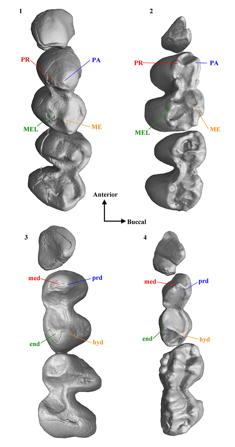
Cusp morphology of P3 to M2 of modern wombats compiled from CT scans of Lasiorhinus latifrons (SAM M4157 and SAM M1924) and Vombatus ursinus (NMV C10928 and NMV C3133). 1, Upper dentition of L. latifrons. 2, Upper dentition of V. ursinus. 3, Lower dentition of L. latifrons. 4, Lower dentition of V. ursinus. Abbreviations as follows: end refers to entoconid, hyd refers to hypoconid, ME refers to metacone, med refers to metaconid, MEL refers to metaconule, PA refers to paracone, PR refers to protocone, prd refers to protoconid.
APPENDIX 5.
List of unambiguous apomorphies. Node numbers correspond to Figure 7.2
