FIGURE 1. Anatomy of the hydrospires of the blastoid Pentremites rusticus. 1.1, Location of one of the five radially distributed hydrospires within the calyx, showing incurrent hydrospire pores, and excurrent spiracle (inferred direction of water flow indicated by the arrows). 1.2, Oblique view of a section of a hydrospire and associated structures. Modified from Schmidtling and Marshall (2010).
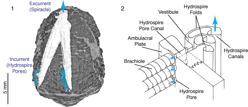
FIGURE 2. Schematic showing hypothesized flow patterns within the hydrospire folds. 2.1, In Hypothesis 1, the flow has an adoral component representing respiratory leakage. 2.2, In Hypothesis 2, the flow is entirely radial, without leakage. See text for further discussion.
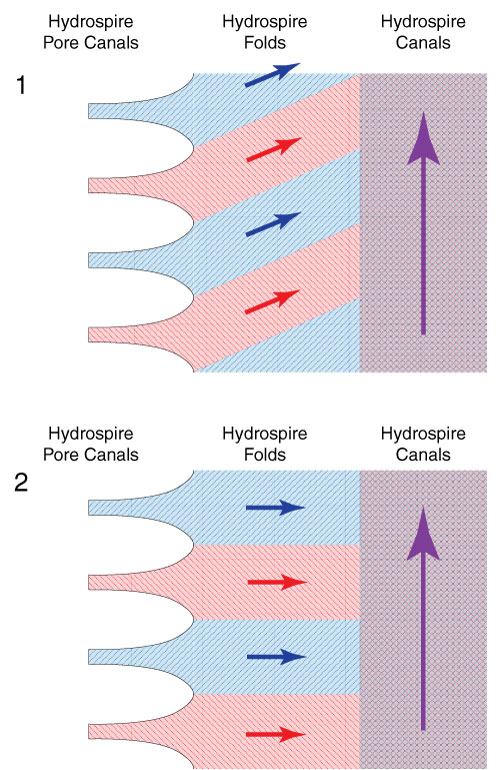
FIGURE 3. Digital and physical models use to visualize fluid flow. 3.1, Digital solid model of approximately the lower quarter of a hydrospire of Pentremites rusticus, using Blender (see text). 3.2, 3D-printed rendering of the digital model, shown with inlet headers connected.
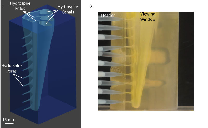
FIGURE 4. Visualization of the flow within the 3D printed model (Re = 0.376, see Table 1). Flow in the folds consists of horizontal bands of distinct red and blue color, indicating no adoral component to flow and no mixing within the folds, consistent with Hypothesis 2 (see text, Figure 2.2). The still used in the print version of this paper is a single frame from the flow pattern observed, showing the steady-state flow pattern after nine minutes of flow. The animation is sped up 16x (for video see supplementary material).