FIGURE 1. Left hyoid apparatus of the ground sloth Paramylodon harlani (modified from Stock, 1925). Shaded in grey is the fossil specimen analyzed in this work.
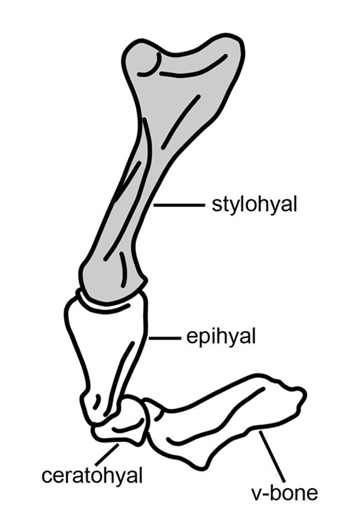
FIGURE 2. Juvenile stylohyal (CAV 476) in 1, medial view; 2, lateral view. The glittering of the cylindrical part is due to its having been bathed in gold for SEM observations; 3, close up of the cut mark analyzed by Fariña et al (2014); 4, close up of the marks in the midshaft. White arrows indicate probable trampling marks; black arrows indicate probable cut marks. Scale bar equals 50 mm.
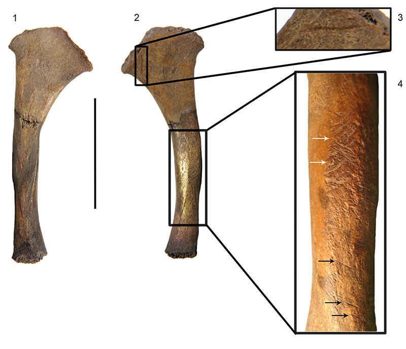
FIGURE 3. Three-dimensional reconstruction of the juvenile ground sloth stylohyal (CAV 476). Medial view (3D object) available in Appendix 2.
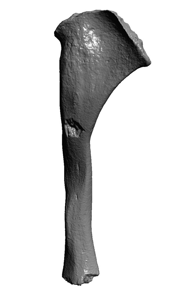
FIGURE 4. Three-dimensional reconstruction of the stylohyal of adult Glossotherium (MNHN 914). Medial view (3D object) available in Appendix 2.
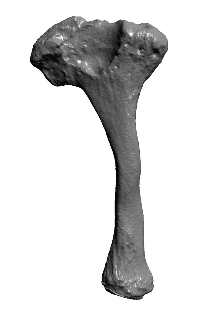
FIGURE 5. Stylohyals of 1, juvenile ground sloth (CAV 476); 2, adult Glossotherium (MNHN 914); 3, adult Scelidotherium .(MLP 3-671); 4, Adult Megatherium (MNHN PAM 297). Scale bar equals 50 mm.
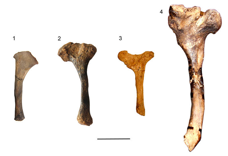
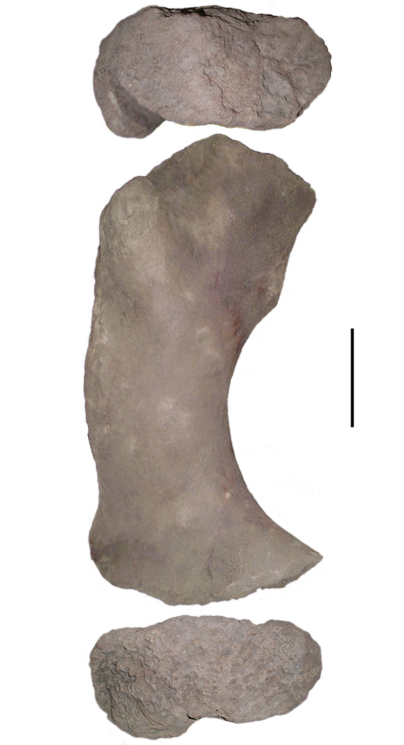
FIGURE 6. Left femur of juvenile Lestodon (CAV 935) in posterior view showing the proximal (top) and distal (bottom) irregular surfaces of the diaphysis. Scale bar equals 100 mm.