FIGURE 1. 1, Location of Drimolen compared to Pretoria in the NE and Krugersdorp in the SW as well as other fossil sites in the UNESCO Cradle of Humankind World Heritage Area (Google Earth). 2, Google Earth image of the Drimolen hominin site showing the relationship of the Main Quarry (DMMQ) site from which the hominins have been recovered and the new Makondo site (DMK) that is the focus of this paper. T represents the location of Andre Keyser's preliminary excavation trench, the white arrow indicates the view in Figure 1.3. 3, the Makondo excavations in progress in 2014 showing the various Makondo features from which the fossils were recovered and looking east. In Figure 1.3 (4) denotes the location of Figure 1.4. 4, In situ fossils during excavation in 2014, showing the heavy manganese staining and concentration of fossils.
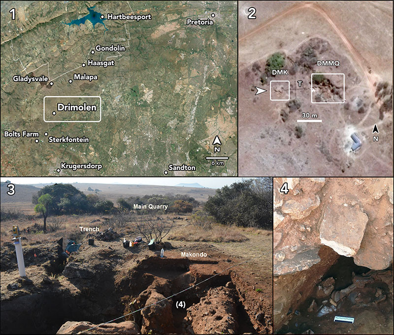
FIGURE 2. The Drimolen Makondo deposit and features at the end of the 2014 excavation season. Fossils were recovered from decalcified deposits adhering to the walls of the Main Makondo in 2013 and mainly from within the northern and eastern portions of the Eastern Makondo in 2014. While the fossils are defined as having come from different makondo features these solution tubes have all been formed within the same depositional unit of the Drimolen Makondo.
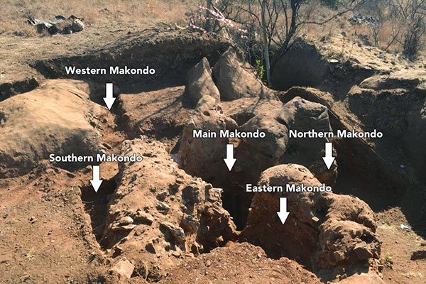
FIGURE 3. Cercopithecoides sp. craniodental specimens from the Drimolen Makondo. 1 and 2, DNM 1, partial mandible; 1, dorsal and 2, right lateral views. 3 and 4, DNM 95, right premaxilla and maxilla; 3,labial and 4, lingual views. Scale bars equal 1 cm.
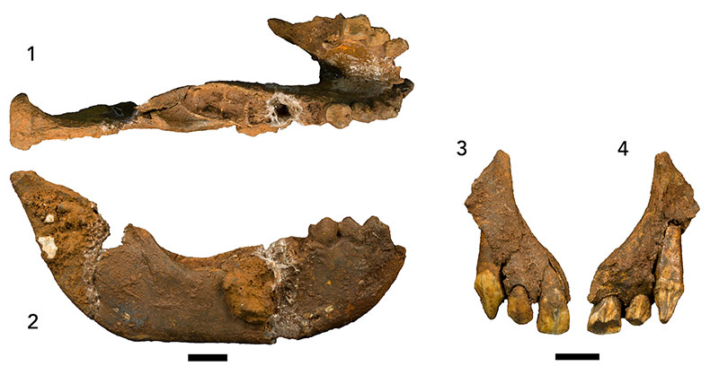
FIGURE 4. Vulpes chama craniodental specimens from the Drimolen Makondo. 1 and 2, DNM 471-1, partial cranium; 1, dorsal and 2, left lateral views. 3 and 4, DNM 471-2, right maxilla; 3, buccal and 4, occlusal views. 5 and 6, DNM 471-3, right mandible; 5, buccal and 6, lingual views. Scale bars equal 1 cm.
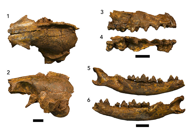
FIGURE 5. Dinofelis sp. postcranial specimens from the Drimolen Makondo. 1-6, DNM 2, left second metatarsal; 1, proximal and 2, distal articular surfaces, 3, medial, 4, dorsal, 5, ventral, and 6, lateral views. 7, DNM 54-1 and 54-2, left calcaneus in articulation; anterior view. Scale bars equal 1 cm.
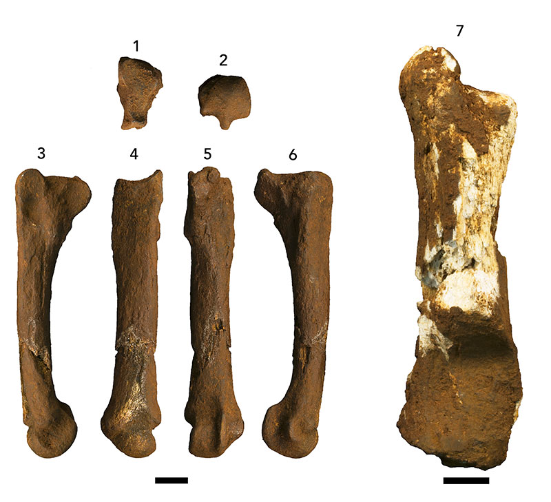
FIGURE 6. Chasmaporthetes ? nitidula postcranial specimens from the Drimolen Makondo. 1-5, DNM 3-4, partial left fourth metatarsal; 1, proximal articular surface, 2, lateral, 3, ventral, 4, dorsal, and 5, medial views. 6-8, DNM 3-3, pedal proximal phalanx; 6, dorsal, 7, lateral, and 8, ventral views. 9-11, DNM 3-2, pedal middle phalanx; 9, dorsal, 10, lateral, and 11, ventral views. 12-14, DNM 3-6, pedal terminal phalanx; 12, dorsal, 13, lateral, and 14, ventral views. Scale bars equal 1 cm.
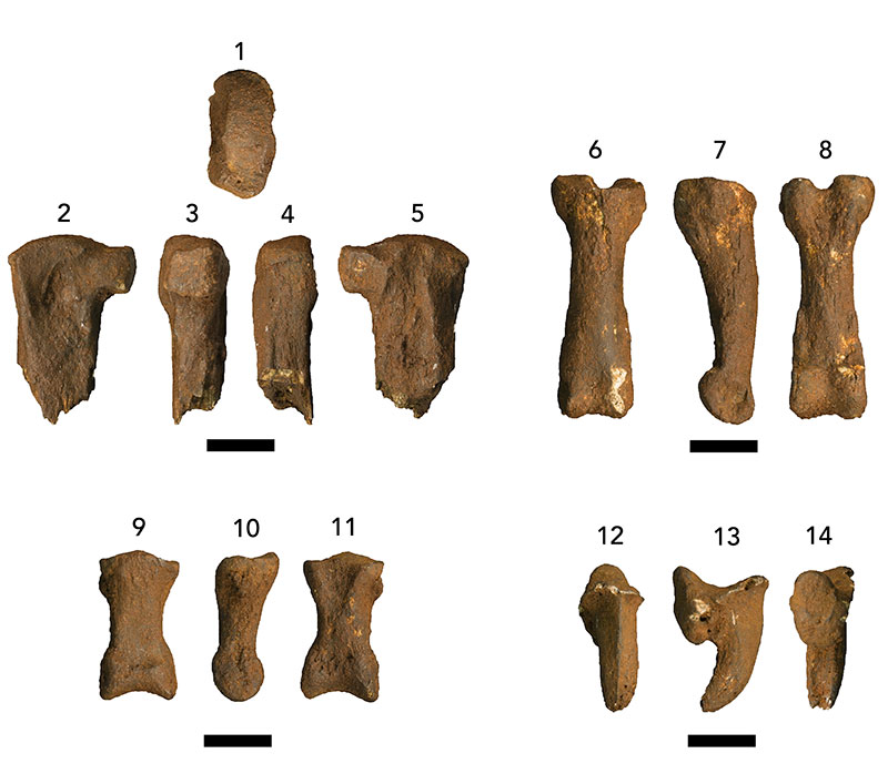
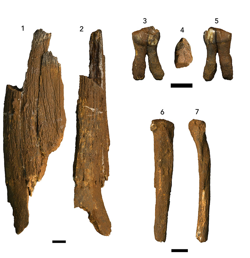
FIGURE 7. Artiodactyla and Perissodactyla from the Drimolen Makondo assemblage. 1 and 2, DNM 143-1, Hippotragus sp. partial horn core; 1, lateral and 2, anterior views. 3-5, DNM 57, Metridiochoerus sp. right maxillary third premolar; 3, lingual, 4, occlusal, and 5, buccal views. 6 and 7, cf. Eurygnathohippus cornelianus left proximal fourth metatarsal; 6, medial and 7, posterior views. Scale bars equal 1 cm.