FIGURE 1. Location of the Howe Ranch quarries in the Bighorn Basin, North-Central Wyoming, USA (grey star). Abbreviations: CO, Colorado; ID, Idaho, MT, Montana, N, north; NE, Nebraska; SD, South Dakota; UT, Utah. Scale bar equals 100 km. Modified from Christiansen and Tschopp (2010).
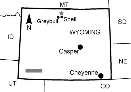
FIGURE 2. General quarry map of the Howe-Stephens Quarry north of Shell, Wyoming. Circle indicates Camarasaurus SMA 0002. Prepared by E. Premru, © by SMA.
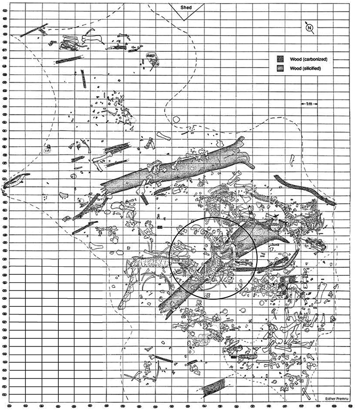
FIGURE 3. Detailed quarry map of the specimen SMA 0002 excavated in 1992-93 at the Howe-Stephens Quarry. Note the preservation of skin impressions in various regions of the skeleton. Modified from Ayer (2000).
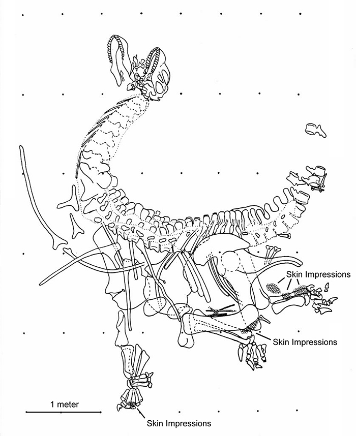
FIGURE 4. The original quarry maps of both manus (1), the right pes (2), and the left pes (3). Note the plaster cast in (1) that does not include all phalanges, the skin impressions in (2), and the bones labeled 'neg' (= negative) in (3). The latter were already separated during the excavation. Black elements are carpal or tarsal bones, dark gray marks metapodials, and light gray phalanges. Rectangles relate to grids on the detailed quarry map of SMA 0002 (see Figure 3). Abbreviations: f, fibula; h, humerus; r, radius; t, tibia; u, ulna.
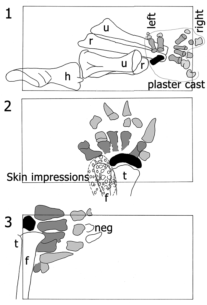
FIGURE 5. The autopodia of SMA 0002 as mounted. The left manus (1) and pes (3) are visible in anterior view, the right manus (2) and pes (4) in posterior view. The mounted unguals of the right manus and pes are casts, as well as the right php IV-2. The original unguals are stored separately at the SMA. Scale bar equals 10 cm.

FIGURE 6. Process of preparing a real 3D model of the left manus and pes of Camarasaurus: skeletal model (1) as a pre-stage, (2) shows the finished model, (3) shows the imprints produced by the model.

FIGURE 7. General drawing of the right manus in posterior view. Phm II-1 and II-2 are probably misidentified (indicated with an (m), phm III-2 might actually be phm II-2 (arrow, see text for more detailed discussion). Scale bar equals 10 cm.
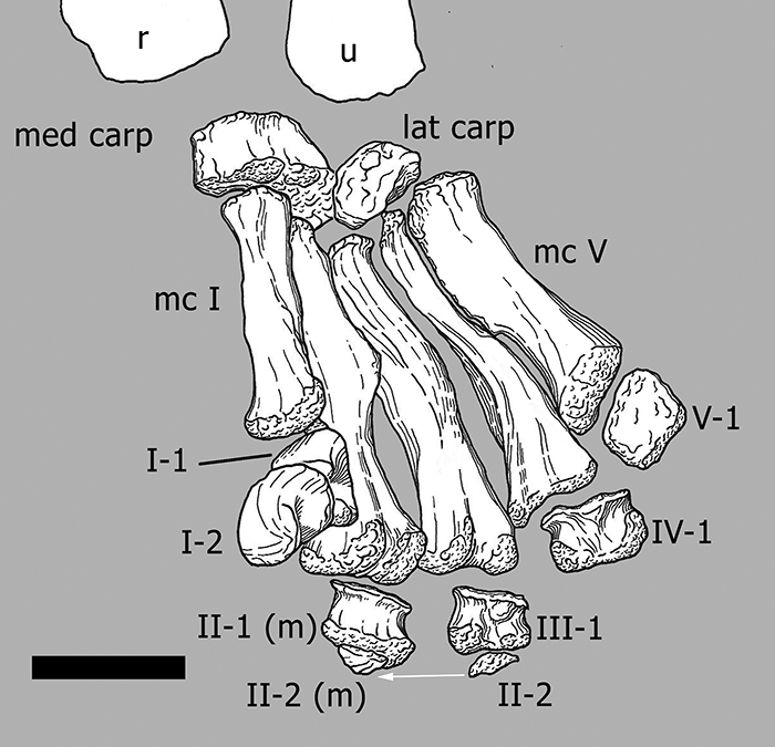
FIGURE 8. Photographs and drawings of the left medial carpal in proximal (1, 6), anterior (2, 7), medial (3, 8), posterior (4, 9), and lateral views (5, 10). Light gray represents adjoining bones, dark gray original matrix. Abbreviations: carp, carpal; lat, lateral; mc, metacarpal. Photographs taken by Rosemarie Roth.

FIGURE 9. Photographs and drawings of the left lateral carpal in proximal (1, 2), anterior (3, 4), and lateral views (5, 6). Light gray represents adjoining bones, dark gray original matrix. Abbreviations: carp, carpal; mc, metacarpal; med, medial. Photographs taken by Rosemarie Roth.
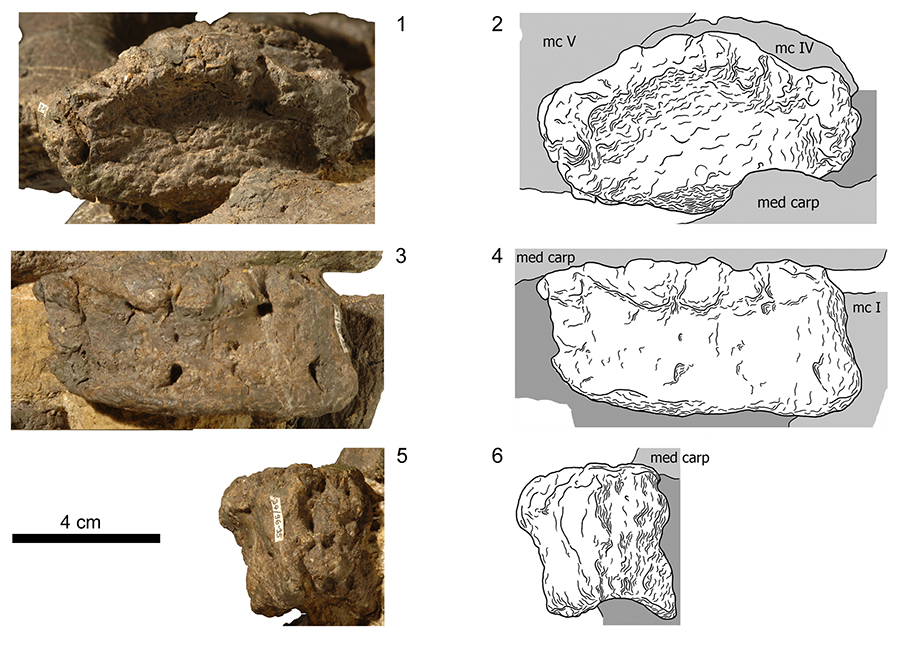
FIGURE 10. Photographs and drawings of the left metacarpal I in medial (1, 2) and anterior views (3, 4). Light gray represents adjoining bones, dark gray original matrix. Abbreviations: carp, carpal; I-1, first phalanx of digit I; mc, metacarpal; med, medial. Photographs taken by Rosemarie Roth.
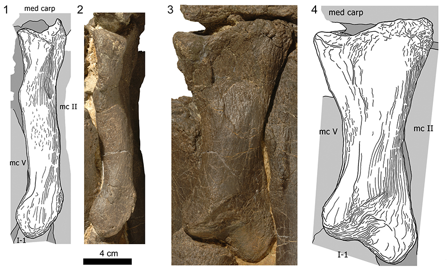
FIGURE 11. Photographs and drawings of the left metacarpal II in anteromedial (1, 2) and anterior views (3, 4). Light gray represents adjoining bones, dark gray original matrix. Abbreviations: carp, carpal; II-1, first phalanx of digit II; mc, metacarpal; med, medial. Photographs taken by Rosemarie Roth.
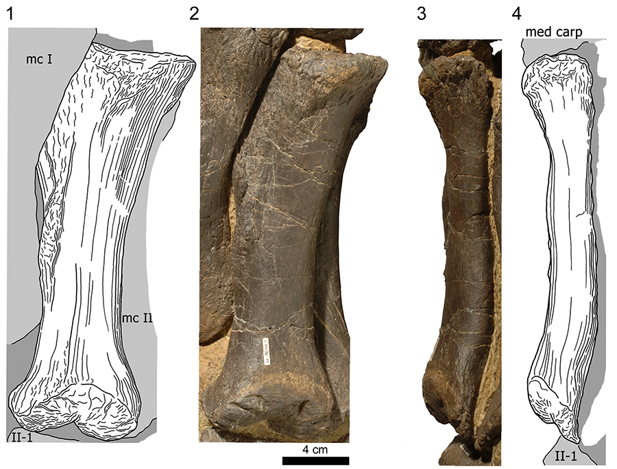
FIGURE 12. Photographs and drawings of the left metacarpal III in medial (1, 2) and anterior views (3, 4). Light gray represents adjoining bones, dark gray original matrix. Abbreviations: carp, carpal; II-1, first phalanx of digit II; lat, lateral; mc, metacarpal. Photographs taken by Rosemarie Roth.
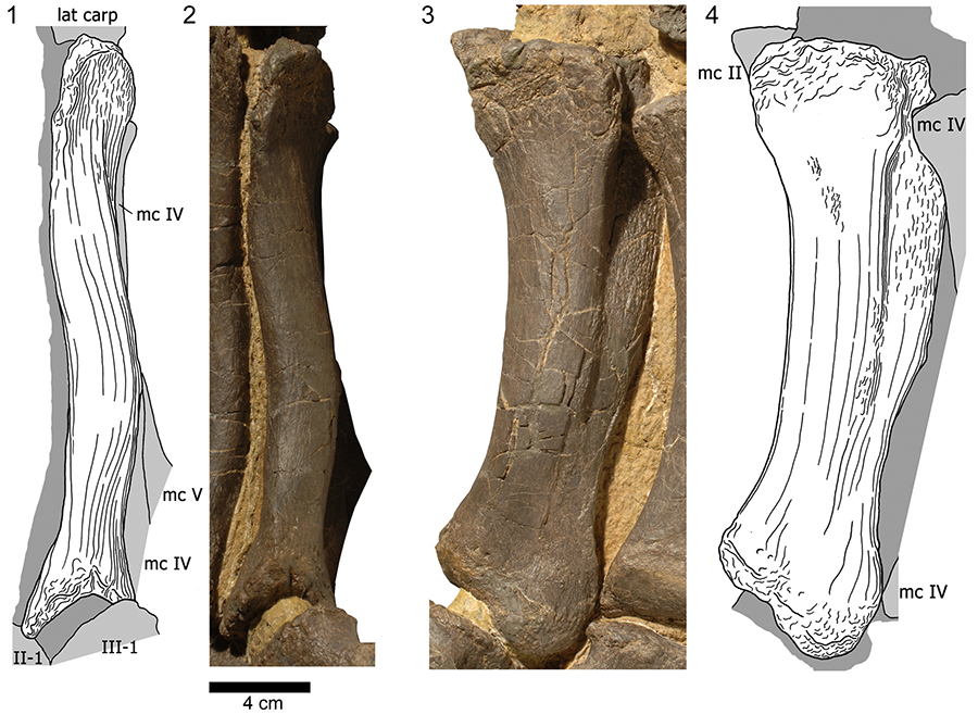
FIGURE 13. Photograph and drawing of the left metacarpal IV in anterior view. Light gray represents adjoining bones, dark gray original matrix. Light gray represents adjoining bones, dark gray original matrix. Abbreviations: mc, metacarpal; V-1, first phalanx of digit V. Photographs taken by Rosemarie Roth.
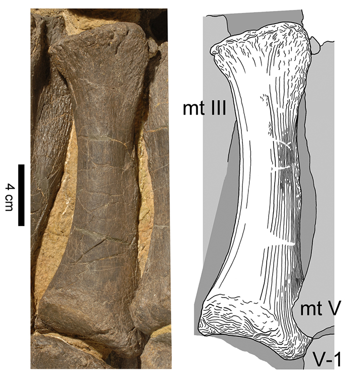
FIGURE 14. Photographs and drawings of the left metacarpal V in anterior (1, 2) and lateral views (3, 4). Light gray represents adjoining bones, dark gray original matrix. Abbreviations: mc, metacarpal; V-1, first phalanx of digit V. Photographs taken by Rosemarie Roth.
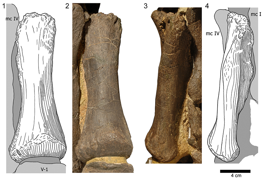
FIGURE 15. Photographs and drawings of the phalanges of the left manual digit I. Manual phalanx I-1 in medial (1, 2) and posterior views (3, 4). Manual ungual (phm I-2) in medial (5, 6), posterior (7, 8), distal (9, 10), and medial views (11, 12). Light gray represents adjoining bones, dark gray original matrix. Abbreviations: mc, metacarpal; V-1, first phalanx of digit V. Photographs taken by Rosemarie Roth.
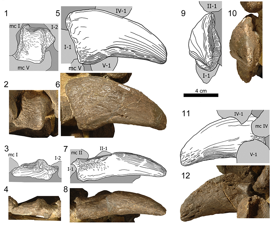
FIGURE 16. Right manual ungual phm I-2 of the Camarasaurus SMA 0002 in medial (left), dorsal, lateral, and palmar (right, top to bottom) views. Schematic drawing indicates location of the claw. The metal bar overlain with semi-transparent white in the photo of the lateral view functions as support for the mount. Abbreviation: mg, medial groove. Scale bar equals 5 cm.
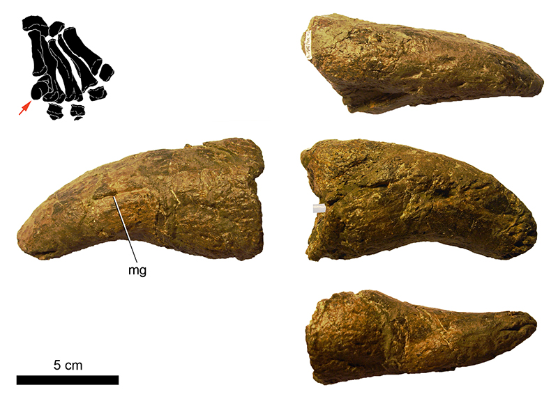
FIGURE 17. Photographs and drawings of the phalanges of the left manual digit II. Manual phalanx II-1 (1-4, 7, 8) and II-2 (3, 5, 6) in medial (1, 2), anterior (3-5), and distal views (6-8). Light gray represents adjoining bones, dark gray original matrix. Abbreviations: II-1, first phalanx of digit II; mc, metacarpal. Photographs taken by Rosemarie Roth.
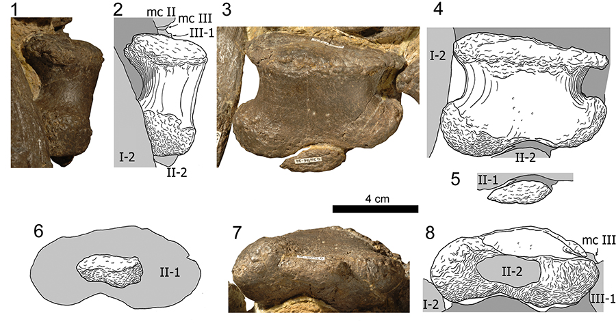
FIGURE 18. Photographs and drawings of the left phm III-1 in anterior (1, 5), lateral (2, 6), posterior (3, 7), and distal views (4, 8). Light gray represents adjoining bones, dark gray original matrix. Abbreviations: IV-1, first phalanx of digit IV; mc, metacarpal. Photographs taken by Rosemarie Roth.
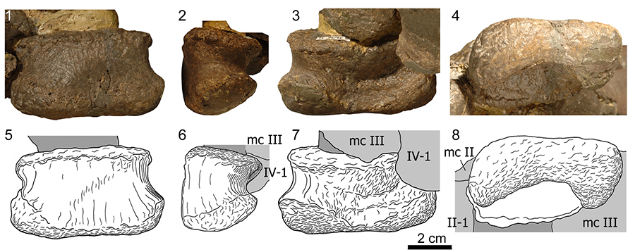
FIGURE 19. Photographs and drawings of the left phm IV-1 in anterior (1, 4), lateral (2, 5), and distal views (3, 6). Light gray represents adjoining bones, dark gray original matrix. Abbreviations: III-1, first phalanx of digit III; mc, metacarpal. Photographs taken by Rosemarie Roth.
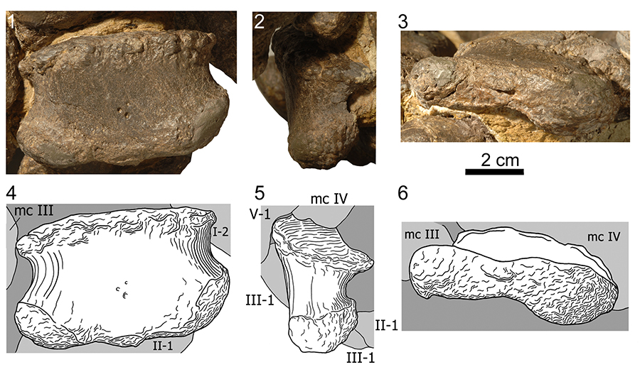
FIGURE 20. Photographs and drawings of the left phm V-1 in anterior (1, 5), lateral (2, 6), posterior (3, 7), and distal views (4, 8). Light gray represents adjoining bones, dark gray original matrix. Abbreviations: IV-1, first phalanx of digit IV; mc, metacarpal. Photographs taken by Rosemarie Roth.
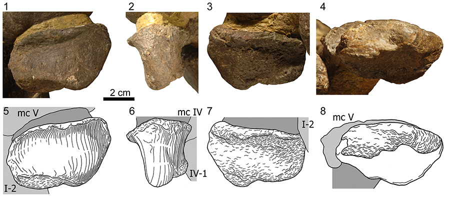
FIGURE 21. General drawing of the left pes in anterior view. Abbreviations: ast, astragalus; calc, calcaneum; f, fibula; IV-1, first phalanx of digit IV; mt, metatarsal; t, tibia. Scale bar equals 10 cm.
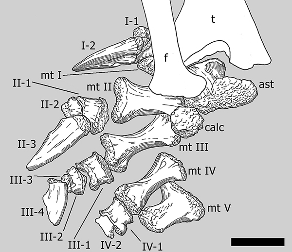
FIGURE 22. Photographs and drawings of the right astragalus (articulated with the tibia) in posterior (1, 2), and distal views (3, 4), and of the right calcaneum (still partly embedded in matrix) in proximal (5, 6), and posterior views (7, 8). Photographs taken by Rosemarie Roth (2) and Esther Premru (4, 5, 7).
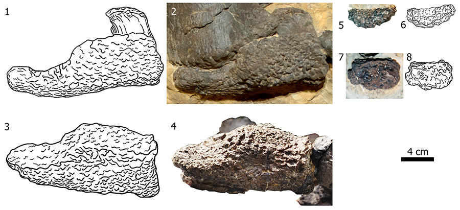
FIGURE 23. Photographs and drawings of the articulated right metatarsals I and II (1, 3, 5, 6), and mt III (2, 4, 7, 8) in posterior (1-4) and distal views (5-8). Posterior surface in 5-8 faces upwards. Photographs taken by Esther Premru.
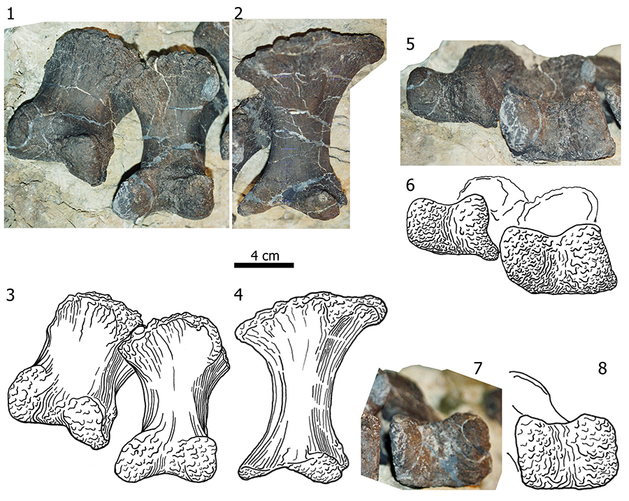
FIGURE 24. Photographs and drawings of the right metatarsal IV in anterior (1), medial (2), posterior (3), lateral (4), proximal (5), and distal views (6). Photographs taken by Esther Premru.
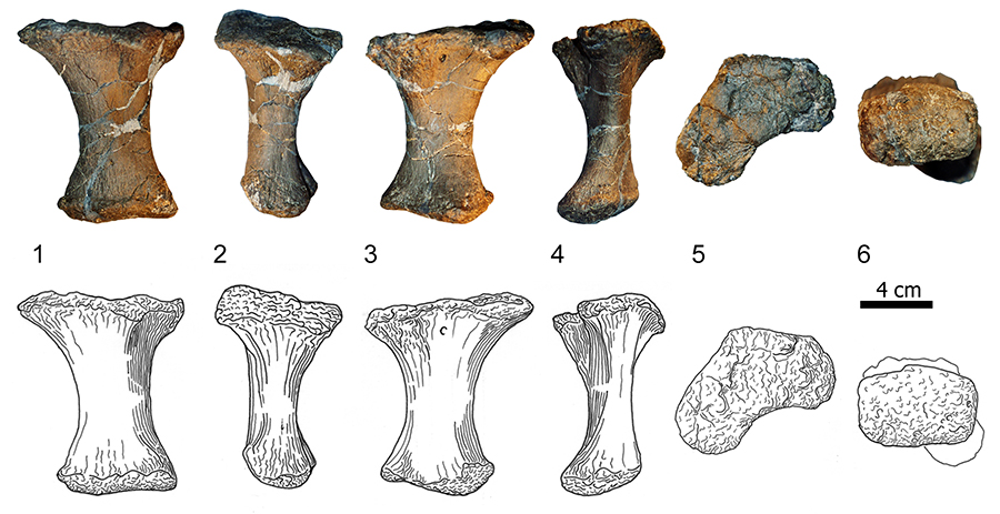
FIGURE 25. Photographs and drawings of the right metatarsal V in anterior (1), medial (2), posterior (3), lateral (4), proximal (5), and distal views (6). Photographs taken by Esther Premru.
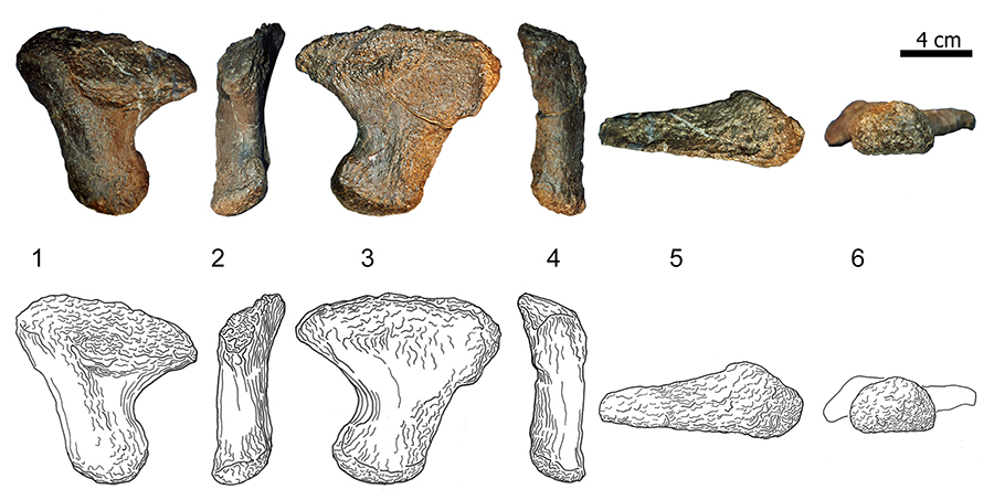
FIGURE 26. Photographs and drawings in anterior view of the phalanges of the right pedal digit I (1, 2), digit II (3, 4), digit III (5, 6), and digit IV (7, 8). Abbreviations: II-1, first phalanx of digit II. Photographs taken by Esther Premru.
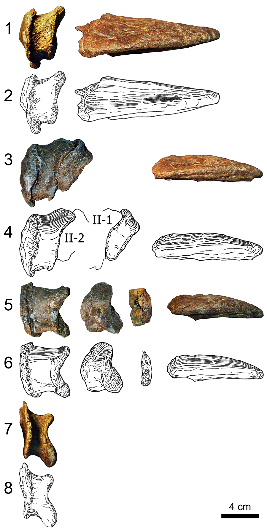
FIGURE 27. Photographs and drawings in medial view of the phalanges of the right pedal digit I (1, 2), digit II (3, 4), digit III (5, 6), and digit IV (7, 8). The oval outlines in (5), (6) indicate an element where no photographs were available in medial view (php III-3). Abbreviations: II-1, first phalanx of digit II. Photographs taken by Esther Premru.
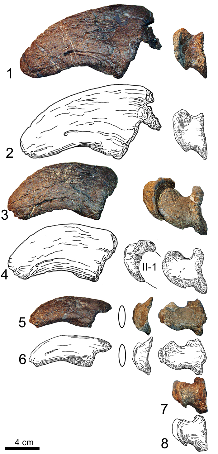
FIGURE 28. Photographs and drawings in lateral view of the phalanges of the right pedal digit I (1, 2), digit II (3, 4), digit III (5, 6), and digit IV (7, 8). The oval outlines in (5), (6) indicate an element where no photographs were available in lateral view (php III-3). Abbreviations: II-1, first phalanx of digit II. Photographs taken by Esther Premru.
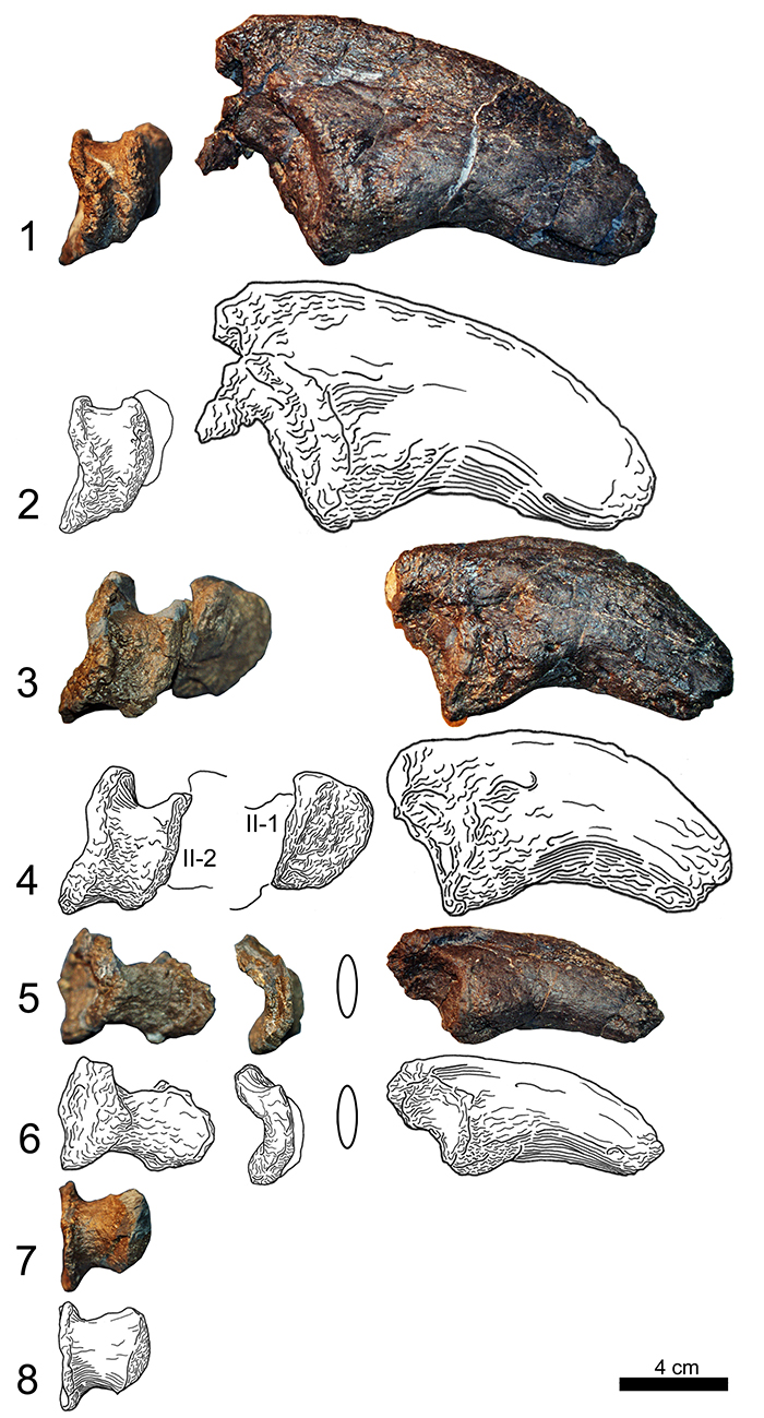
FIGURE 29. Photographs and drawings in posterior view of the phalanges of the right pedal digit I (1, 2), digit II (3, 4), digit III (5, 6), and digit IV (7, 8). Abbreviations: II-2, second phalanx of digit II. Photographs taken by Esther Premru.
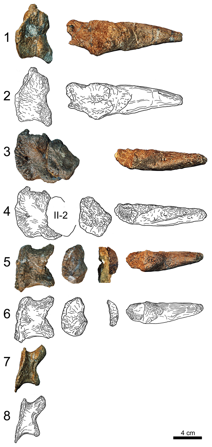
FIGURE 30. Photographs and drawings in proximal view of the phalanges of the right pedal digit I (1, 2), digit II (3, 4), digit III (5, 6), and digit IV (7, 8). The oval outlines in 3-6 indicate an element where no photographs were available in proximal view (php II-2 and III-3). Photographs taken by Esther Premru.
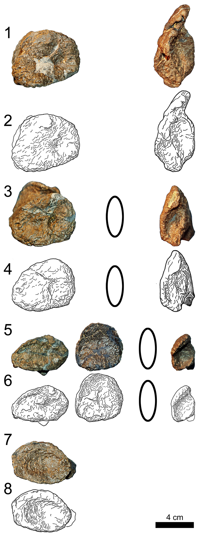
FIGURE 31. Photographs and drawings in distal view of the phalanges of the right pedal digit I (1, 2), digit II (3, 4), digit III (5, 6), and digit IV (7, 8). The oval outlines in (3), (4) indicate an element where no photographs were available in distal view (php II-2). Photographs taken by Esther Premru.
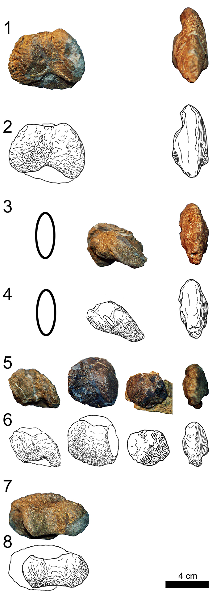
FIGURE 32. Photographs of the manual skin impression preserving also the negatives of the distal tips of the phalanges in proximal (1), lateral (2), and distal views (3). Photographs taken by Rosemarie Roth.
FIGURE 33. Photographs of the two different skin structures found in the region of the hindlimb and the pes of Camarasaurus sp. SMA 0002: (1) shows the large polygonal structure, (2) the positive and negative impressions of the smaller texture. Scale bars equal 5 cm. Photographs taken by Rosemarie Roth.
FIGURE 34. X-ray CT images of the left metacarpals in longitudinal (1) and transverse (2) cross section. Note the absence of hollow medullary cavities.
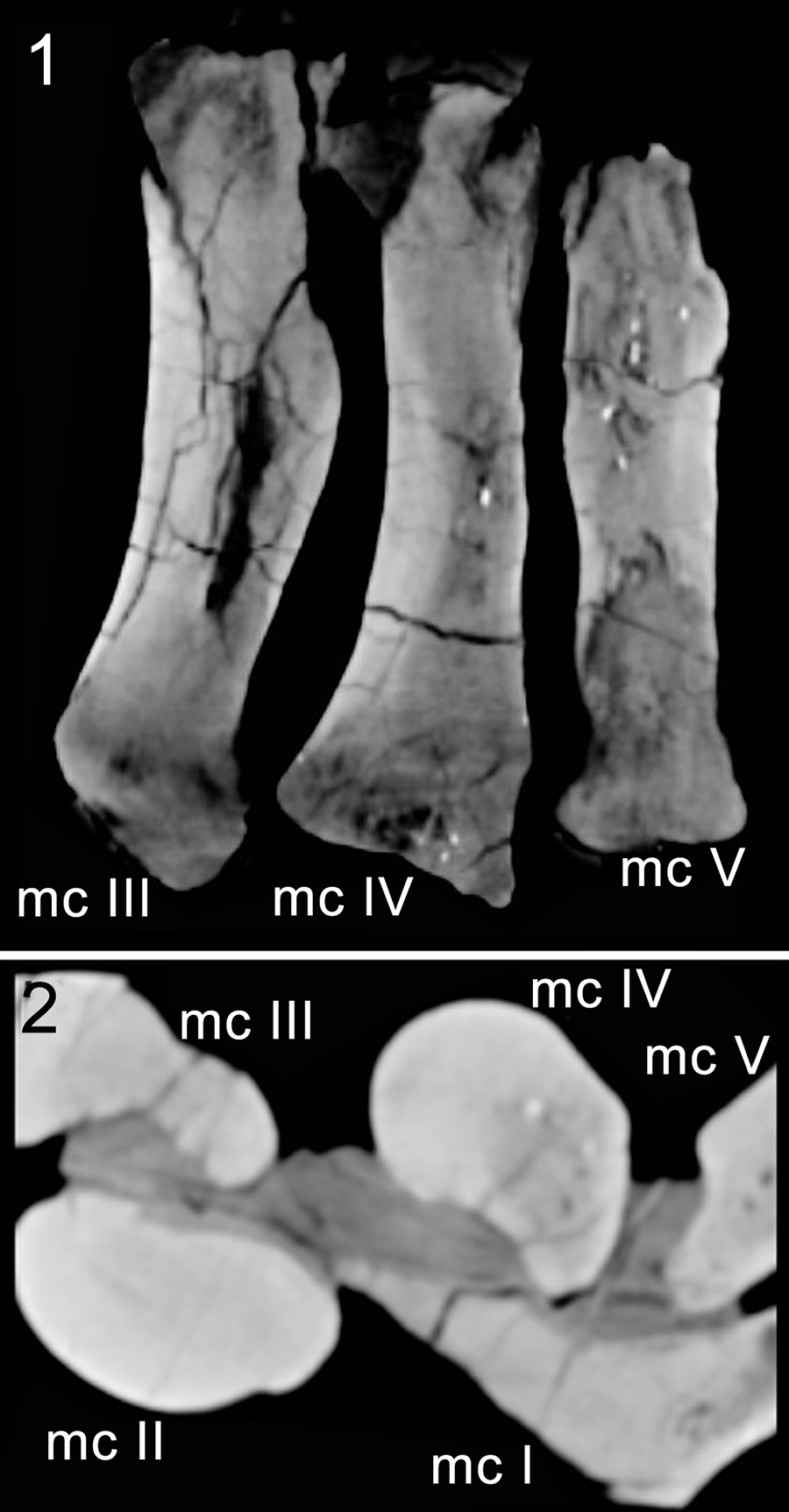
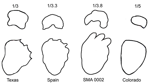
FIGURE 35. Comparison of the supposed footprint of SMA 0002 with published tracks from different locations. Reported (Texas, Colorado) or calculated (Spain, SMA 0002) heteropody rate is indicated on top. Modified from Lockley and Meyer (2000; Texas, Colorado), Castanera et al. (2012; Spain, left print reversed).