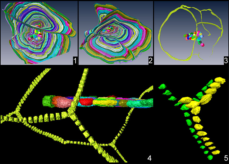
FIGURE 1. Specimen A1: 1) equatorial and axial sections of one specimen.; 2) equatorial view of the test; 3) lateral view of the test; 4) close-up to the nepiont and the first chambers in equatorial section; 5) segmentation of the entire nepiont. For more information, refer to text.
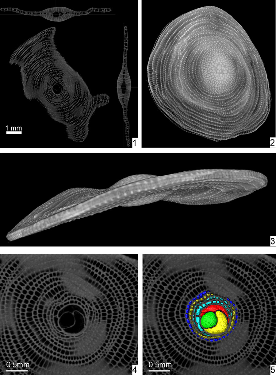
FIGURE 2. Segmentation and equatorial sections of specimens possessing multiple nepionts: 1) specimen A2; 2) specimen A3; 3) specimen A17; 4) specimen A10; 5) specimen A18; 6) specimen A5; 7) specimen A6. Scale bar equals 0.5 mm.
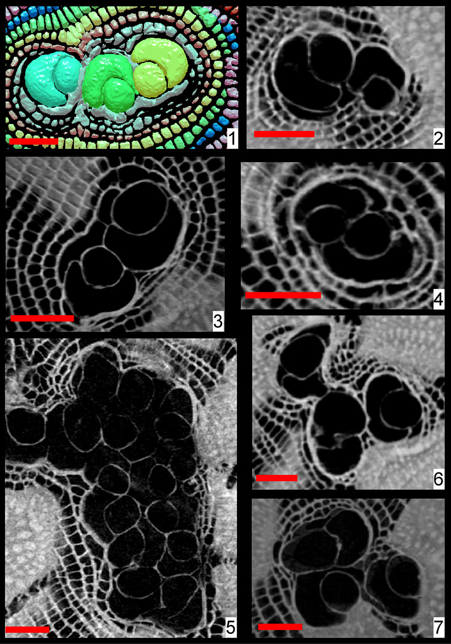
FIGURE 3. Proloculus diameters. 1) proloculus diameter for all individuals in the presented population, except for A18. Multiple bars indicate the presence of several proloculi. 2) diameters of all proloculi identified within the specimen A18.
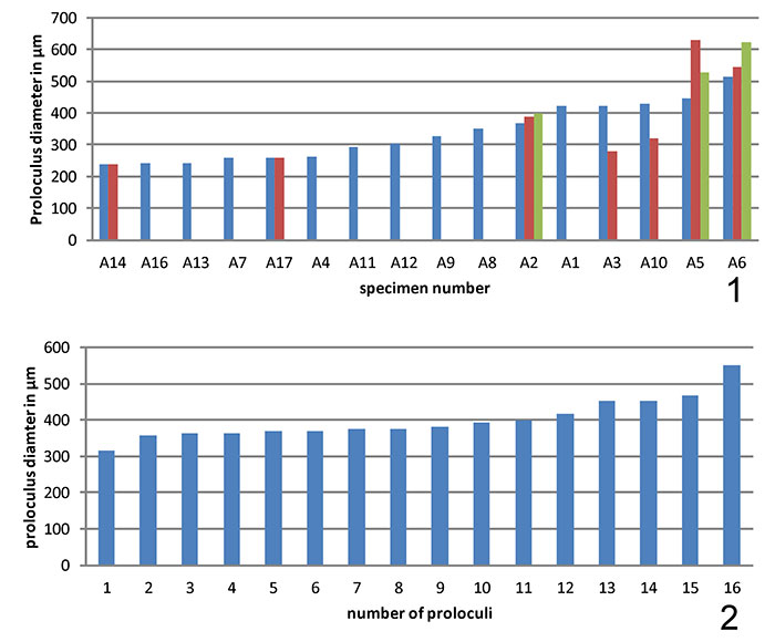
FIGURE 4. Relative position of embryos and secondary equatorial layers: 1) Specimen A2; 2) specimen A3; 3) specimen A6; 4) specimen A14.
FIGURE 5. Multiple equatorial layers and T-connection in specimen A18.