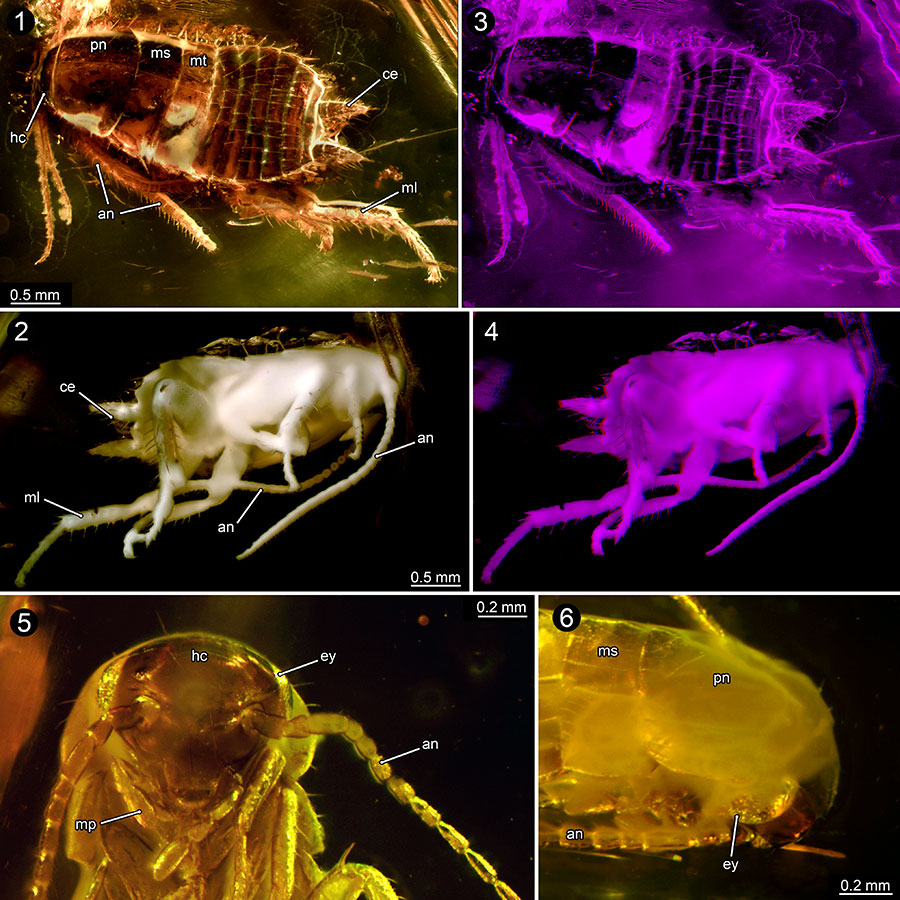
FIGURE 1. Overview of BSPG 1967 XX, part of the Bayerische Staatssammlung für Paläontologie und Geologie, Munich. Germany; documented with composite imaging. Single piece of Baltic amber with inclusion of 13 cockroach nymphs.
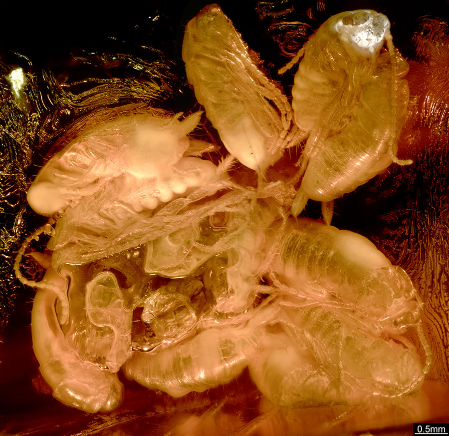
FIGURE 2. Close-ups of nymphs in amber (BSPG 1967 XX). 1, Overview of a single nymph (red-coloured specimen in Figure 4). 2, Head with antennae and maxillary palp equipped with setae of specimen in 1, compound eyes not observable. 3, Cerci equipped with setae (blue-coloured specimen in Figure 4). 4, Close-up of antenna equipped with setae (yellow-coloured specimen in Figure 4), arrows point to setae. Abbreviations: a4-a6, abdominal segments 4-6; an, antenna; ce, cercus; hc, head capsule; mp, maxillary palp; ms, mesonotum; mt, metanotum; pn, pronotum; pl, coxa of prothoracic leg; ta, terminal article of the antenna.
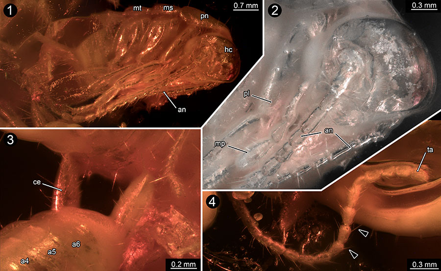
FIGURE 3. Overview of BSPG 1967 XX, documented with stereo imaging from all available sides of the specimen (1-3). Images presented as red-cyan stereo-anaglyphs; please use red-cyan glasses to view.
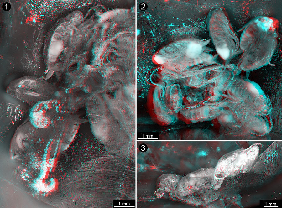
FIGURE 4. Overview images of BSPG 1967 XX. 1-3, Colour-marked versions of Figure 3 (different individuals marked in different colours, same individuals marked in same colour in different viewing angles). 4-5, Volume renderings based on micro-CT data (Amira) (different viewing angles in 4 and 5). 6, Tomographic section from micro-CT image stack (red-coloured specimen in 1 and 2), inner structures are not preserved.
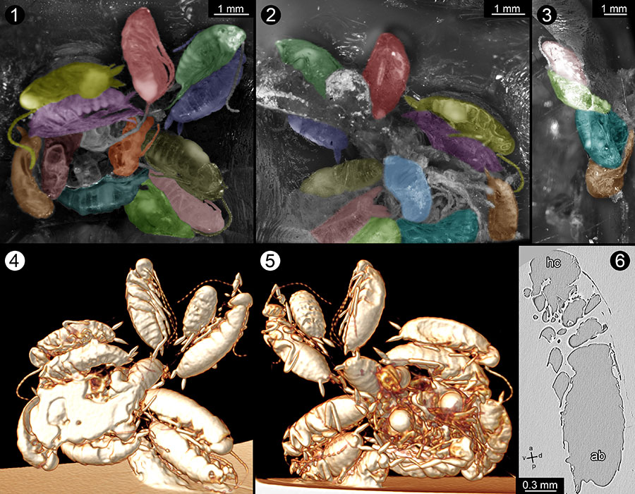
FIGURE 5. Volume renderings of one nymph of BSPG 1967 XX (red-coloured specimen in Figure 4) in about lateral (1 - 2), ventral (3 - 4), and dorsal (5) views generated with Drishti 2.4 based on micro-CT data. Abbreviations: cl, clypeus; co I, coxa of prothoracic leg; fe I, femur of prothoracic leg; lr, labrum; md, mandible; pt I, praetarsus of prothoracic leg (tibial claw); ta I, tarsus of prothoracic leg; ti I, tibia of prothoracic leg; a1-a7, abdominal segments 1-7. Other abbreviations are the same as in Figure 2.
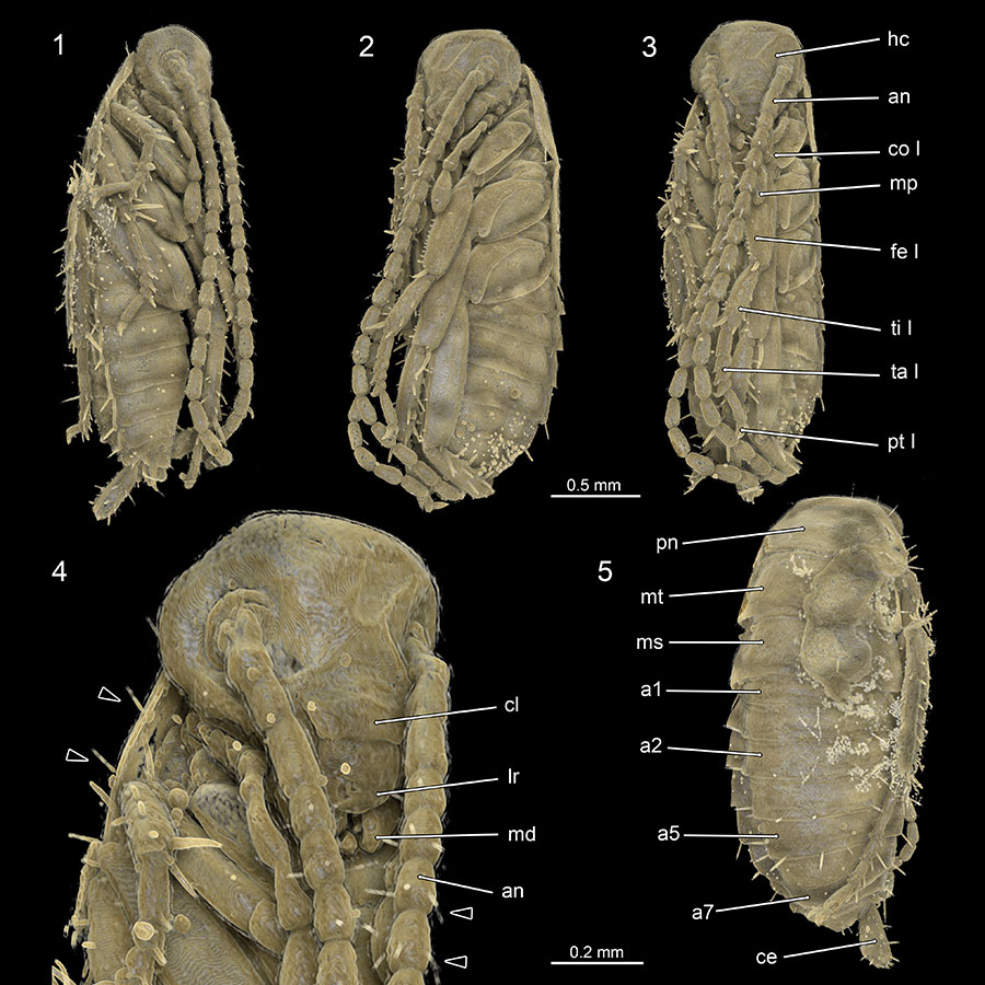
FIGURE 6. Nymphs of the extant species Periplaneta americana (1-3) and Blaberus craniifer (4-5). 1-3, Hatchling of P. americana in dorsal (1), lateral (2), and ventral (3) views documented with compound microscope. 4-5, Early instar nymph of Blaberus craniifer in ventral (4) and dorsal (5) views documented with composite autofluorescence microscopy. Abbreviations: co III, coxa of metathoracic leg; fe III, femur of metathoracic leg; ta III, tarsus of metathoracic leg; ti III, tibia of metathoracic leg. Other abbreviations are the same as in Figure 2 and Figure 4.
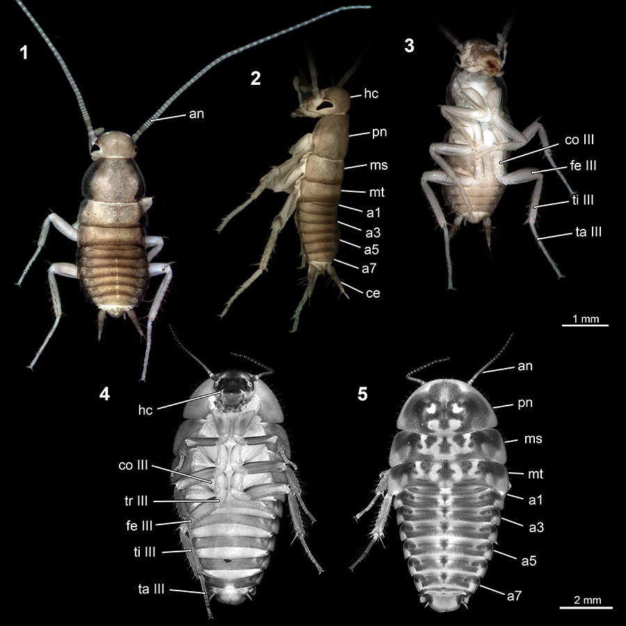
FIGURE 7. Comparison of mouthparts of one nymph of BSPG 1967 XX and first instar of Periplaneta americana . 1-3, Surface reconstruction of the mouthparts of one nymph in amber; based on micro-CT data. Same individual as in Figure 2, about lateral (1 and 3) and frontal (2) views. 4-5, Head with mouthparts of first instar nymph of P. americana ; frontal view of the head (4) and colour-marked close-up image of the mouthparts (5), composite autofluorescence image. Abbreviations: hp, hypopharynx; lb, labium; lp, labial palp; mx, maxilla. Other abbreviations are the same as in Figure 2 and Figure 4.
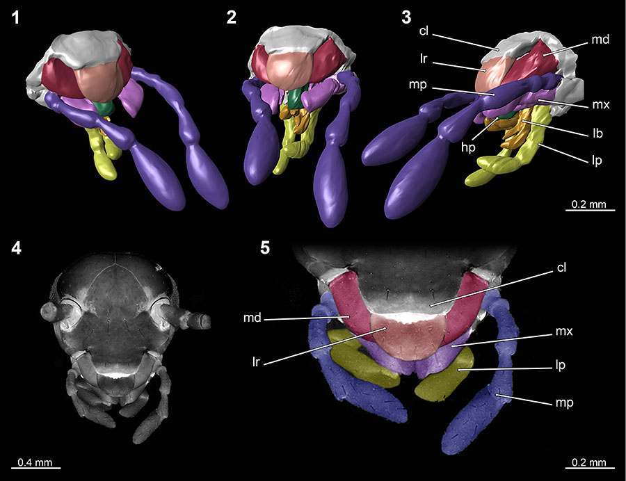
FIGURE 8. Close-ups of maxillary palp of one nymph of BSPG 1967 XX. 1, Palp of red-coloured nymph in Figure 4 documented with stereo imaging. Images presented as red-cyan stereo-anaglyphs; please use red-cyan glasses to view, red left, cyan right. 2-4, Close-ups of volume rendering of maxillary palp and part of antenna of red-coloured nymph in Figure 4; documented at three different grey values. 5, Combined version of 2-4, arrows point to setae. Abbreviations are the same as in Figure 2 and Figure 4.
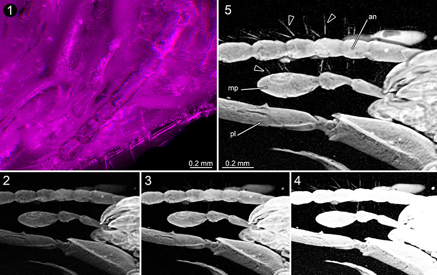
FIGURE 9. Comparison of volume renderings of one nymph of BSPG 1967 XX generated by Amira 5.6 (left) and Drishti 2.4 (right) (images with system based pseudocolour). Note especially the differences of the level of details in the marked areas (arrows).
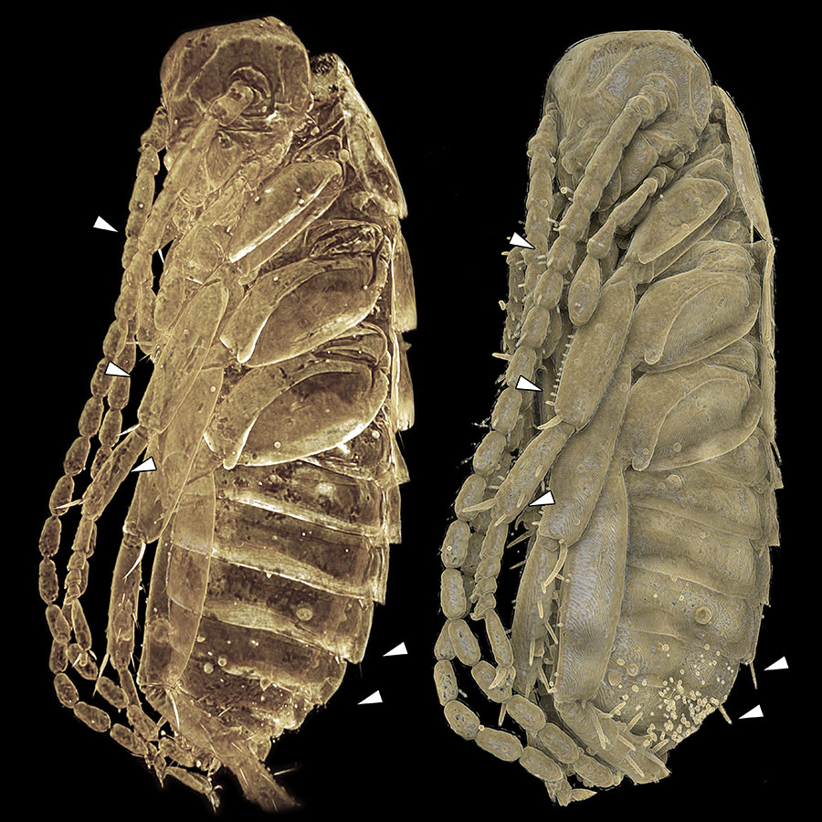
FIGURE 10. Blattodean nymphs in Baltic amber. 1-4, Specimen PE 61065, dorsal view (1), distinct pigmentation of cuticula visible; ventral view (2); and stereo-images of PE 61065 (3-4). Images presented as red-cyan stereo-anaglyphs; please use red-cyan glasses to view. 5-6, Specimen ZMUC 901795, close-up image of the head (5) and close-up image of the head lateral view (6). Abbreviation: ml, femur of metathoracic leg. Other abbreviations are the same as in Figure 2 and Figure 4.