FIGURE 1. Diagrammatic sections through the walls of fossil clitellate annelid cocoons showing the general structure and representation of layers. 1, Burejospermum. 2, Pilothylakos. 3, Dictyothylakos. 4, Pegmatothylakos.
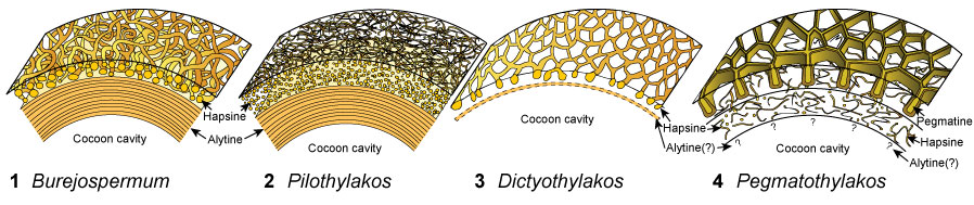
FIGURE 2. Locality maps and stratigraphic column of the studied succession. 1, Map of Antarctica showing the location of the study area. 2, Map of the northern Antarctic Peninsula showing the location of Seymour Island. 3, Geological sketch map of Seymour Island, Antarctic Peninsula, showing the positions of fossil localities IAA 1/90 and IAA 2/95. 4, Stratigraphic column of the La Meseta Formation on Seymour Island (from Reguero et al., 2013). Strontium date values from Dingle and Lavelle (1998), Dutton et al. (2002), Reguero et al. (2002), and Ivany et al. (2008).
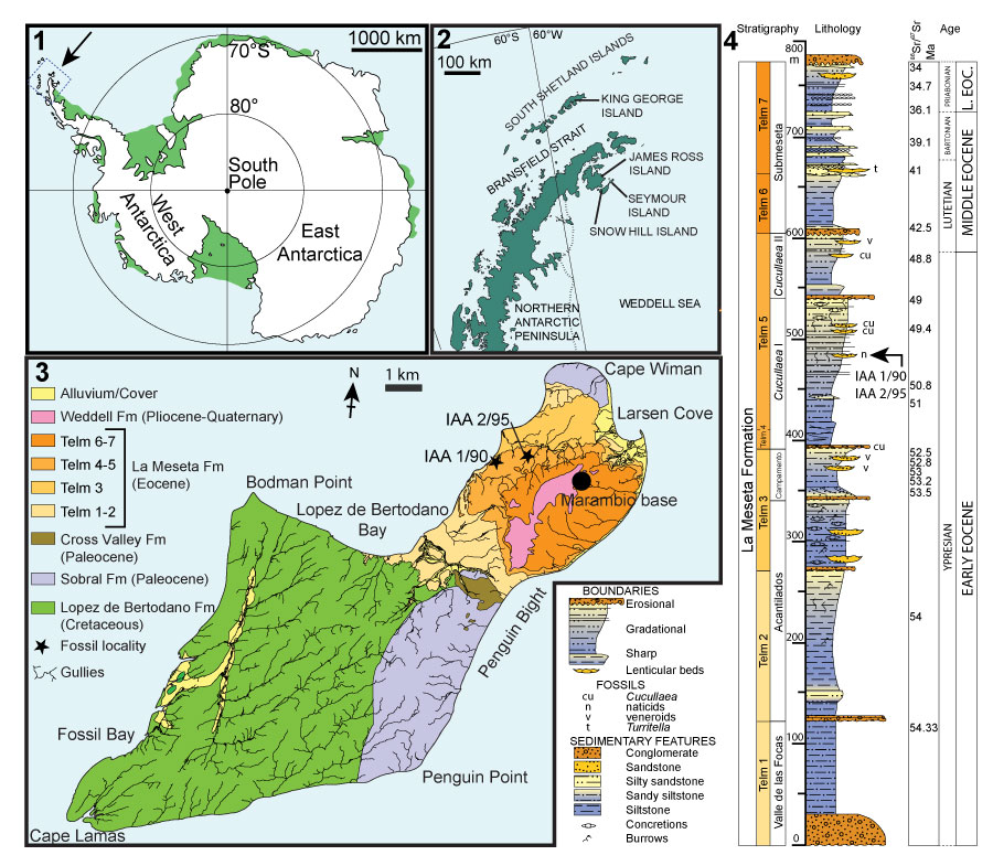
FIGURE 3. Photographs of sampling localities. 1, Aerial view of ‘Ungulate site’ (IAA 1/90, 64°14'04.67"S, 56°39'56.38"W) with ‘ Natica horizon’ marked by arrow. 2, Detail of section between Cucullaea 1 bed (below quad bike) and ‘ Natica horizon’ at IAA 1/90 (arrowed) showing dominance of poorly consolidated sandstones, mudstones and shell-rich conglomerates. 3, Panoramic view of ‘Marsupial site’ (IAA 2/95: 64°13'58"S, 56°39'06"W) with ‘ Natica horizon’ exposed along foreground ridge, and Cockburn Island in background. 4, ‘ Natica horizon’ near site IAA 2/95 showing incised base and lenticular character of the bed. Photographs by T. Mörs (1, 2), F. Degrange (3), and J. Hagström (4).
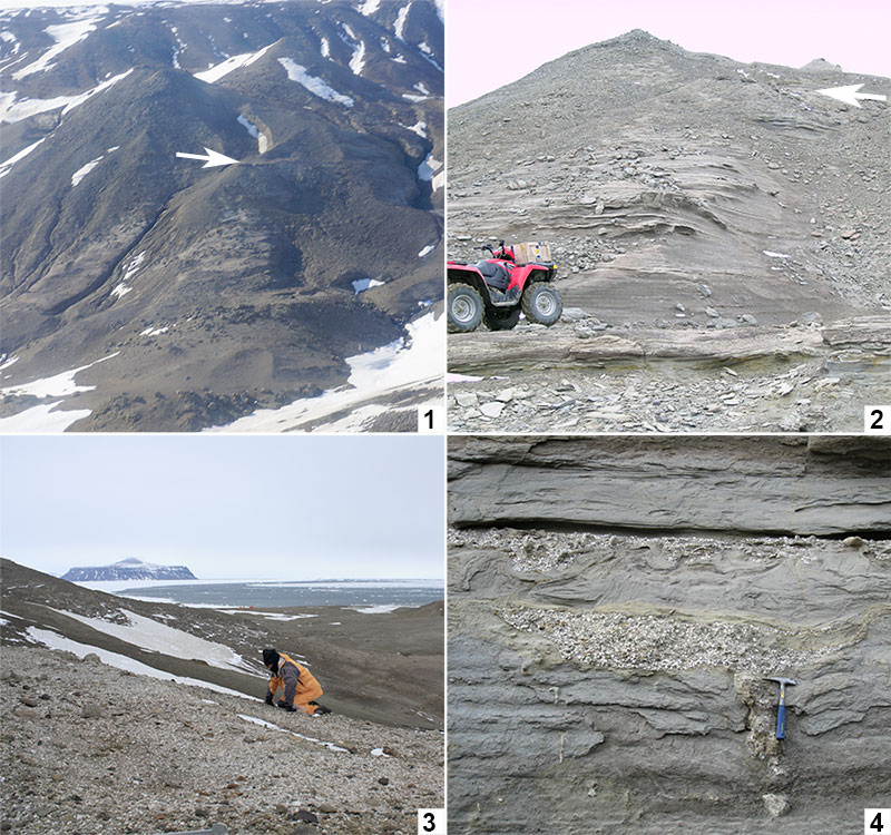
FIGURE 4. Light micrographs of early Eocene clitellate annelid cocoons from Seymour Island. 1-3, Burejospermum seymourense sp. nov. (NRMS089727). 4-6, Burejospermum punctatum sp. nov. (NRMS089728). 7-9, Pegmatothylakos manumii sp. nov. (NRMS089730). 1, 4, 7, Lateral views of cocoons (posterior to left); g = equatorial girdle. 2, 5, 8, Views of posterior end of cocoons showing opening left by detachment of operculum. 3, 6, 9, Enlargements of cocoon wall exterior showing pale felt-like hapsine covering in B. seymourense (3), relatively smooth surface in B. punctatum ( 6) and reticulate pegmatine of P. manumii (9). Scale bars represent 1 mm in 1, 2, 4, 5, 7, 8; 100 µm in 3, 6, 9.
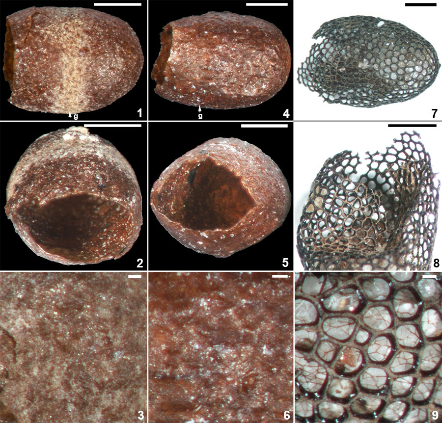
FIGURE 5. Scanning electron micrographs of Burejospermum seymourense sp. nov. (NRMS089727). 1, Cocoon in lateral view. 2, Details of typical hapsine threads. 3, Detail of dense hapsine threads over girdle region. 4, Elliptical secreted plate-like structures on exterior of hapsine. 5, Broken cocoon wall in cross-section showing solid alytine (lower) and thread-like hapsine (upper). 6, Detail of plate-like secretion on exterior of hapsine. Scale bars represent 1 mm in 1; 100 µm in 2; 50 µm in 3, 4; 25 µm in 5, 6.
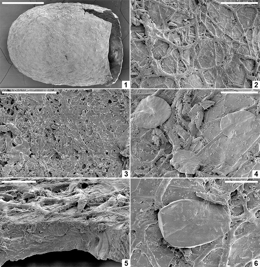
FIGURE 6. Computed tomographic images of a Burejospermum seymourense cocoon wall (NRMS089729) generated from Synchrotron-radiation-based X-Ray tomographic microscopy (SRXTM) at the TOMCAT beamline of the Swiss Light Source at the Paul Scherrer Institute, Switzerland. 1, Isosurface rendering of cocoon’s external surface showing mineral grains embedded in hapsine. 2, Isosurface rendering of cocoon’s internal surface showing creasing caused by alytine threads. Arrow indicates the position of a hollow tube within the alytine. 3, 4, Longitudinal orthoslice sections, at different focal planes, of a tube within the alytine showing pseudosegmentation. 5-8, Translucent volume rendering of a tube within the alytine imaged at different focal planes and magnifications to highlight the transverse to chevron-shaped thread arrangement lining the tube wall. 9, Isosurface rendering of hapsine outer surface showing details of the complex arrangement of hirudoin threads. 10, Transverse orthoslice section of the cocoon wall showing the near-solid alytine layer (bottom) and mesh-like hapsine layer (top). 11, Isosurface rendering of interior alytine wall showing an embedded spermatozoal nuclear region. 12, Colour-inverted rendering of Figure 6.11. 13, Volume rendering of the cocoon wall showing numerous mineral (white-yellow) inclusions. 14, Oblique translucent volume rendering of the cocoon wall showing the distribution of mineral grains mainly in the outer hapsine layer. Scale bars represent 100 µm in 1, 2, 10, 13, 14; 50 µm in 9; 25 µm in 3-7; 10 µm in 8; 5 µm in 11, 12.
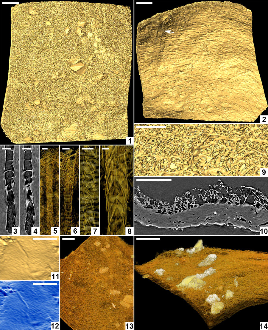
FIGURE 7. Scanning electron micrographs of Burejospermum punctatum sp. nov. (NRMS089728). 1, Cocoon in lateral view. 2, Details of sparse hapsine threads embedded in alytine surface. 3, Detail of typical cocoon surface showing pitting. 4, Detail of girdle region showing granular microtexture and a single surface pit. 5, Detail of outer alytine surface showing cleft and fold structures (arrowed). 6, Cross-section of cocoon wall showing laminated architecture. Scale bars represent 1 mm in 1; 100 µm in 3, 5; 10 µm in 2, 4, 6.
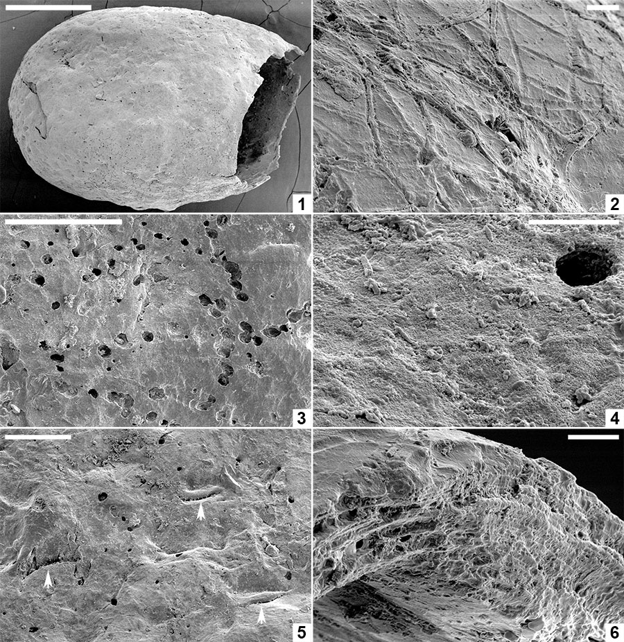
FIGURE 8. Scanning electron micrographs of Pegmatothylakos manumii sp. nov. (NRMS089730). 1, Cocoon in lateral view. 2, Enlargement of robust reticulate pegmatine covering thread-like hapsine. 3, Enlargement of areole in pegmatine reticulum showing underlying mesh-like hapsine. 4, Enlargement of girders forming pegmatine showing hackly inner zone and smoothly welded threads lining girder margins. 5, Detail of hackly and smooth threads in pegmatine girders. 6, Broken pegmatine girder showing irregular internal structure. 7, Enlargement of interior surface of pegmatine girder showing smooth threads with irregular branching and orientation. 8, Enlargement of hapsine threads. Scale bars represent 1 mm in 1; 250 μm in 2; 100 μm in 3, 4; 25 μm in 5-8.
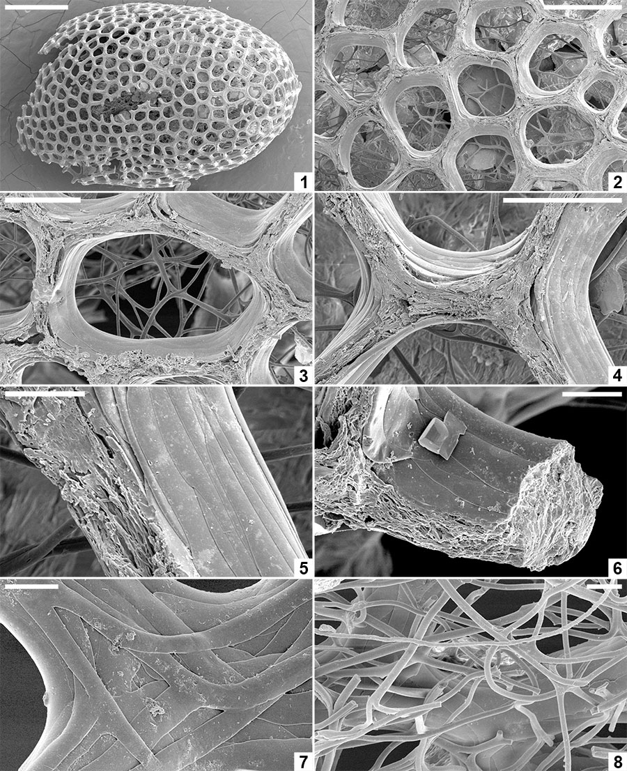
FIGURE 9. Spermatozoa and bacteria preserved embedded in the innermost layer of the alytine wall of Burejospermum seymourense sp. nov., NRMS089729 ( 1-12); Burejospermum punctatum sp. nov., NRMS089728 ( 13); and Pegmatothylakos manumii sp. nov., NRMS089730 (14). 1, Overview of cocoon inner surface showing a range of elongate spermatozoa and elliptical-reniform bacteria. 2, Spermatozoon acrosome (white arrow) with swollen tip (probably containing the acrosome vesicle) and apical button (black arrow). 3, Enlargement of a spirally organized detached acrosome. 4, Thin sheet-like innermost layer of alytine draped over mineral inclusion (top) and entombing numerous elliptical bacteria. 5, Several spermatozoal nuclear regions, one with an attached whip-like flagellum (white arrow). A pair of dimpled elliptical bacteria are also present in the upper left (black arrow). 6, Enlargement of a portion of a spermatozoon flagellum showing slightly beaded structure. 7, Enlargement of the nuclear and mitochondrial region of a spermatozoon showing granular ornamentation and swellings in the anterior portion of the attached flagellum. Reniform bacterium also present in upper right (white arrow). 8, Several spermatozoal flagella showing indistinct spiral architecture. 9, Nuclear (white arrow), mitochondrial (black arrow) and anterior flagellar (hollow arrow) regions of a spermatozoon. 10, Enlargement of a flagellum showing beaded structure that probably reflects helical architecture. 11, Enlargement of Figure 9.9 showing details of nuclear (n), mitochondrial (m) and anterior flagellar (f) regions. 12, Overlapping elliptical bacterium (b) and flagellate spermatozoon. 13, Elongate coiled acrosome (a) and slender flagellum (f) of separate spermatozoa. 14, Nuclear (n) and flagellar (f) regions of a spermatozoon. Scale bars represent 10 µm in 1, 4, 5, 13, 14; 2 µm in 2, 3, 7-9; 1 µm in 6, 10-12.
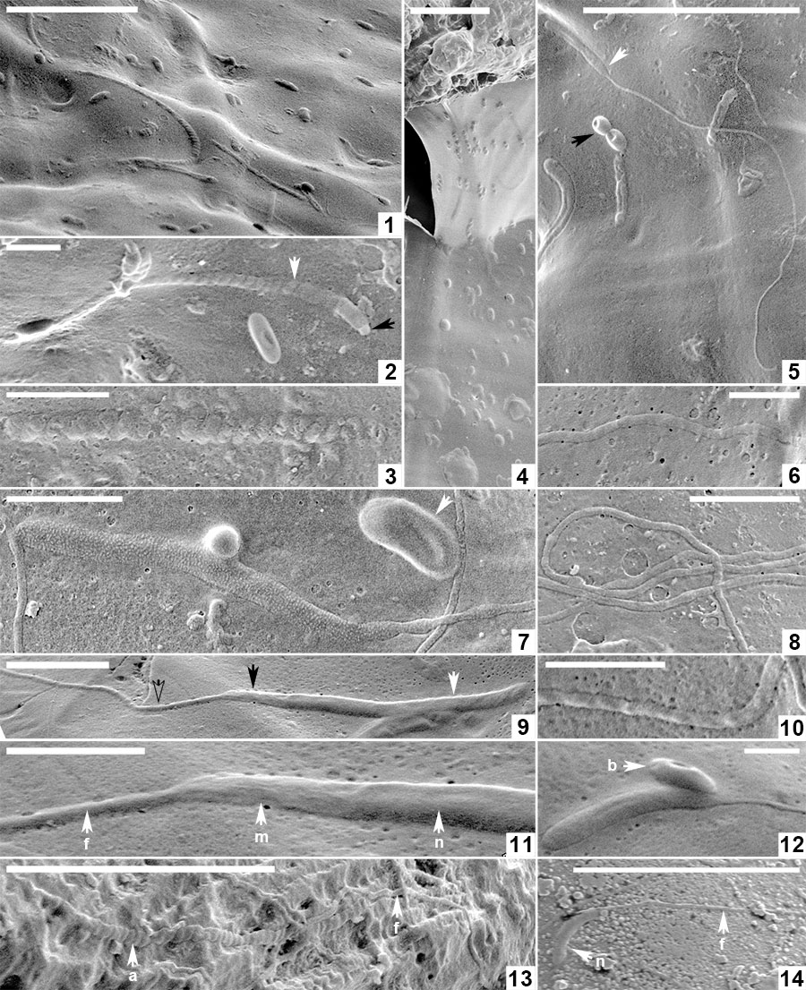
FIGURE 10. Organic inclusions embedded in the innermost layer of the alytine wall of Burejospermum seymourense sp. nov. (NRMS089729) b = bacteria in a chain, n = detached spermatozoal nuclear regions, a = detached spermatozoal coiled acrosome regions, u = unidentified vermiform bodies. Scale bar represents 5 µm.
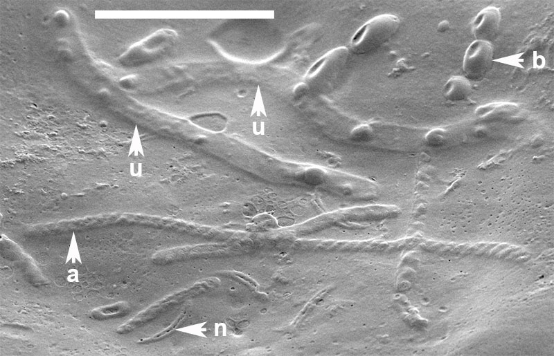
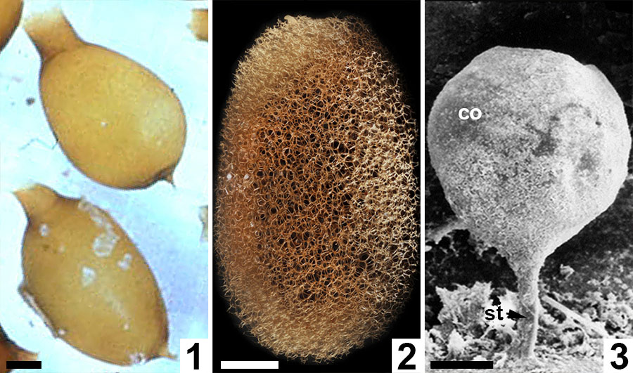
FIGURE 11. Comparative morphologies of extant clitellate annelid cocoons. 1, Light micrograph of two earthworm (Oligochaeta: Lumbricus terrestris) cocoons with prominent polar extensions and walls constructed almost entirely of the solid alytine layer (from Clive A. Edwards, The Ohio State University, Columbus, USA, Public domain via Wikimedia Commons). 2, Light micrograph of a true leech (Hirudinea: ? Macrobdella decora) cocoon with a wall dominated by a Dictyothylakos -like meshed hapsine layer (courtesy of Macroscopic Solutions, LLC. www.macroscopicsolutions.com). 3, Scanning electron micrograph of a crayfish worm (Branchiobdellida: Cambarincola macrocephelus or C. fallax) cocoon with a thin scabrate hapsine layer overlying solid alytine (courtesy of Dr Naglaa M SH Geasa, Tanta University, Egypt; after Geasa, 2014, figure 6G); co = cocoon body lacking an operculum; st = attachment stalk. Scale bars represent 1 mm in 1, 2; 50 µm in 3.