FIGURE 1. Map showing the main Tertiary basins of the Iberian Peninsula with the location of Les Saleres (LS) fossil site (modified from Antunes et al., 1997).
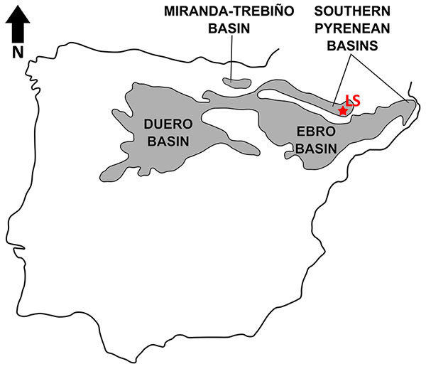
FIGURE 2. Agerinia roselli from Les Saleres. IPS-2543, left mandible fragment with P3 and P4 in occlusal (1 ), buccal (3), lingual (5), and mesial (7) views; enlarged images of mesial roots of the same specimen in occlusal (2 ), buccal (4), lingual (6), and mesial (8) views; white arrows indicate the position of the most mesial root; black arrows indicate the position of the root immediately mesial with respect to the P3. Scale bar represents 3 mm in both cases.
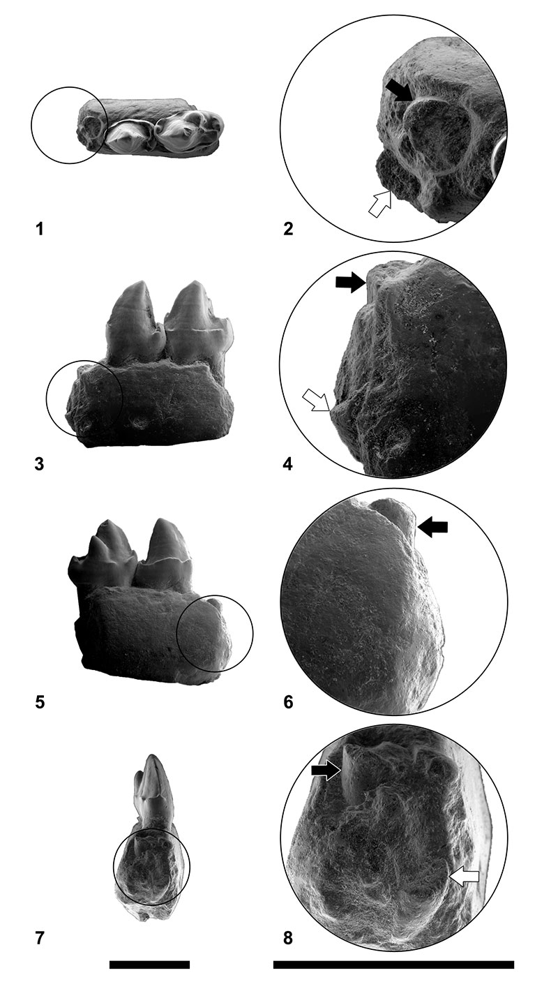
FIGURE 3. Agerinia roselli from Les Saleres. IPS-82793, isolated right M1 in occlusal (1), buccal (2) and lingual (3) views. IPS-82816, isolated right M1 in occlusal (4), buccal (5) and lingual (6) views. IPS-2542, left mandible fragment with M2 in occlusal (7), buccal (8) and lingual (9) views. IPS-82794, right mandible fragment with M2 in occlusal (10), buccal (11) and lingual (12) views. IPS-1981, holotype, left mandible fragment with M2 and M3 in occlusal (13), buccal (14) and lingual (15) views. IPS-2541, right mandible fragment with M2 and M3 in occlusal (16 ), buccal (17) and lingual (18) views. IPS-82795, right mandible fragment with M3 in occlusal (19), buccal (20) and lingual (21) views. IPS-82790, left mandible fragment preserving part of the ramus mandibularis and a fragment of the M3 in occlusal (22), buccal (23) and lingual (24) views. Scale bar represents 3 mm.
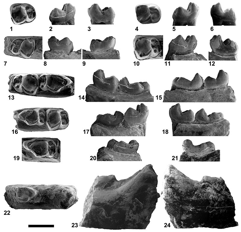
FIGURE 4. Body mass estimate regressions for Agerinia roselli. 1, derived from the area of the M1; 2, derived from the area of the M2. Black dots represent different molars of A. roselli. Black line indicates regression based on extant prosimian data from Egi et al. (2004).
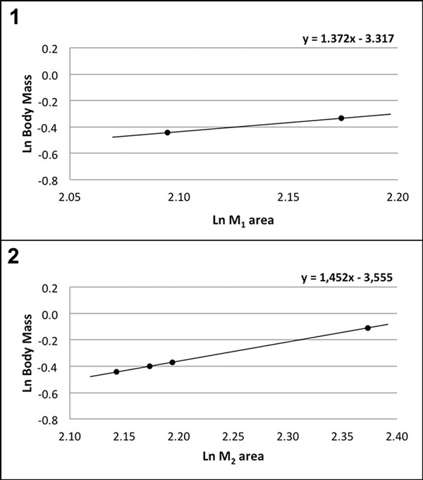
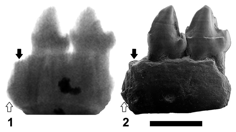
FIGURE 5. Radiograph represented with inverted colours (1) and ESEM micrograph (2) of the left mandible fragment of Agerinia roselli (IPS-2543) in buccal view. White arrows indicate the position of the most mesial root; black arrows indicate the position of the root immediately mesial with respect to the P3. Scale bars represent 3 mm.