FIGURE 1. Tanytarsini - inclusions in Baltic amber from the Hoffeins collection (for details see Table 1 and Material examined).
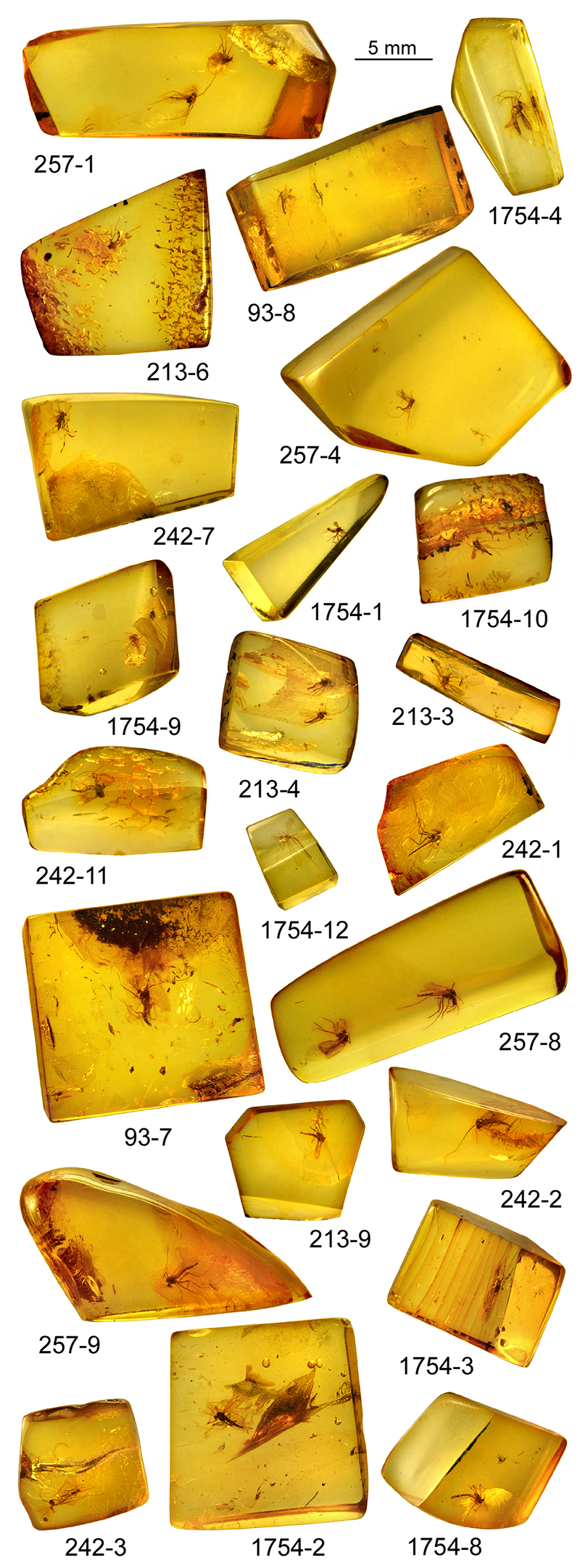
FIGURE 2. Rheotanytarsus hoffeinsorum sp. nov., adult male, holotype. 1, as syninclusion in amber (at left); 2, habitus.
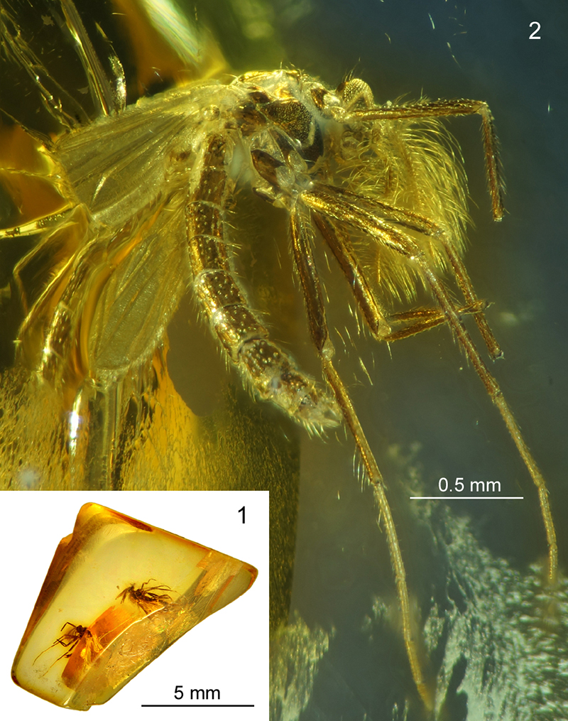
FIGURE 3. Rheotanytarsus hoffeinsorum sp. nov., adult male, holotype. Hypopygium and its structures in dorsal (1-3) and ventral aspect (4-6), photographed in reflected light (1, 4, 5) and drawn (2, 3, 6); 3 , superior volsella; 5-6, median volsella (6 magnified ca. 2 times relative to 5 and ca. 5 times relative to 4).
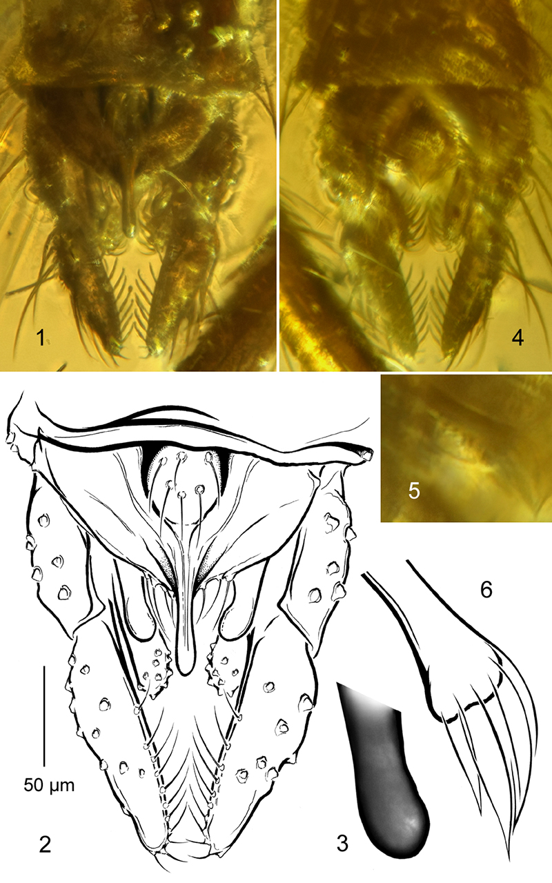
FIGURE 4. Tanytarsus crocota sp. nov., adult male, holotype. 1, inclusion in amber; 2, habitus; 3-4, wing photographed in transmitted (3) and reflected light (4); 5, spur of fore leg tibia.
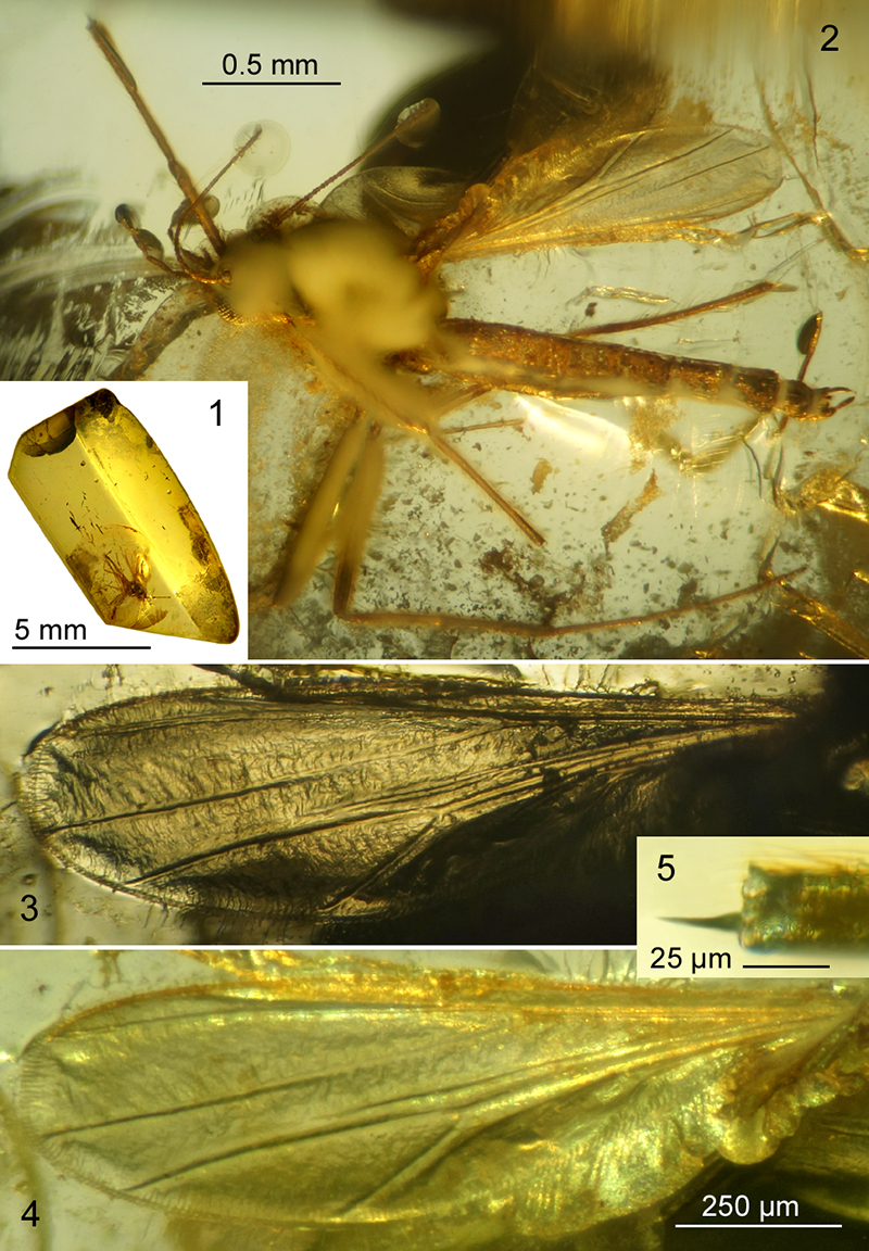
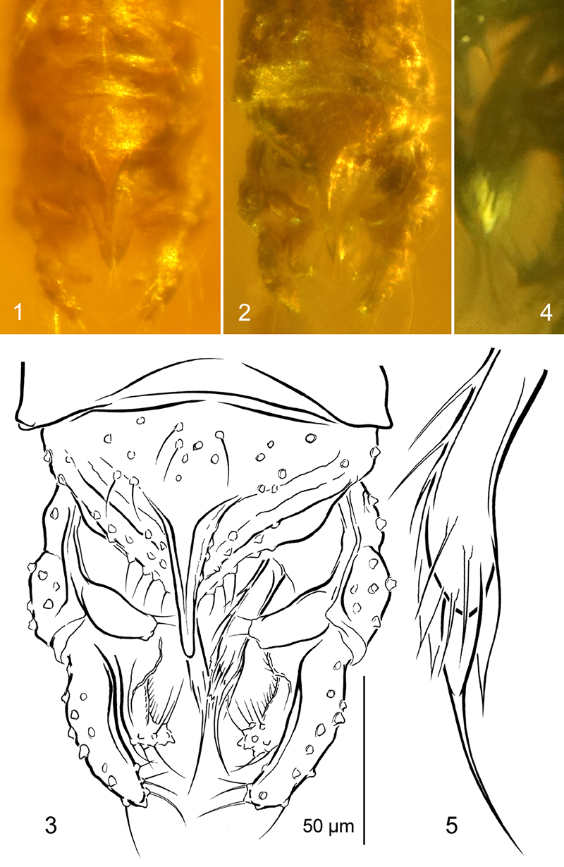
FIGURE 5. Tanytarsus crocota sp. nov., adult male, holotype. Hypopygium and its structures in dorsal (1, 2) and ventral aspect (3, 4), photographed in reflected light (1), transmitted light (3) and drawn (2, 4); 3-4, median volsella (4 magnified ca. 3 times relative to 2 and 3).
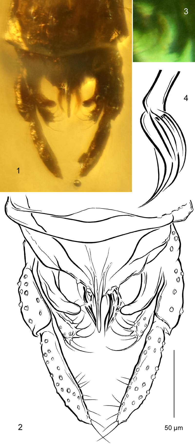
FIGURE 6. Variations of diagnostic structures in males of Tanytarsus protogregarius (1, 2) and T. serafini (3-6). Hypopygium in dorsolateral aspect (1, 3, 5) and its structures magnified ca. 3-4 times (below): anal point (2, 4) and superior volsella (6).
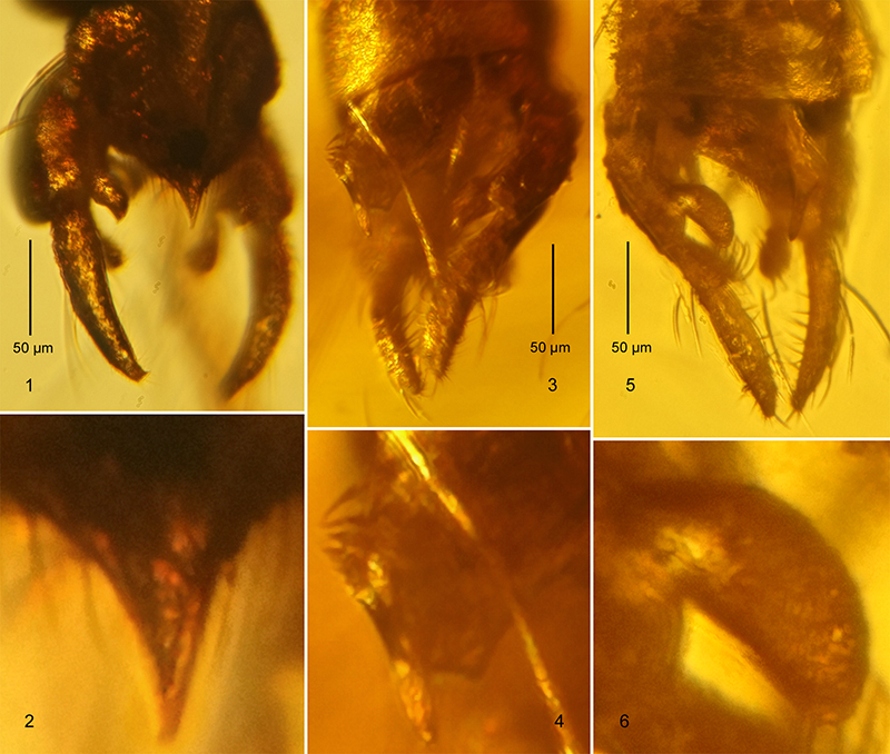
FIGURE 7. Stempellinella fibra sp. nov., adult male, holotype. 1, inclusion in amber; 2, habitus; 3, antenna (white arrows: borders between well discernible flagellomeres; grey arrow: incomplete fusion); 4, frontal tubercle (black arrow), antennal pedicel and eye.
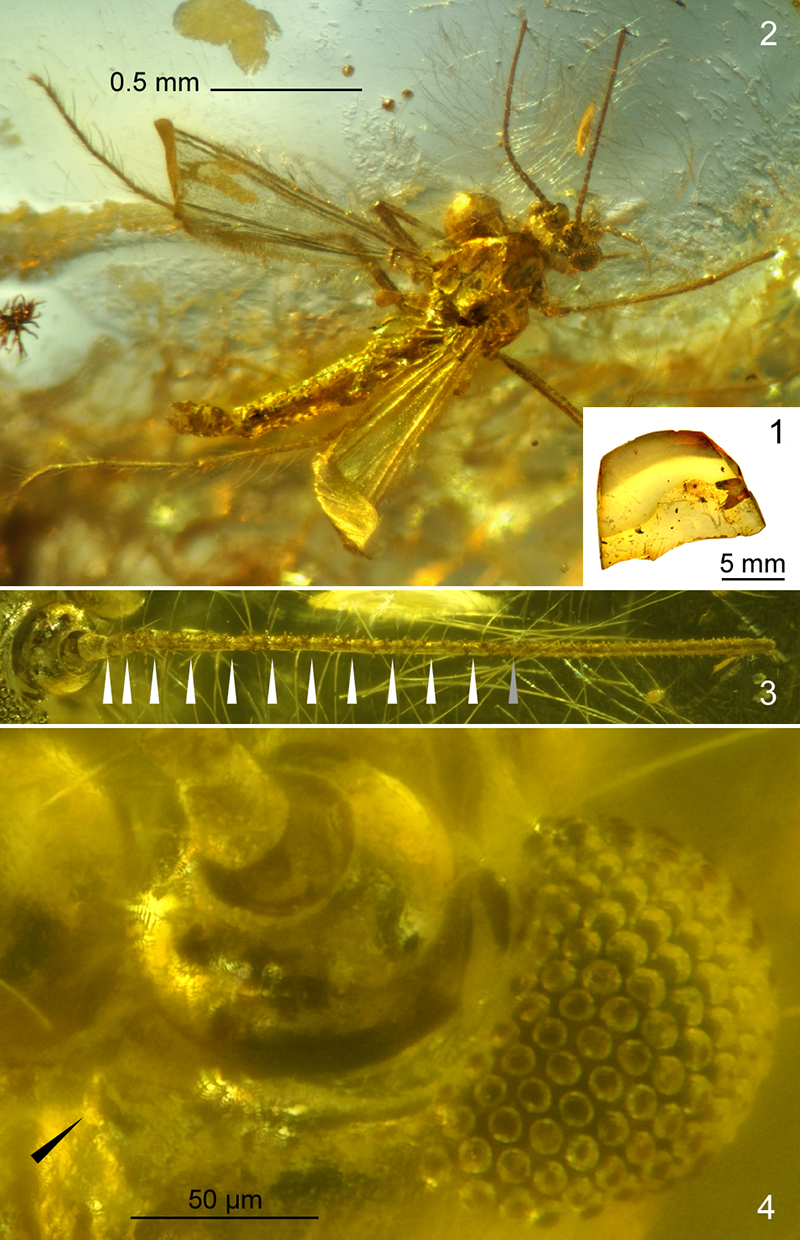
FIGURE 8. Stempellinella fibra sp. nov., adult male, holotype. Hypopygium and its structures in dorsal aspect, photographed in reflected light (1, 2), in transmitted light (4) and drawn (3, 5); 4-5, median volsella (5 magnified ca. 2 times relative to 4).