FIGURE 1. The Dockum Group exposures (highlighted in grey) in western Texas and eastern New Mexico (modified after Lehman, 1994a). Fossil localities are spotted (yellow-filled circles) within the corresponding counties of Garza, Howard and Randall (marked in red).
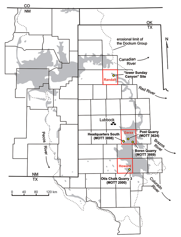
FIGURE 2. Generalized columnar sections of the Dockum Group in Texas in Randall, Garza and Howard counties, including the stratigraphic positions of the cited quarries in bold (modified after Ash, 1976; Lucas and Anderson, 1993b; Martz, 2008; Martz et al., 2013; and Bill Mueller, personal commun., 2014, 2015). Unit numbers and lithostratigraphic correlatives for the Garza County section are taken from Martz et al. (2013).
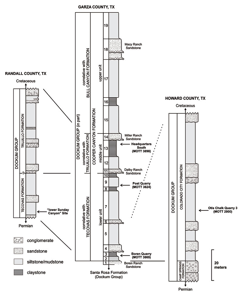
FIGURE 3.1, Location map of the "lower Sunday Canyon" Site within the Palo Duro Canyon State Park (bordered in red on the map, taken from Google Earth 2016); 2, Field photograph of the "lower Sunday Canyon" Site, indicated by the red arrow (Photo credit: Bill Mueller).
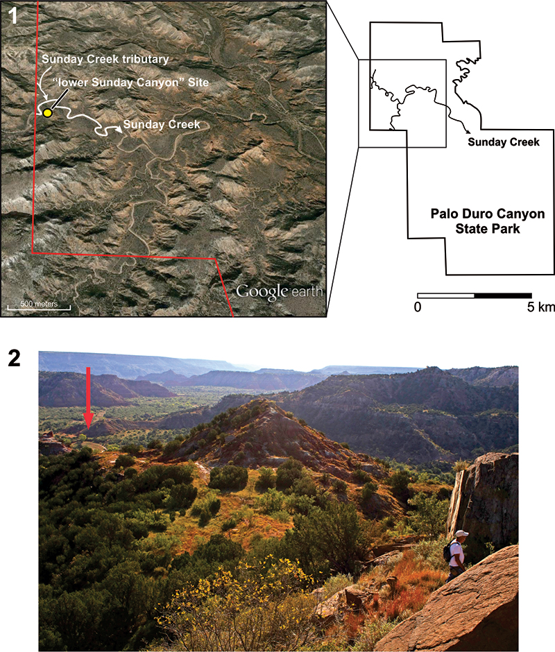
FIGURE 4. Complete listing of hitherto published Dockum non-dinosaurian dinosauromorph specimens from the Dockum Group of Texas with voucher information and representing elements, and their stratigraphic distribution. Lithologic thicknesses are not to scale.
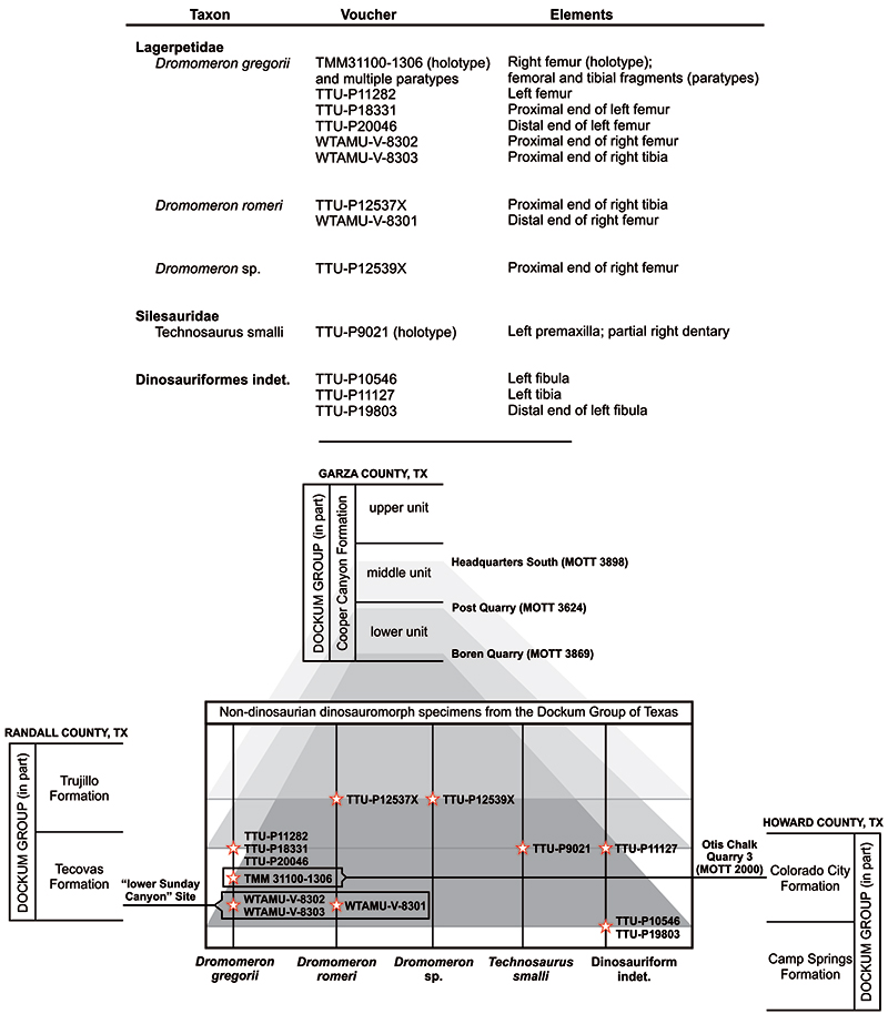
FIGURE 5.Dromomeron gregorii (TTU-P18331), proximal end of left femur in 1, anterior view; 2, medial view; 3, posterior view; 4, lateral view; 5, proximal view. Abbreviations: at, anterior trochanter; fh, femoral head (note that the femoral is broken); ft, fourth trochanter; pmt, posteromedial tuber; tf, trochanteric fossa; ts, trochanteric shelf. Hatching indicates the damaged areas. Arrow indicates anterior side.
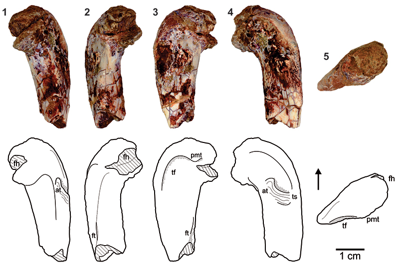
FIGURE 6.Dromomeron gregorii (TTU-P20046), distal end of left femur in 1, anterior view; 2, medial view; 3, posterior view; 4, lateral view; 5, distal view. Abbreviations: fc, fibular condyle; ig, intercondylar groove; lc, lateral condyle; mc, medial condyle; r, ridge; s, scar. Hatching indicates the damaged areas. Arrow indicates anterior side.
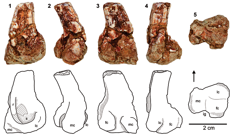
FIGURE 7.Dromomeron gregorii (WTAMU-V-8302), proximal end of right femur in 1, anterior view; 2, medial view; 3, posterior view; 4, lateral view; 5, proximal view. Abbreviations: at, anterior trochanter; fh, femoral head; ft, fourth trochanter; pmt, posteromedial tuber; tf, trochanteric fossa; ts, trochanteric shelf; ve, ventral emargination. Hatching indicates the damaged areas. Arrow indicates anterior side.
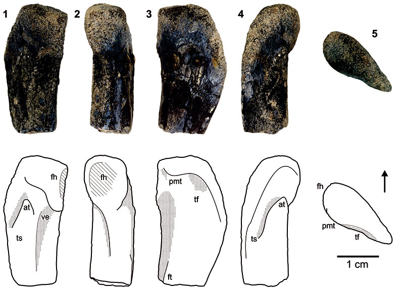
FIGURE 8.Dromomeron gregorii (WTAMU-V-8303), proximal end of right tibia in 1, lateral view; 2, anterior view; 3, medial view; 4, posterior view; 5, proximal view. Abbreviations: cc, cnemial crest; lc, lateral crest; mc, medial crest; r, ridge. Arrow indicates anterior side.
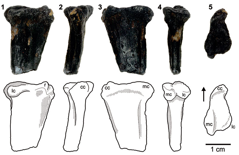
FIGURE 9.Dromomeron romeri (TTU-P12537X), proximal end of right tibia in 1, anterior view; 2, lateral view; 3, posterior view; 4, medial view; 5, proximal view. Abbreviations: cc, cnemial crest; lc, lateral condyle; mc, medial condyle. Hatching indicates the damaged areas. Arrow indicates anterior side.
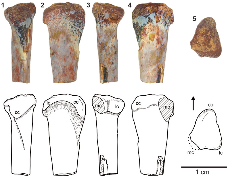
FIGURE 10.Dromomeron romeri (WTAMU-V-8301), distal end of right femur in 1, anterior view; 2, medial view; 3, posterior view; 4, lateral view; 5, distal view. Abbreviations: amr, anteromedial ridge (sensu Irmis et al., 2007b); fc, fibular condyle; ig, intercondylar groove; lc, lateral condyle; mc, medial condyle; mlr, mediolateral ridge. Arrow indicates anterior side.
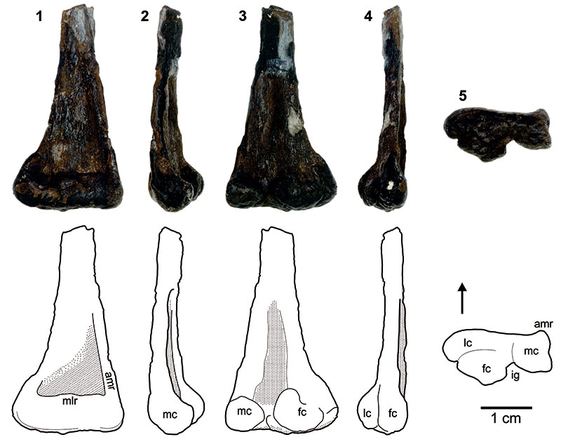
FIGURE 11.Dromomeron sp. (TTU-P12539X), proximal end of right femur in 1, anterior view; 2, medial view; 3, posterior view; 4, lateral view; 5, proximal view. Abbreviations: fh, femoral head (note that the femoral head is broken); pmt, posteromedial tuber; tf, trochanteric fossa; ve, ventral emargination. Hatching indicates the damaged areas. Arrow indicates anterior side.
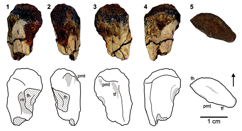
FIGURE 12. Dinosauriformes gen. and sp. indet. (TTU-P10546), left fibula in 1, anterior view; 2, lateral view; 3, posterior view; 4, medial view; 5, proximal view; 6, distal view. Abbreviations: bs, beveled surface; da, damaged anterior part; dm, damaged posterior part; lf, lateral facet; mf, medial facet; pp, posterior process; py, pyramidal process. Arrow indicates anterior side.
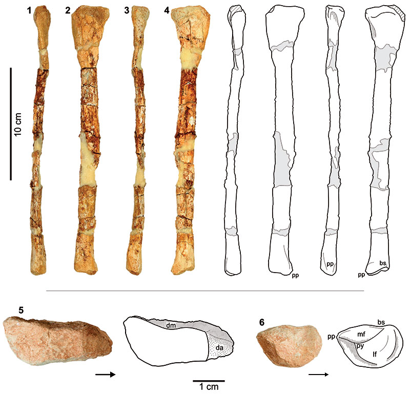
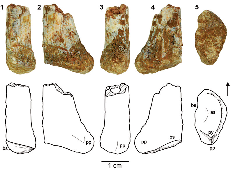
FIGURE 13. Dinosauriformes gen. and sp. indet. (TTU-P19803), distal end of left fibula in 1, anterior view; 2, lateral view; 3, posterior view; 4, medial view; 5, distal view. Abbreviations: as, articular surface for astragalus and calcaneum; bs, beveled surface; pp, posterior process; py, pyramidal process. Hatching indicates the damaged areas. Arrow indicates anterior side.