FIGURE 1. Elephantomyia (Elephantomyia) grata Podenas and Poinar, 2001, No. 5546 (sex unknown). 1, a drawing of the head (reconstructed) in latero-dorsal view; 2, a schematic representation of the relation between the length of palpi (p), antenna (a), rostrum (r) and head (h); 3, a drawing of the left wing venation; and 4, a drawing of the right antenna in latero-dorsal view.
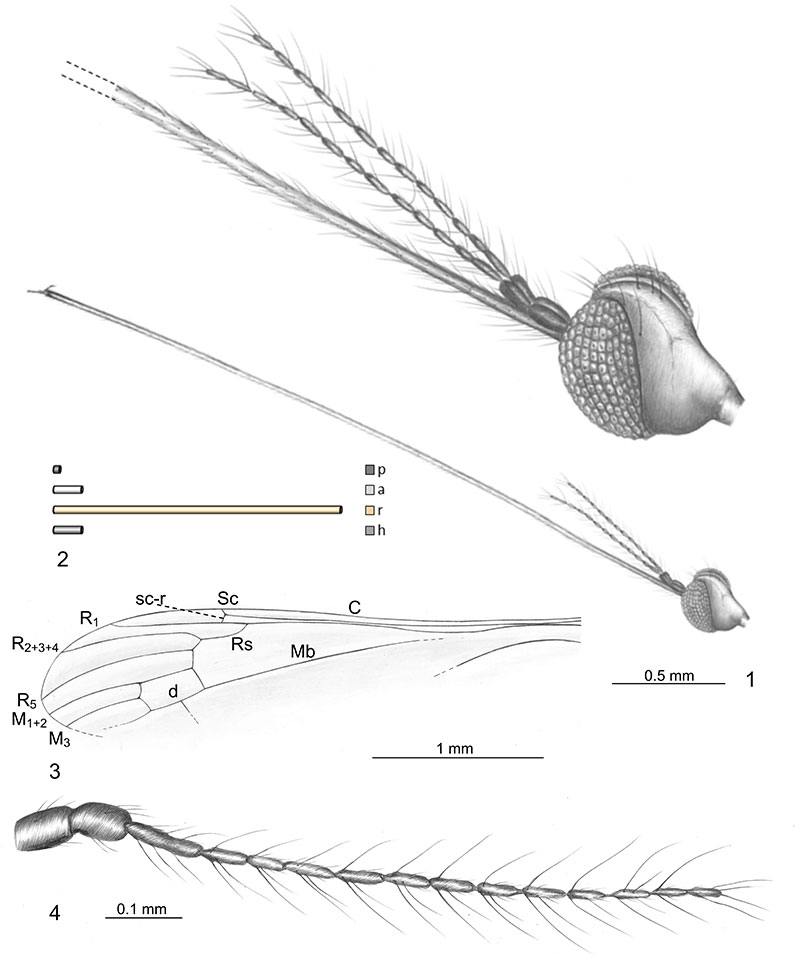
FIGURE 2. Elephantomyia (Elephantomyia) grata Podenas and Poinar, 2001, No. 5546 (sex unknown). 1, a photograph of the body in lateral view; and 2, a photograph of the right antenna in latero-dorsal view.
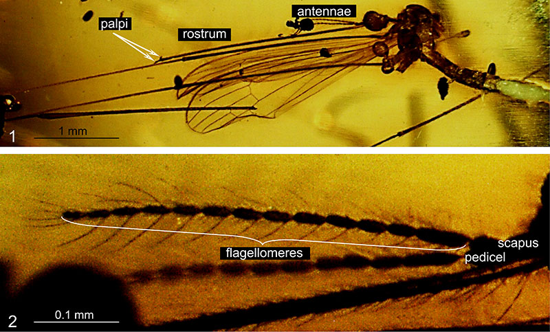
FIGURE 3. Helius (Helius) neali n. sp., No. 10938 (male). 1, a drawing of the head (reconstructed) in fronto-dorsal view; 2, a schematic representation of the relation between the length of antenna (a), rostrum (r) and head (h); 3, a drawing of the left antenna in ventral view; 4, a drawing of the left wing venation; and 5, a drawing of the right gonocoxite and gonostyles in ventral view. Abbreviation: gx, gonocoxite; ing, inner gonostylus; oug, outer gonostylus.
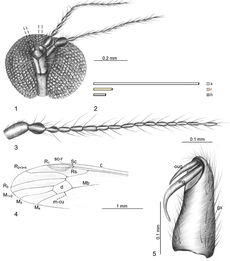
FIGURE 4. Helius (Helius) neali n. sp., No. 10938 (male). 1, a photograph of the body in fronto-dorsal view; and 2, a photograph of the head in fronto-dorsal view and the part of thorax in dorsal view.
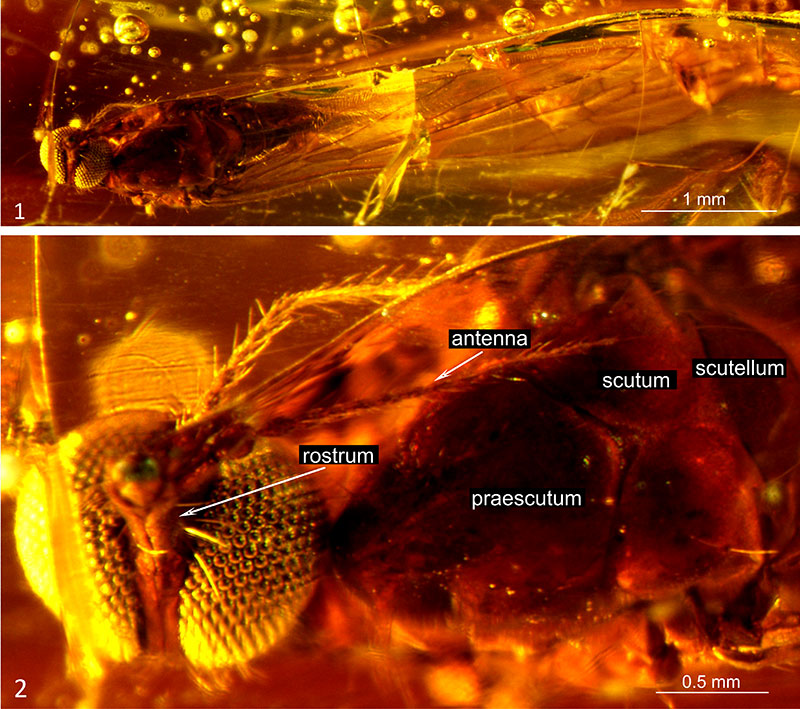
FIGURE 5. Helius (Helius) oosterbroeki n. sp., No. 10189 (male). 1, a drawing of the head (reconstructed) in fronto-ventral view; 2, a drawing of the right antenna in fronto-dorsal view; 3, a drawing of the right palpus in fronto-ventral view; 4, a schematic representation of the relation between the length of palpi (p), antenna (a), rostrum (r) and head (h); 5, a drawing of right gonocoxite and gonostyles in ventral view; and 6, a drawing of the right wing. Abbreviation: gx, gonocoxite; ing, inner gonostylus; oug, outer gonostylus.
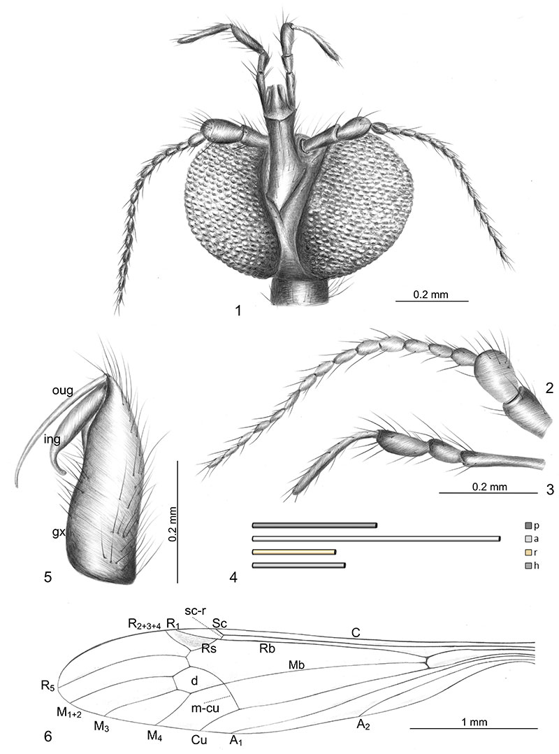
FIGURE 6. Helius (Helius) oosterbroeki n. sp., No. 10189 (male). 1, a photograph of the body in ventral view; 2, a photograph of the head in fronto-ventral view; and 3, a photograph of hypopygium in ventral view.
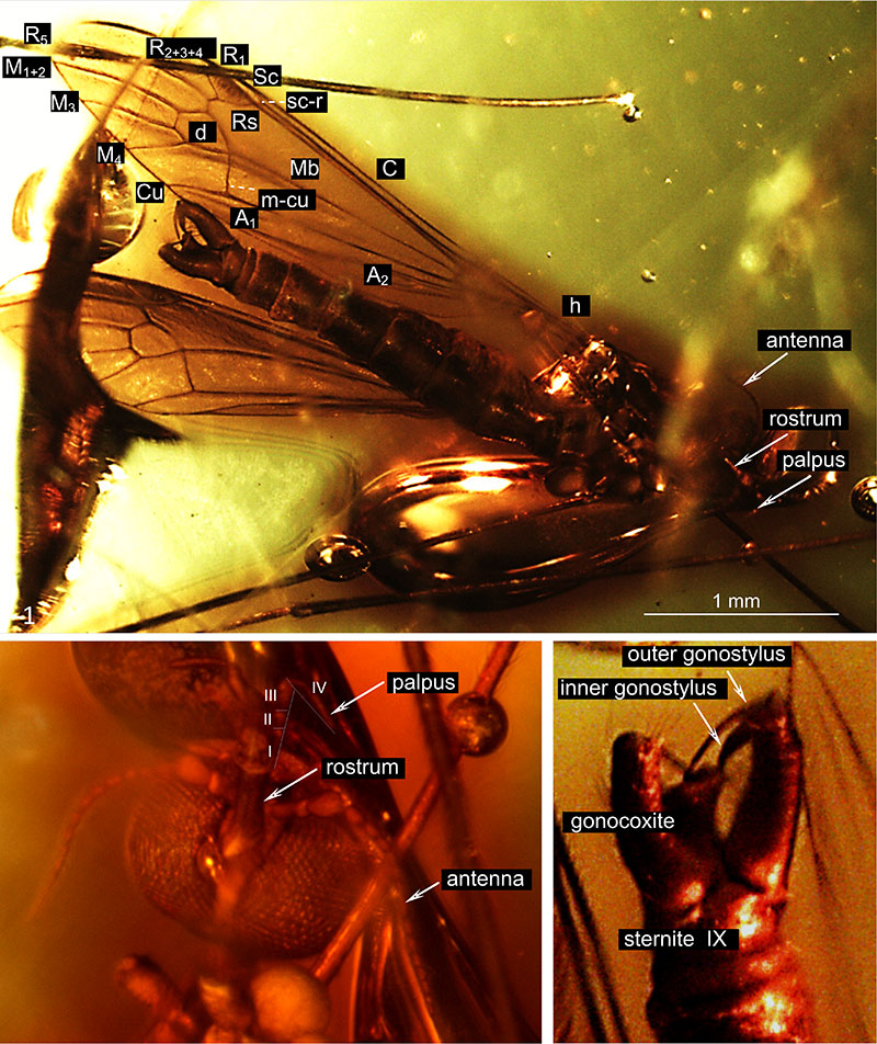
FIGURE 7. Toxorhina (Ceratocheilus) mexicana n. sp., No. PI II 1870 (female). 1, a drawing of the head (reconstructed) in lateral view; 2, a schematic representation of the relation between the length of palpi (p), antenna (a), rostrum (r) and head (h); 3, a drawing of the left wing venation; and 4, a drawing of the ovipositor in latero-ventral view.
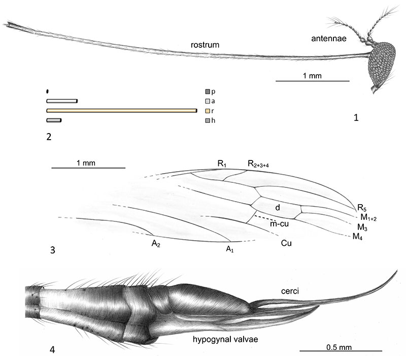
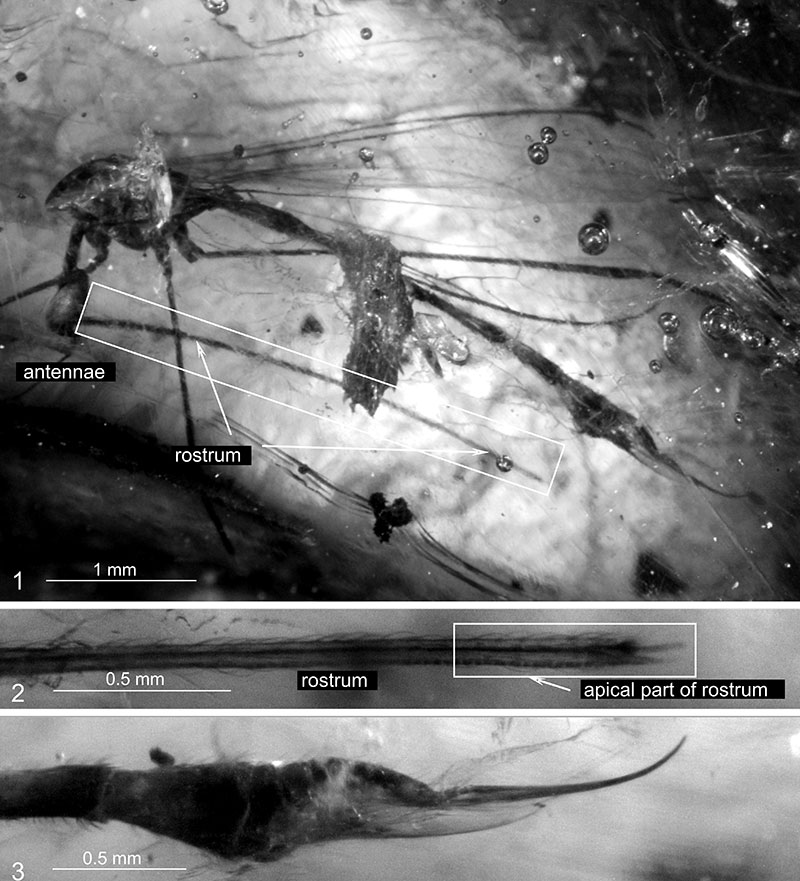
FIGURE 8. Toxorhina (Ceratocheilus) mexicana n. sp., No. PI II 1870 (female). 1, a photograph of the body in lateral view; 2, a photograph of the apical part of rostrum in lateral view; and 3, a photograph of the ovipositor in latero-ventral view.