FIGURE 1. Geographic and stratigraphic position of the locality where the new theropod was found. 1, Overview map of Germany, indicating the area of the locality of the new theropod in north-eastern Northrhine-Westphalia. 2, locality of the disused Pott quarry at Lutternsche Egge in the Wiehengebirge. 3, Simplified stratigraphic column of the rocks that crop out in the Wiehengebirge (E = east; W = west). Modified from Riegraf (1994). 4, Geological map of the area between Bünde and Minden. Middle Jurassic units, including the Ornatenton, are represented by the dullish dark green that follows the course of the Wiehengebirge; light blue-grey colours represent Upper Jurassic units; light green marks the Lower Cretaceous (‘Wealden’) outcrops; orange colours represent Upper Triassic rocks. From the Northrhine-Westphalian Geological Survey (www.gd.nrw.de).
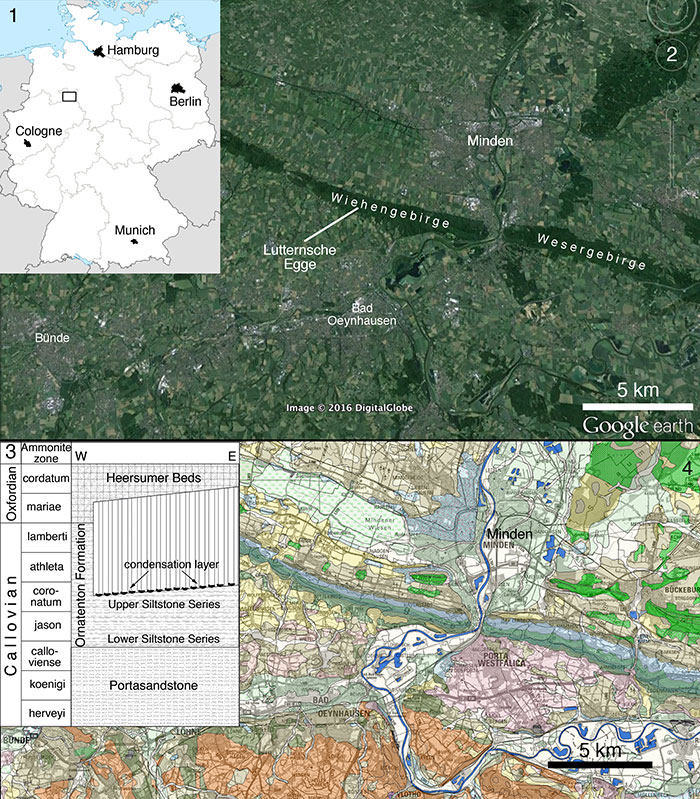
FIGURE 2. Quarry map of the excavation at Lutternsche Egge, showing the position of the different elements of the new taxon in situ. Numbers refer to the specimen numbers of the separate elements (see text). Scale is in 50 cm increments.
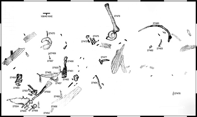
FIGURE 3. Outline reconstruction of Wiehenvenator albati n. gen., n. sp., indicating recovered elements. Based on the reconstruction of Torvosaurus by Scott Hartman; used with permission. Scale bar equals 1 m.
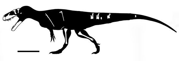
FIGURE 4. Tentative reconstruction of the skull of Wiehenvenator albati, with the recovered elements shown in their approximate relation to each other. Scale bar equals 10 cm.
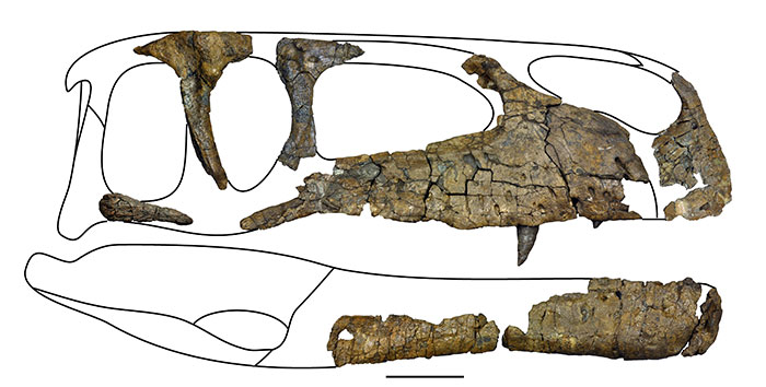
FIGURE 5. Right premaxilla of Wiehenvenator albati in lateral (1), medial (2), anterior (3) and ventral (4) views. Abbreviations: en, external nares; f, foramen; ips, interpremaxillary suture facet; np, nasal process; numbers A1 to A4 indicate alveoli. Scale bar equals 50 mm.
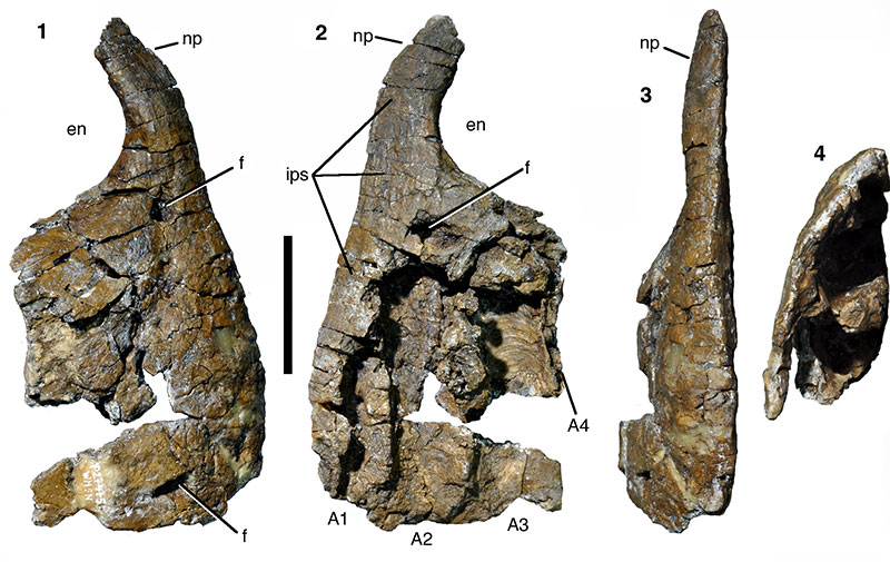
FIGURE 6. Right maxilla of Wiehenvenator albati in lateral (1) and medial (2) views. Abbreviations: af, antorbital fossa; afo, alveolar foramen; amp, base of anteromedial process; ap, ascending process; ep, excavatio pneumatica; f, foramen; iv, invertebrate; jf, jugal facet; pdr, paradental ridge; pmr, promaxillary recess; pro, promaxillary foramen. Scale bar equals 100 mm.
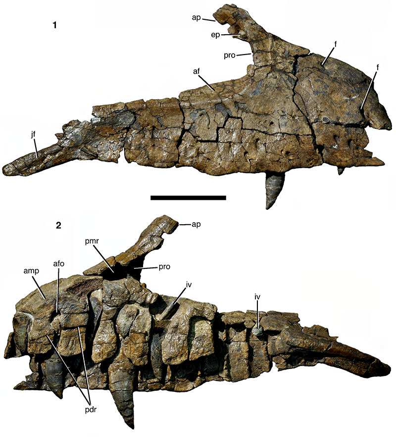
FIGURE 7. Stereophotographs of the right maxilla of Wiehenvenator albati in medial (1) and lateral (2) views.
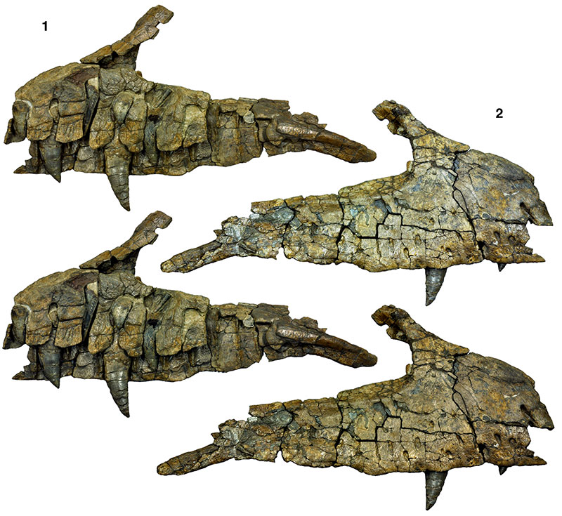
FIGURE 8. Stereophotographs of the promaxillary foramen in the right maxilla of Wiehenvenator albati in posterodorsal view. Abbreviations as in Figure 6. Scale bar equals 50 mm.
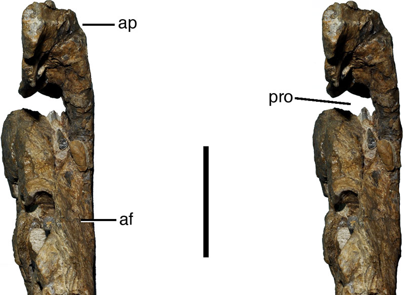
FIGURE 9. Right lacrimal of Wiehenvenator albati in lateral (1; stereophotographs), medial (2), anterior (3; stereophotographs), posterior (4; stereophotographs), and dorsal (5) views. Abbreviations: af, antorbital fossa; d, depression; fo, foramen; lf, lacrimal fenestra; prf, facet for prefrontal; r, ridge. Scale bar equals 50 mm.
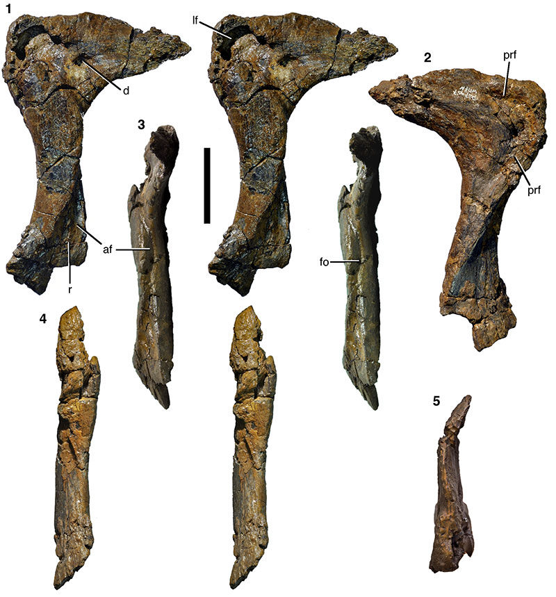
FIGURE 10. Right postorbital of Wiehenvenator albati in lateral (1), anterior (2), medial (3), posterior (4), and dorsal (5) views (all stereophotographs). Abbreviations: d, depression, ff, frontal facet; jf, jugal facet; lf, laterosphenoid facet; of, orbital facet; sob, supraorbital brow; stf, supratemporal fossa. Scale bar equals 100 mm.
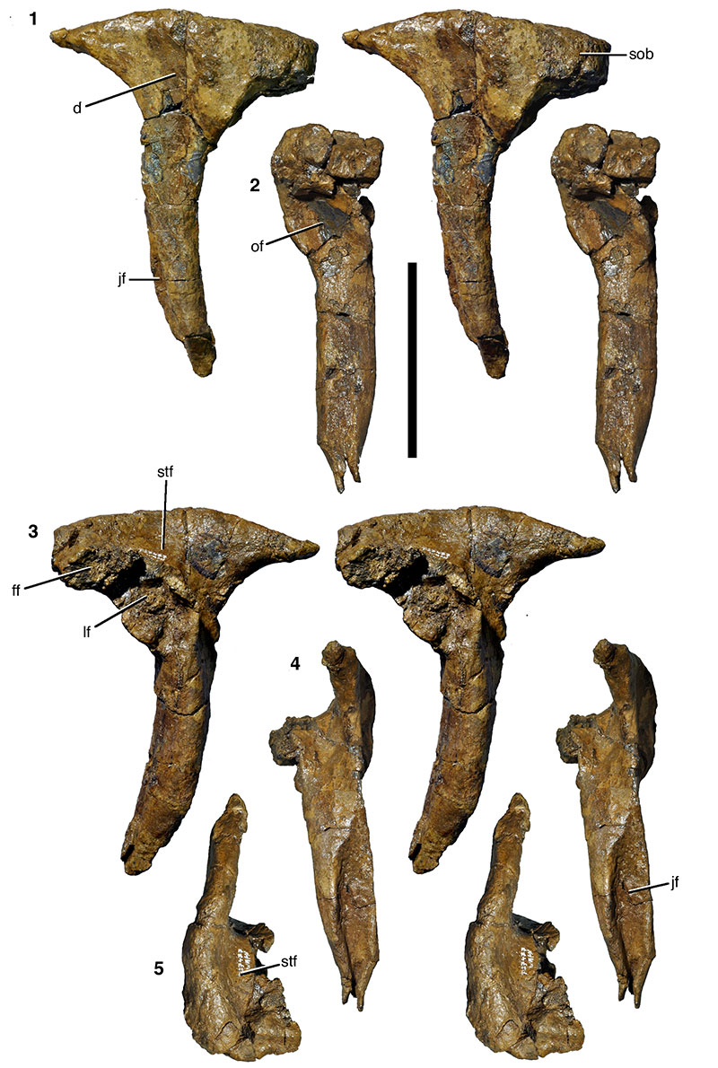
FIGURE 11. Anterior (jugal) process of the right quadratojugal(?) of Wiehenvenator albati in lateral (1), medial (2), dorsal (3), and ventral (4) views. Abbreviations: djp, facet for the dorsal posterior prong of the jugal; vjp, facet for the ventral posterior prong of the jugal. Scale bar equals 100 mm.
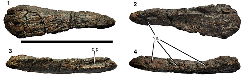
FIGURE 12. Right dentary of Wiehenvenator albati in lateral (1), ventral (2), and medial (3) views. Scale bar equals 100 mm.
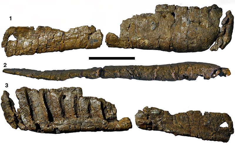
FIGURE 13. Dentition of Wiehenvenator albati. 1, replacement tooth in the 2nd maxillary alveolus; 2, functional tooth in the 4th maxillary alveolus; 3, probable maxillary tooth with partially preserved root (WMN P27459) in lingual view; 4-6, probable maxillary tooth with complete root (WMN P27483) in distal (4), lingual (5) and mesial (6) views; 7, mesial (posterior premaxillary or anterior dentary) tooth (WMN P27467) in labial(?) view; 8, maxillary or dentary tooth (WMN P27473) in labial(?) view; 9, detail of crown apex of WMN P27373, showing the carina that is continuous across the tip (mesial is to the left); 10-12, mesial (probably premaxillary) tooth (WMN P27456) in distal (10), labial (11) and mesial (12) views. Abbreviations: idp, interdental plates; pdl, paradental lamina. Scale bars equal 10 mm (1, 2) and 50 mm (3-12; not 9).
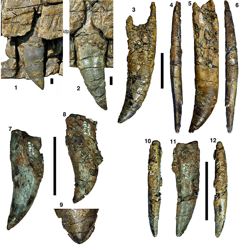
FIGURE 14. Anterior mid-caudal vertebra of Wiehenvenator albati in lateral (1; stereophotographs), dorsal (2), anterior (3), and ventral (4) views. Abbreviations: nc, neural canal; ns, neural spine; pcd, pleurocentral depression; poz, postzygapophysis, prz, prezygapophysis; r, ridge; tp, transverse process. Scale bar equals 100 mm.
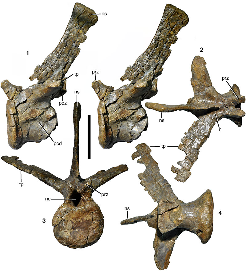
FIGURE 15. Mid-caudal vertebra of Wiehenvenator albati in anterior (1) and lateral (2; stereophotographs) views. Abbreviations as in Figure 14, and: as, anterior spur. Scale bar equals 100 mm.
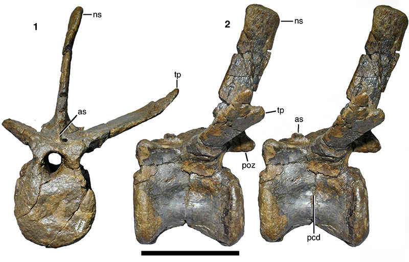
FIGURE 16. Posterior mid-caudal vertebra of Wiehenvenator albati in anterior (1), lateral (2), and ventral (3; stereophotographs) views. Abbreviations as in Figures 14 and 15, and: vg, ventral groove. Scale bar equals 100 mm.
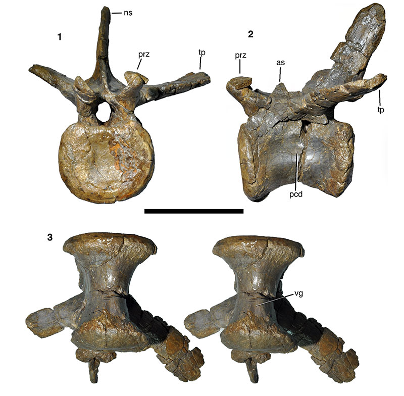
FIGURE 17. Dorsal ribs and gastralia of Wiehenvenator albati. 1, thoracic rib. 2, abdominal rib. 3-5, fused posterior medial gastralia in dorsal (3), ventral (4; stereophotographs), and anterior (5) views. Scale bar equals 100 mm.
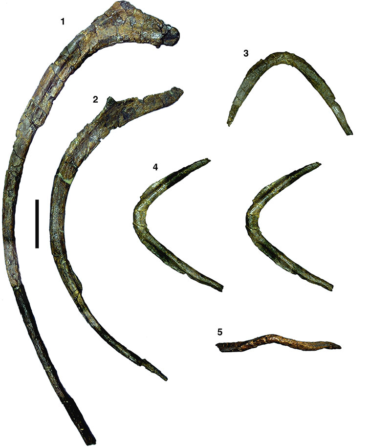
FIGURE 18. Manual phalanx (probably right phalanx III-1) of Wiehenvenator albati in medial (1), dorsal (2) and distal (3) views. Scale bar equals 50 mm.
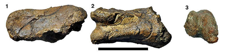
FIGURE 19. Left fibula of Wiehenvenator albati in lateral (1), proximal (2), medial (3), anterior (4), and distal (6) views. 5, detail of proximal shaft of the fibula in medial view (stereophotographs). Abbreviations: d, depression; or, oblique ridge; re, raised edge; sw, swelling; tu, tubercle. Scale bar equals 100 mm (not for 5).
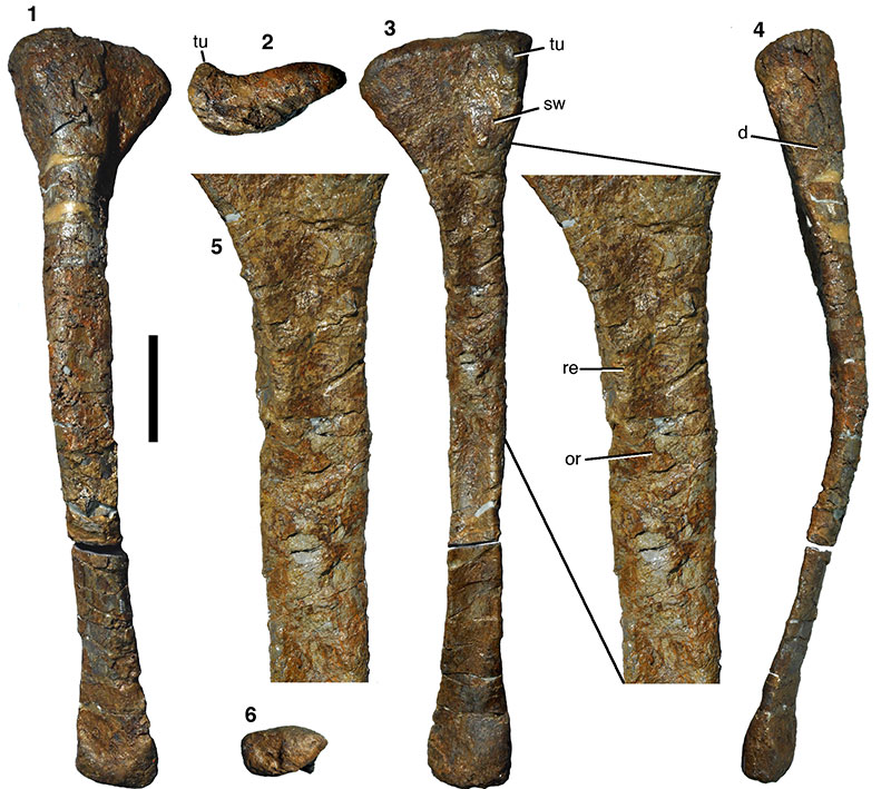
FIGURE 20. Right astragalus of Wiehenvenator albati in anterior (1; stereophotographs), medial (2), proximal (3; stereophotographs), and posterior (4) views. Abbreviations: af, articular facet for ridge on anterior side of tibia; ap, ascending process; d, depression; ff, fibular facet; tf, facet for articulation with tibia. Scale bar equals 50 mm.
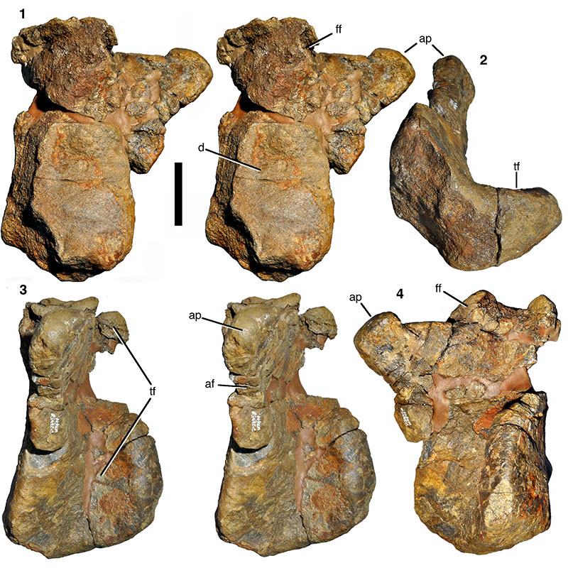
FIGURE 21. Detail of the right astragalus of Wiehenvenator albati: Base of the ascending process and fibular facet in anteroproximal view, showing large foramen anterior to the ascending process. Abbreviations as in Figure 20, and: f, foramen.
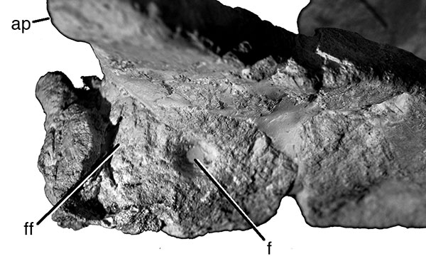
FIGURE 22. Right calcaneum of Wiehenvenator albati in anterior (1) and lateral (2) views. Scale bar equals 50 mm.
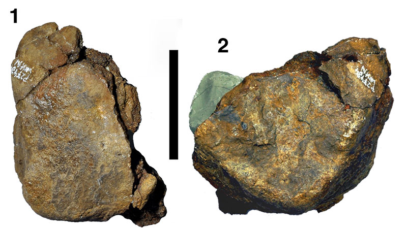
FIGURE 23. Overview of transverse thin section of the left fibula of Wiehenvenator albati. Most of the primary tissue is secondarily remodelled, but growth marks are still preserved anteriorly and posteriorly (white arrows). Abbreviations: lat, lateral; post, posterior. Scale bar equals 5 mm. Inset: Slightly clockwise rotated and magnified image of the posterior outer bone cortex (see frame in overview image). Growth marks are marked by black arrows. Scale bar equals 400 µm.
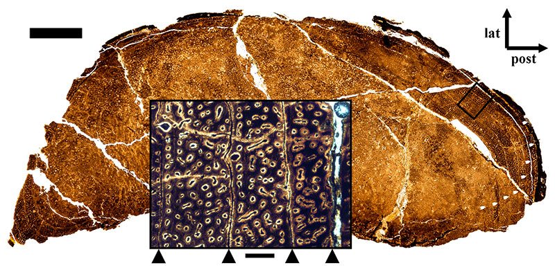
FIGURE 24. Phylogenetic position of Wiehenvenator albati, based on an analysis of 62 taxa and 351 characters (see text for details). 1, strict consensus tree. 2, reduced consensus tree after the a posteriori exclusion of Streptospondylus.
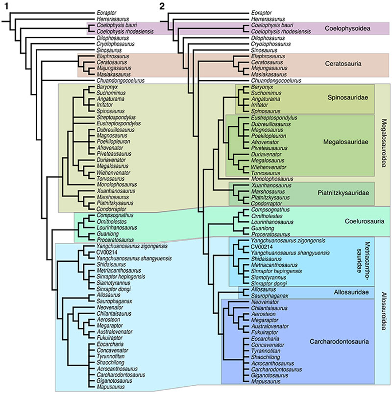
FIGURE 25. Time-calibrated cladogram of early tetanuran relationships, showing the earliest records of the distinct clades, and the rapid evolution of tetanurans in the early Middle Jurassic.
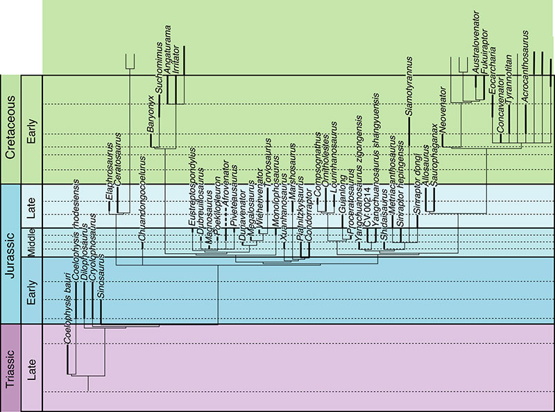
FIGURE 26. Time-calibrated informal supertree of theropod relationships, showing the explosive radiation of the clade in the latest Early and Middle Jurassic. Theropod interrelationships are based on Ezcurra (2012) and Langer et al. (2014) for non-averostran taxa, Rauhut and Carrano (2016) for Ceratosauria, the present study for basal tetanurans, Brusatte and Carr (2016) for tyrannosauroids, and Brusatte et al. (2014) and Foth et al. (2014) for other coelurosaurs.
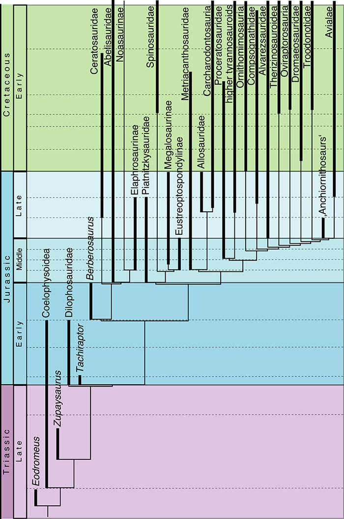
FIGURE 27. Representation of different clades of Middle (1) and Late Jurassic (2) nominal theropod taxa. Figures 3 and 4 show the same for large theropod taxa (body weight > 200 kg) only.

FIGURE 28. Composition of Middle and Late Jurassic theropod faunas, as percentage of localities in which certain clades have been recorded.
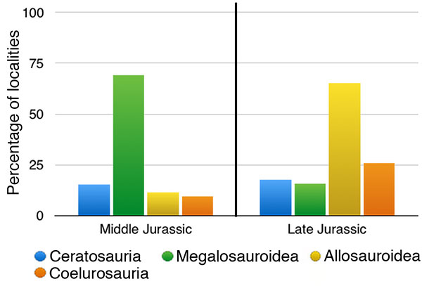
FIGURE 29. Number of Late Jurassic localities in which theropods have been recorded. The green portion of the column indicates how many of these occurrences are accounted for by the Morrison Formation.
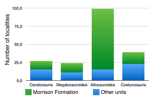
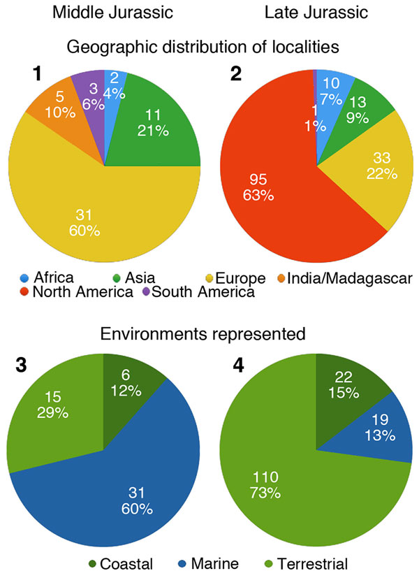
FIGURE 30. Possible biases that influence interpretations of the Jurassic theropod fossil record. Geographic distribution of Middle (1) and Late Jurassic (2) localities with identifiable theropod remains, and environments represented by Middle (3) and Late Jurassic (4) theropod localities.