FIGURE 1. Fossil diplostracan remains from Koonwarra. 1, An image of a fossil ephippium belonging to Ceriodaphnia sp. from the Koonwarra Fossil Beds (photographed with obliquely directed light), NMV P332637, 2, Interpretive drawing of Figure 1.1, scale bar for 1-2 equals 0.1 mm. 3, Diplostracan indet, NMV P109450 (figure 70G and 71C in Jell and Duncan, 1986), See Figure 1.4 for scale bar. 4, Diplostracan indet, NMV P109462 (figure 70H and 71E in Jell and Duncan, 1986). Scale bar in 1.4 applies to 1.3 as well. Scale bar for 1.3-4 equals 1 mm. Abbreviations: aII - antenna II; hd - head; tl - thoracic limbs; vv - valves.
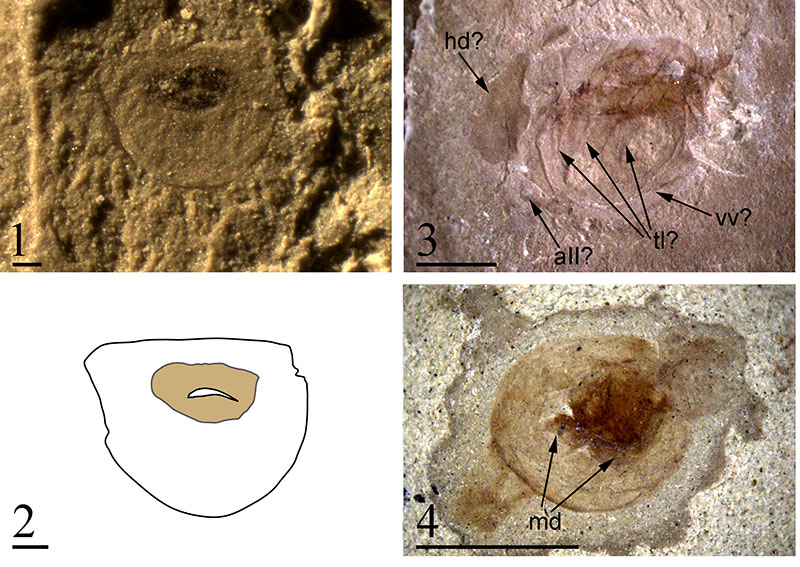
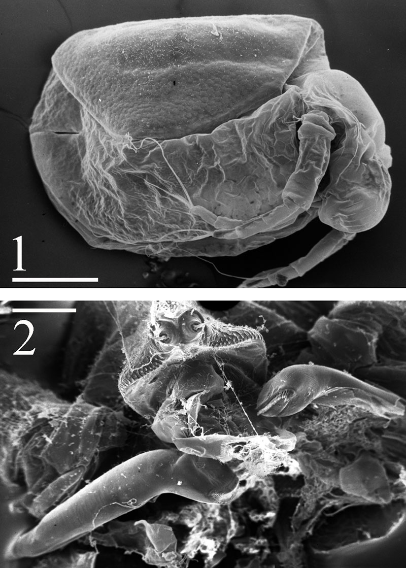
FIGURE 2. Modern Ceriodaphnia sp. 1, Scanning electron microscope image of a modern ephippium-bearing Ceriodaphnia sp. female (collected from Glubokoe Lake, European Russia) prior to releasing the ephippium during molting, ephippium tilted slightly away from angle of photograph, scale bar equals 0.1 mm. 2, Scanning electron microscope image of the mandibles of Daphnia magna , scale bar equals 0.1 mm.