FIGURE 1. Extant representative of Tanyderidae: Araucoderus gloriosus Alexander, 1920 in natural habitat. Photograph: R. Isai Madriz.
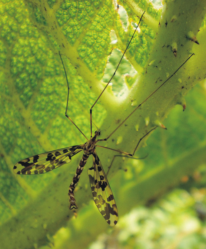
FIGURE 2. Localities of fossil outcrops (other than “Se”) and extant (“Se”) members of Nannotanyderinae subfam. nov. Abbreviations: B, Baltic region (this study); Do, Dorset of UK ( Nannotanyderus oliviae); Db, Dobbertin of Germany ( N. krzeminskii); G, Grimmen of Germany (N. krzeminskii and N. grimmensis); K, Karatau of Kazakhstan (N. kubekovensis); L, Lebanon ( N. ansorgei); M, Myanmar (Dacochile microsoma); S, Shar-Teg of Mongolia ( N. incertus); and Se, South Ethiopian Region (Peringueyomyina barnardi). Modified from Soszyńska-Maj and Krzemiński (2013, figure 1).
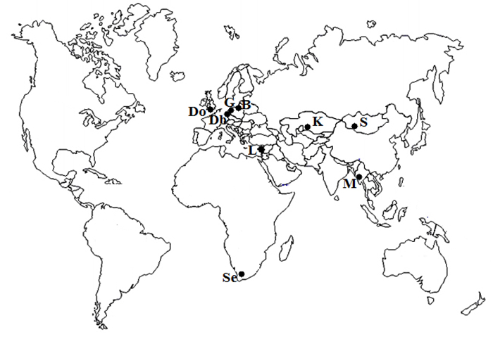
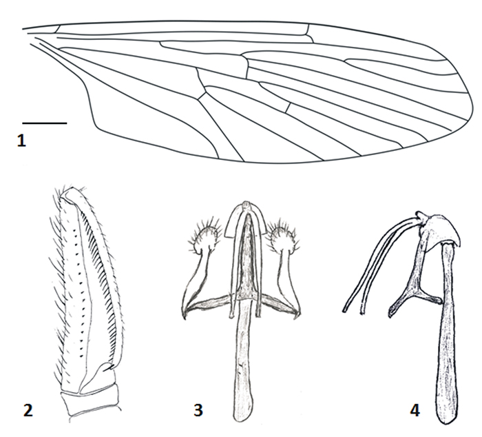
FIGURE 3. Comparison of external structure of male genitalia (1 and 2; in dorsal views) and wing venation (3 and 4; in dorsal views) in subfamilies. 1, Nannotanyderus ansorgei (JG. 385/2B), Nannotanyderinae. 2, Mischoderus annuliferus Hutton, 1900 (ISEZ PAN collection), Tanyderinae. 3, a new genus of Nannotanyderinae described below (holotype, MP/3376). 4, Mischoderus annuliferus (ISEZ PAN collection), Tanyderinae. Scale equals 0.5 mm.
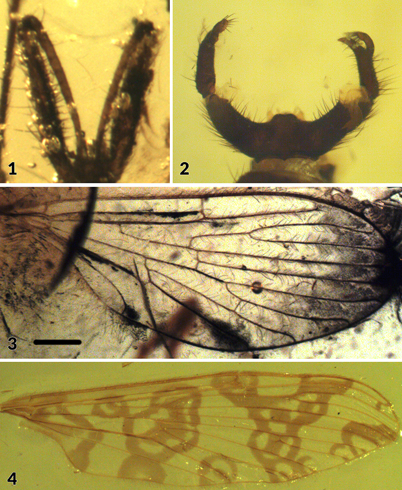
FIGURE 4. Drawing of Nannotanyderus oliviae wing venation (I-F/MP/1/1600/12) in dorsal view. Scale equals 0.5 mm.
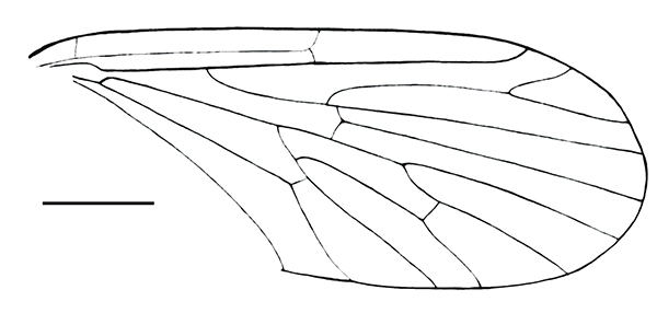
FIGURE 5. Drawing of Nannotanyderus krzeminskii wing venation (LGA 1145) in dorsal view. Scale equals 0.5 mm.
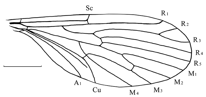
FIGURE 6. Drawing of Nannotanyderus grimmenensis wing venation (LGA 2222) in dorsal view. Scale equals 0.5 mm.
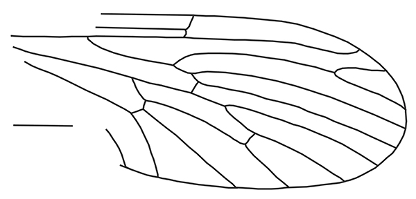
FIGURE 7. Drawing of Nannotanyderus incertus wing venation (No. 4270/2075) in dorsal view. Scale equals 0.5 mm.
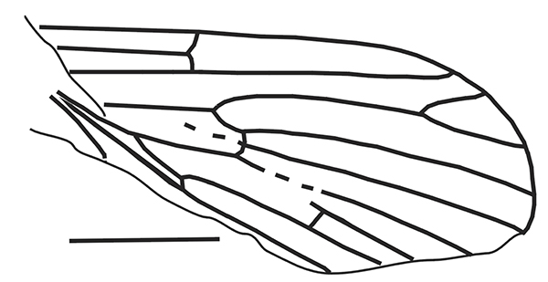
FIGURE 8. Drawing of Nannotanyderus kubekovensis wing venation (No. 2066/2182) in dorsal view. Scale equals 0.5 mm.
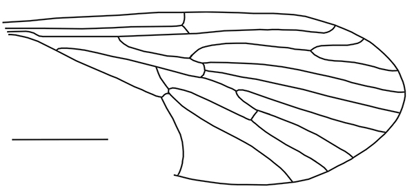
FIGURE 9. Photographs of Nannotanyderus ansorgei (JG. 385/2B). 1, whole specimen. 2, male genitalia in ventral view. Scale equals 0.5 mm.
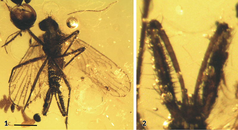
FIGURE 10. Drawings ofNannotanyderus ansorgei (JG. 385/2B). 1, wing venation. 2, male genitalia in dorsal view. Scale equals 0.5 mm.
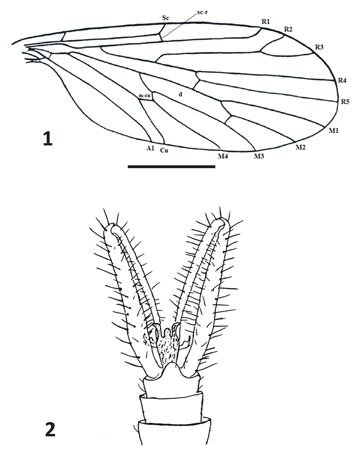
FIGURE 11. Photographs of Dacochile microsoma (Bu 1262). 1, whole specimen. 2, male genitalia in lateral view. Scale equals 1 mm.
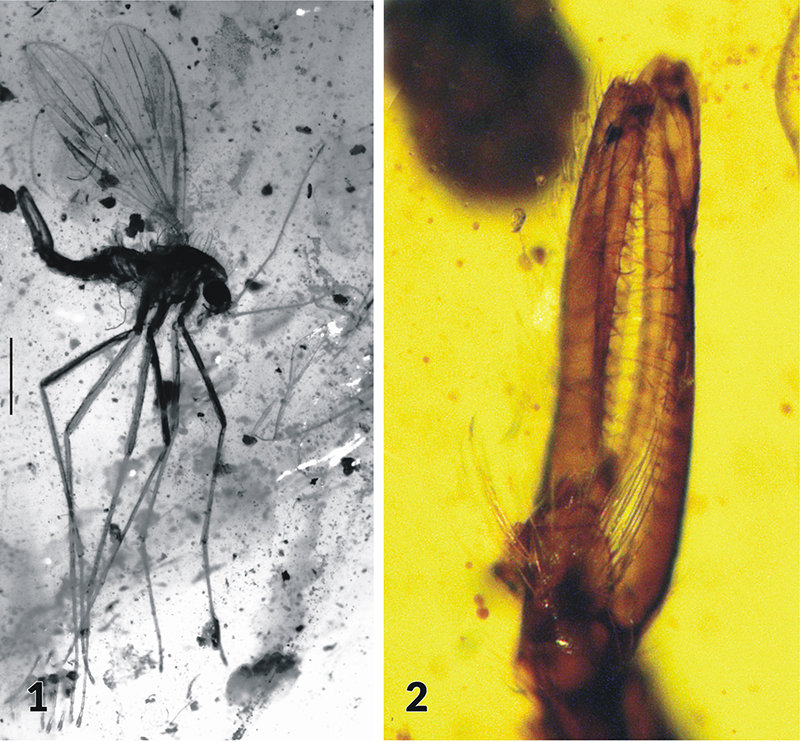
FIGURE 12. Drawings of Dacochile microsoma. (Bu 1262). 1, wing venation. 2, male genitalia in lateral view. Scale equals 0.5 mm.
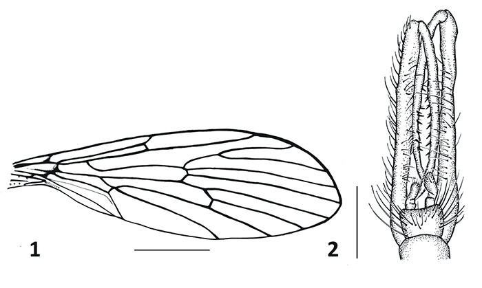
FIGURE 13. Photographs of Coramus gedanensis gen. et sp. nov. 1, whole specimen (paratype, MP/3377). 2, wing venation (holotype, MP/3376). 3, close-up of head (holotype, MP/3376). 4, close-up of legs (holotype, MP/3376) in lateral view. Scale equals 0.5 mm.
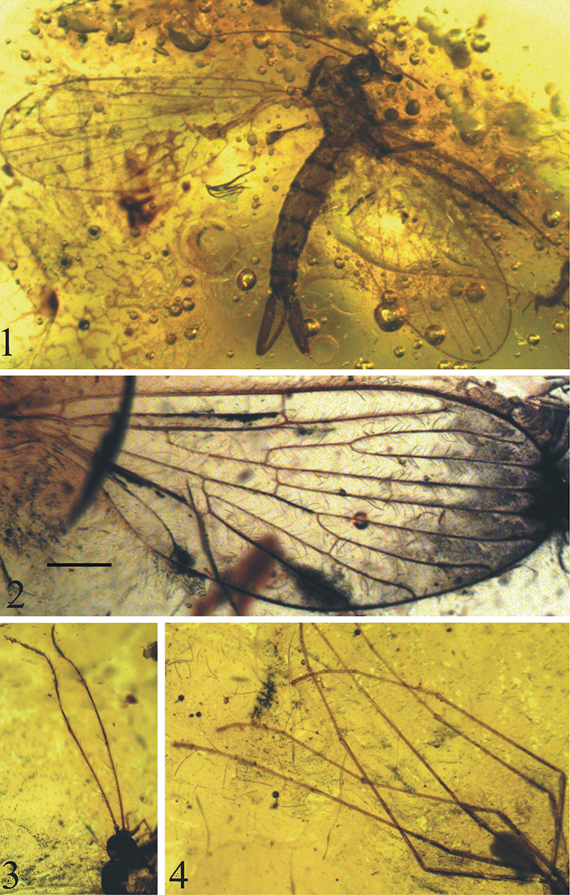
FIGURE 14. Drawings of Coramus gedanensis gen. et sp. nov. (MP/3376). 1, wing venation. 2, male genitalia in dorsal view. Scale equals 0.5 mm.
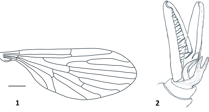
FIGURE 15. Photograph of Peringueyomyina barnardi (ISEZ PAN collection) whole specimen with elongated rostrum in lateral view. Scale equals 1mm.
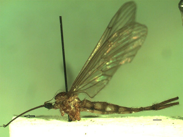
FIGURE 16. Drawings of Peringueyomyina barnardi (ISEZ PAN collection). 1, wing venation in dorsal view. 2-4, male genitalia, external built in dorsal view ( 2), aedagal complex in dorsal (3) ventral-lateral (4) views. Scale equals 1 mm.