FIGURE 1. 1, Metapodials of Crocuta spelaea in anatomical semi-connection. 2, Cranium of Crocuta spelaea. 3, Filtering the excavation grid. 4, Excavation of various Crocuta spelaea remains from the hyena level.
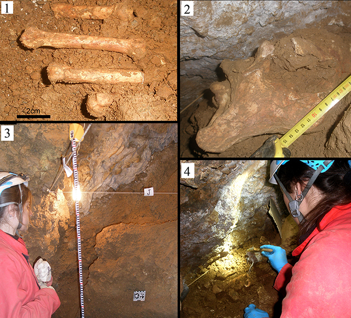
FIGURE 2. 1, Geographical situation of Los Aprendices Cave. 2, Panoramic view of the western slopes of the Peñas del Cabo. 3, Mouth of Los Aprendices Cave. 4, Main gallery of the cave.
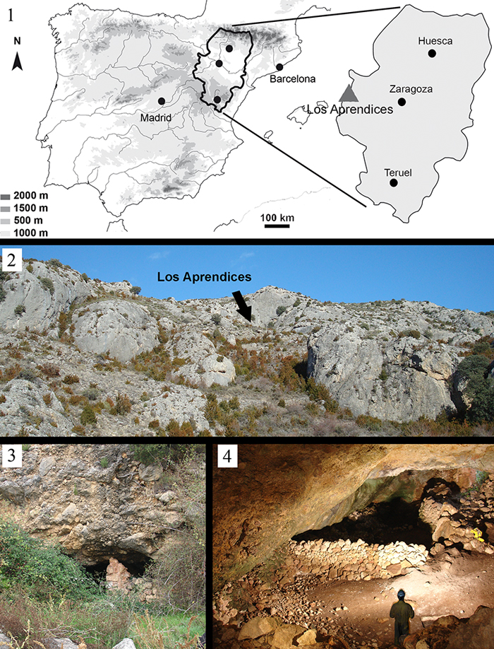
FIGURE 3. 1, Elevation view of Los Aprendices Cave. 2, Plan view of Los Aprendices Cave. 3, Detailed elevation view of the hyena gallery showing the excavation grid. 4, Detailed plan view of the hyena gallery showing the excavation grid as well as how the squares that were excavated were coloured. 5, Sediment cone that blocks off the former entrance to the hyena gallery. Within it one can see the hyena level, from which the skeleton of Crocuta spelaea was recovered.
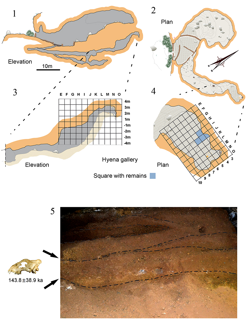
FIGURE 4. Panoramic photo of the skeleton of Crocuta spelaea from Los Aprendices.
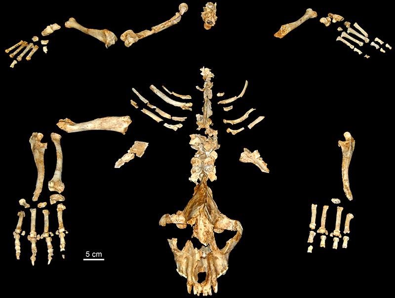
FIGURE 5. Photograph of the cranium (1-4, MPZ 2014/657) and hemi-mandible (5-7, MPZ 2014/593) of Crocuta spelaea: 1-2, lateral view; 3, dorsal view; 4, occlusal view; 5, buccal view; 6, lingual view; 7, occlusal view.
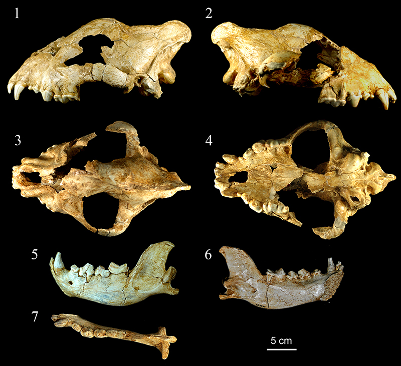
FIGURE 6. Crocuta spelaea forelimb remains from Los Aprendices. 1, left scapula in dorsal view (MPZ 2014/584). 2, left humerus in cranial view (MPZ 2014/672). 3, right radius in palmar view (MPZ 2014/611). 4, left ulna in medial view (MPZ 2014/610). 5, right ulna in medial view (MPZ 2014/676). 6, right Mtc V in dorsal view (MPZ 2014/618). 7, right Mtc IV in dorsal view (MPZ 2014/617). 8, right Mtc III in dorsal view (MPZ 2014/616), 9, right Mtc II in dorsal view (MPZ 2014/615). 9, left Mtc II in dorsal view (MPZ 2014/685). 10, left Mtc III in dorsal view (MPZ 2014/684). 11, left Mtc IV in dorsal view (MPZ 2014/ 581). 12, left Mtc V in dorsal view (MPZ 2014/575). 13, right scapholunate in proximal view (MPZ 2014/613). 14, right pisiform in proximal view (MPZ 2014/614). 15, trapezoid (MPZ 2014/619). 16, right cuneiform in dorsal view (MPZ 2014/659). 17, unciform (MPZ 2014/678) 18-26, first phalanx in dorsal view (MPZ 2014/576, 577, 578, 583, 621, 622, 626, 631). 27-34, second phalanx in lateral view (MPZ 2014/594, 599, 600, 601, 627, 639, 654, 655). 35-37, third phalanx in dorsal vier (MPZ 2014/590, 603, 680).
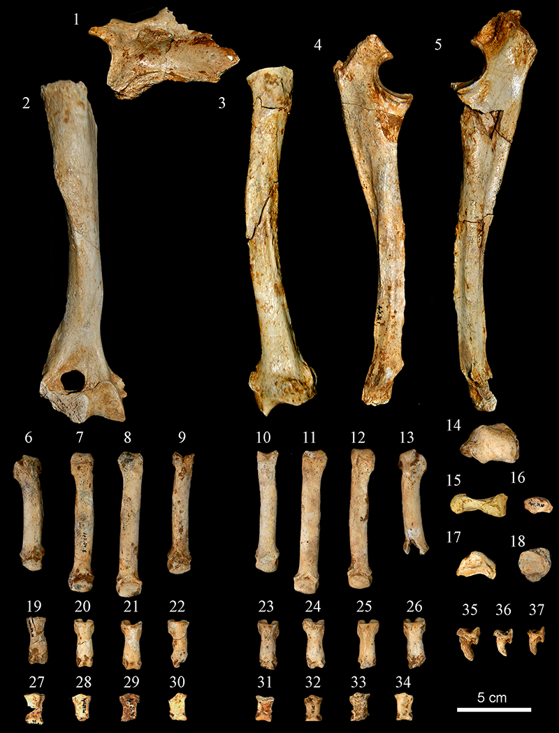
FIGURE 7. Crocuta spelaea hind limb remains from Los Aprendices. 1, right femur in caudal view (MPZ 2014/587). 2, left tibia in cranial view (MPZ 2014/681). 3, right tibia in cranial view (MPZ 2014/671). 4, left calcaneus in dorsal view (MPZ 2014/677). 5, right calcaneus in dorsal view (MPZ 2014/632). 6, left astragalus in dorsal view (MPZ 2014/682). 7, left navicular in lateral view (MPZ 2014/625). 8, right cuboid in proximal view (MPZ 2014/ 623). 9, left cuboid in proximal view (MPZ 2014/ 629). 10, left first cuneiform in proximal view (MPZ 2014/62). 11-14, first phalanx in dorsal view (MPZ 2014/579, 580, 620, 624). 15, right Mtt V in dorsal view (MPZ 2014/636). 16, right Mtt IV in dorsal view (MPZ 2014/635). 17, right Mtt III in dorsal view (MPZ 2014/634). 18, right Mtt II in dorsal view (MPZ 2014/633). 19, left Mtt II in dorsal view (MPZ 2014/642). 20, left Mtt III in dorsal view (MPZ 2014/643). 21, left Mtt IV in dorsal view (MPZ 2014/644). 22, left Mtt V in dorsal view (MPZ 2014/645).
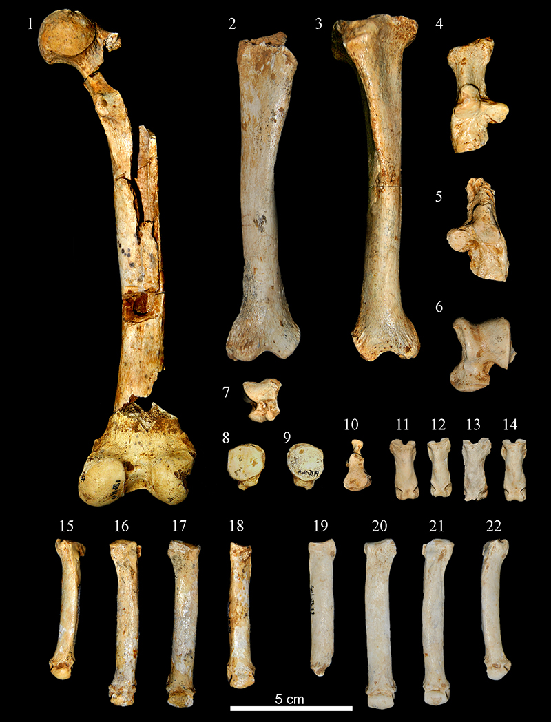
FIGURE 8. Bivariate graph representing the width of the condyles in relation to the total length of the cranium. Abbreviations:Ba, Badel Cave; BW, Bad Wildungen; Do, Döbritzer Cave; Ja, Jaurens; Ke, Ketsch; Pe, Perick Caves; Ro, Rösenbecker Cave; Th, Thiede; Oa, Oase Cave; Sr, Srbsko Cave; Vy, Výpustek Cave (Diedrich, 2011a); Sl, Sloup Cave (Diedrich, 2012b); Ga, Gargas (Cardoso, 1993); Lu, Lunel Viel (Bonifay, 1971). The measurements of the crania of extant C. crocuta specimens were taken by J.M.-M of the Royal Museum for Central Africa (RMCA) of Belgium.
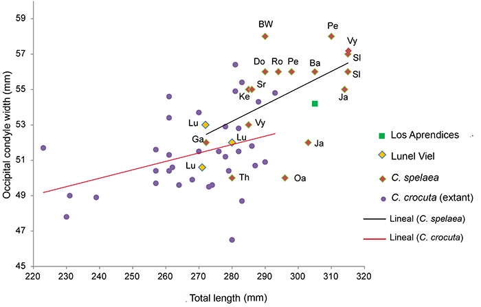
FIGURE 9. Graph representing total length versus total width of P4 of extant and cave hyaena from several sites. The comparative measurements have been extracted from, Reynolds, 1902; Bonifay, 1971; Ballésio, 1979; Clot, 1980; Argant, 1991; Cardoso, 1993; Blasco and Montes, 1997; Fosse, 1997; Castaños, 1987; Baryshnikov, 1999; Altuna and Mariezkurrena, 2000; García, 2003; Testu, 2006; Fourvel, 2012; and J.M.-M personal commun., 2015. Abbreviations: ATA, Atapuerca; Ag, Aguilón P-7; Ap, Los Aprendices; Mo, Mollet; Ol, Olopte B; L’Or Cova de l’Or; SI, Sima I. Confidence ellipses with confidence interval (0.95) are figured.
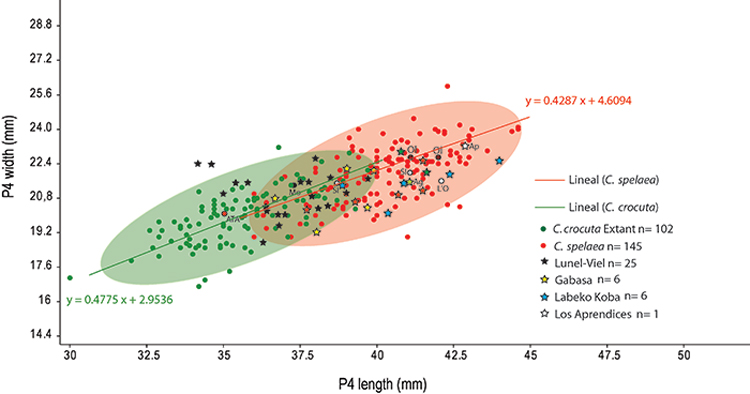
FIGURE 10. Graph representing total length versus total width of m1 of extant and cave hyaena from several sites. The comparative measurements (in mm) have been extracted from, Reynolds, 1902; Bonifay, 1971; Ballésio, 1979; Clot, 1980; Argant, 1991; Cardoso, 1993; Blasco and Montes, 1997; Fosse 1997; Castaños, 1987; Baryshnikov, 1999; Altuna and Mariezkurrena, 2000; García, 2003, Testu, 2006; Fourvel, 2012 and J.M.-M personal commun., 2015. Abbreviations: ATA, Atapuerca; Ap, Los Aprendices; Ar, Arbreda; Fo, Fontainhas; IPS, Institut de Paleontologia M. Crusafont de Sabadell; Mo, Mollet; To, El Toll; Va, Valdegoba. Confidence ellipses with confidence interval (0.95) are figured.
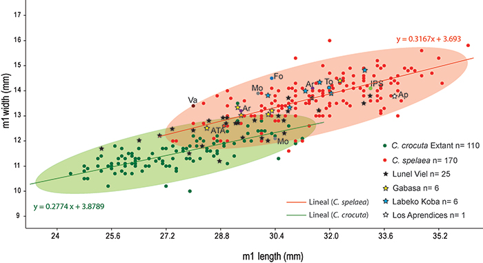
FIGURE 11. 1, Graph representing total length of radius versus distal width of extant and cave hyaena from several sites. The comparative measurements (in mm) have been extracted from Ehrenberg, 1940; Cardoso, 1993; García, 2003; Diedrich, 2011e; and J.M.-M personal commun., 2015. 2, Total length of third metacarpus versus smallest breadth of the diaphysis (SD) of extant and cave hyaena from several sites. The comparative measurements (in mm) have been extracted from Ehrenberg, 1940; Cardoso, 1993; García, 2003; Fourvel, 2012, and J.M.-M personal commun., 2015. Confidence ellipses with confidence interval (0.95) are figured.
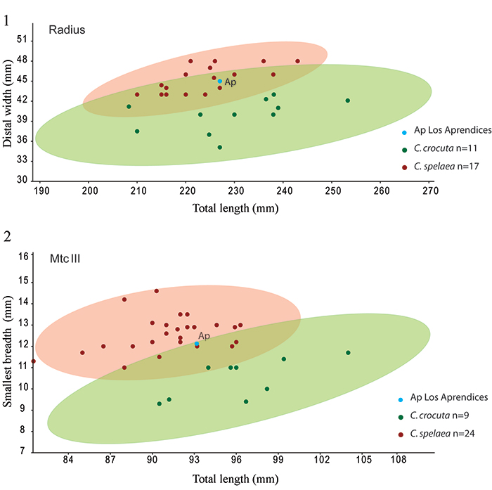
FIGURE 12. 1, Graph representing total length of femur versus distal width of extant and cave hyaena from several sites. The comparative measurements have been extracted from Ehrenberg, 1940; Cardoso, 1993; Altuna and Mariezkurrena, 2000; Diedrich, 2011e. 2, Total length of tibia versus distal width of extant and cave hyaena from several sites. The comparative measurements have been extracted from Reynolds, 1902; Ehrenberg, 1940; Cardoso, 1993; Diedrich, 2011e; Fourvel, 2012. 3, Total length of third metatarsus versus smallest breadth (SD) of the diaphysis of extant and cave hyaena from several sites. The comparative measurements have been extracted from Ehrenberg, 1940; Cardoso, 1993; García, 2003; Fourvel, 2012. Confidence ellipses with confidence interval (0.95) are figured.
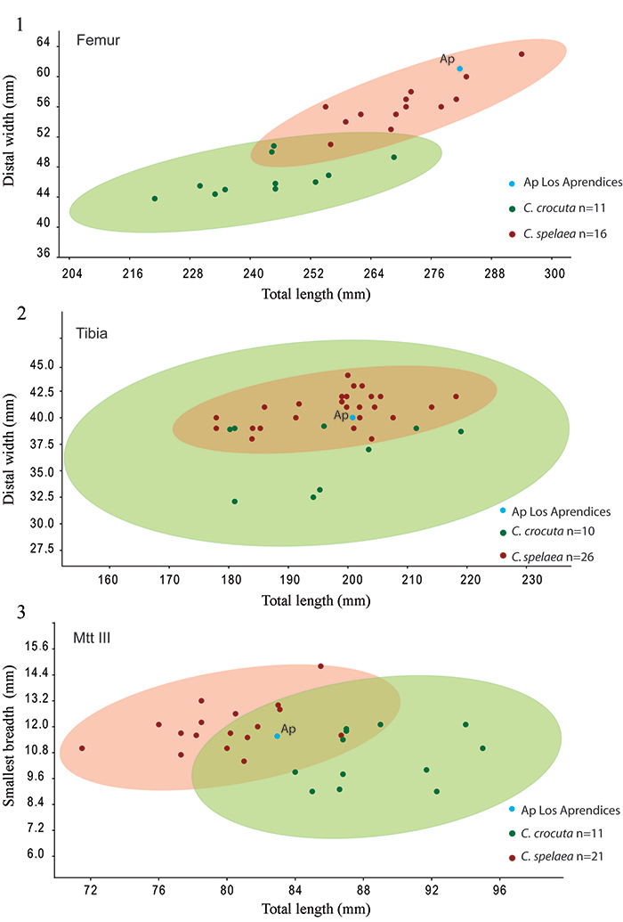
FIGURE 13. 1, Analysis of the breakage of the long bones from the skeleton of Crocuta spelaea from Los Aprendices, showing the abundance of each type of fracture according to the criteria analysed: angle, delineation and edge of the breaks. 2, Analysis of the breakage of the diaphyses in terms of circumference (C1, C2, C3) and length (L1, L2, L3, L4) for the skeleton of Crocuta spelaea from Los Aprendices.
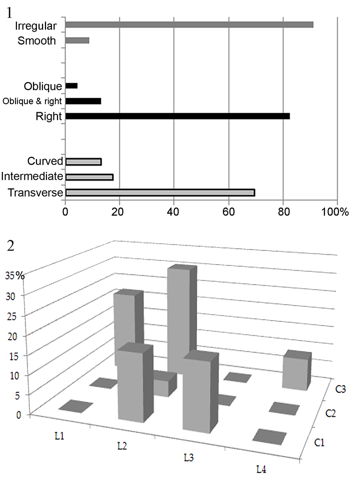
FIGURE 14. Photographs of the various types of marks (indicated by white arrows) present on the skeleton of Crocuta spelaea from Los Aprendices. 1, detailed image of the proximal epiphysis of the right humerus (MPZ 2014/672). 2, humerus in cranial view (MPZ 2014/672). 3, humerus in lateral view (MPZ 2014/672). 4, right ulna (MPZ 2014/676) showing “furrowing” on the proximal epiphysis, as well as a “puncture”. 5, left ulna (MPZ 2014/610) showing “furrowing” on the proximal epiphysis. 6, right tibia (MPZ 2014/681) showing “furrowing” on the proximal epiphysis. 7-8, lateral and dorsal views of a left calcaneus (MPZ 2014/677) showing “furrowing” and grooves on the calcaneal tuberosity. 9, rib (MPZ 2014/591) showing a “puncture”. 10, lumbar vertebra (MPZ 2014/582) in caudal view showing “furrowing” on the vertebral body. 11, lumbar vertebra (MPZ 2014/582) in ventral view showing three pits, corresponding to the incisors of a medium-sized carnivore. 12-13, lateral and caudal views of a dorsal vertebra (MPZ 2014/674) showing how the vertebral body has been torn off by a bite.
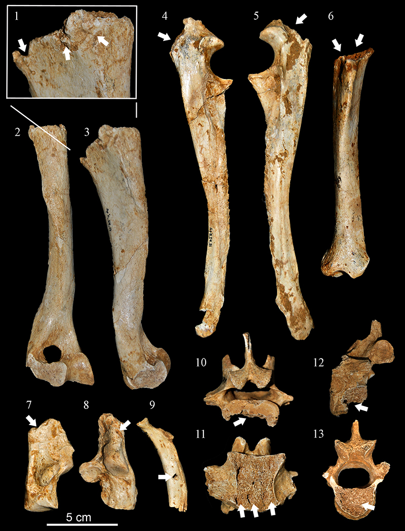
FIGURE 15. Palaeogeographic and diachronic distribution of the Crocuta in the Iberian Peninsula. The numbers refer to the sites listed in Appendix 7.
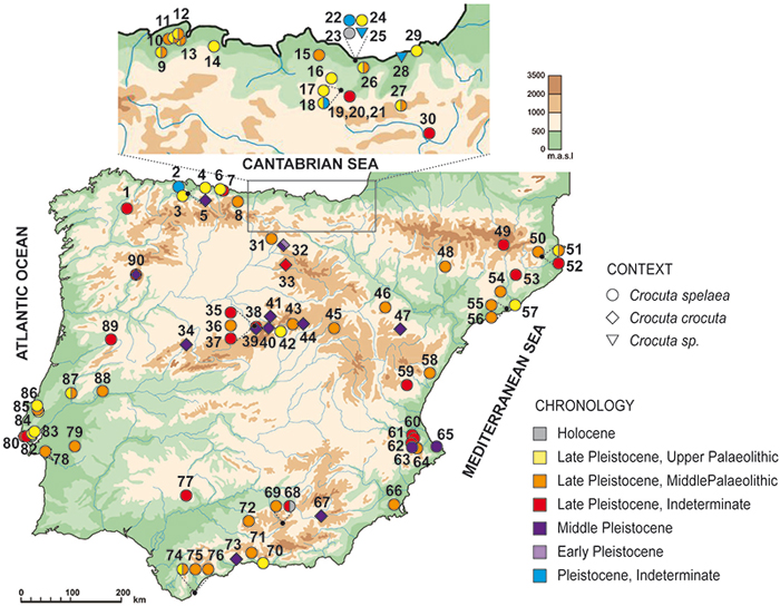
FIGURE 16. Graph representing total length of femur versus total length of third metatarsal in the same individual of extant Crocuta crocuta and the three unique complete skeletons of Crocuta spelaea from three sites Koněprusy Cave (Diedrich, 2012a); Gargas (Cardoso, 1993) and Los Aprendices this work. Comparative measurements (in mm) have been extracted from Ehrenberg, 1940; Cardoso, 1993; and J.M.-M personal commun., 2015. Confidence ellipse with confidence interval (0.95) is figured.
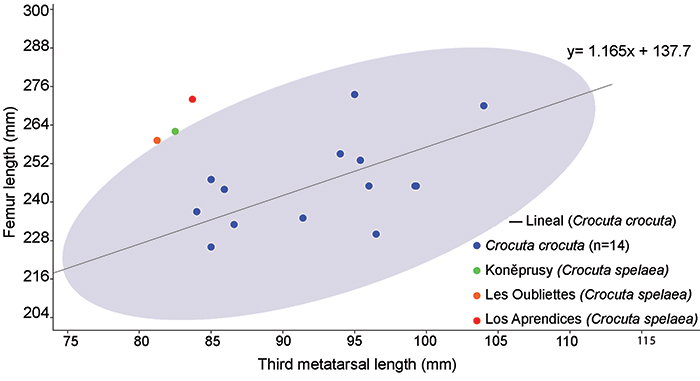
FIGURE 17. Means (square) and standard deviations (DS) of the values for the width (1) and the length (2) of pits on the epiphyses of long bones for various modern-day carnivores, together with the data from Los Aprendices (arrows). Key to the data: “Selvaggio and Wilder, 2001; ‘ Domínguez-Rodrigo and Piqueras, 2003; ^ Delaney-Rivera et al., 2009; * Saladié, 2009.
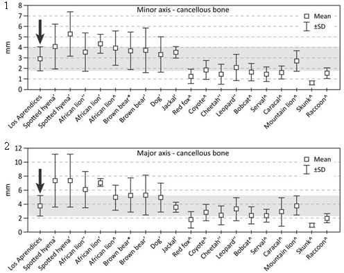
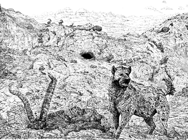
FIGURE 18. Reconstruction of Crocuta spelaea in the vicinity of Los Aprendices Cave, feeding on the carcass of a specimen of Capra pyrenaica (illustration by Gianfranco Mensi).