FIGURE 1. CT scan slice showing an approximately mediolateral view (i.e., longitudinal section) of an Asian lion’s forelimb. 1, Dark grey is adipose and connective tissues, lighter grey is muscles, white is bone. Bottom right corner white is a density calibration phantom (1.69 g cm -3; “cortical bone”). 2, Segmentation of the lion forelimb with select muscles highlighted. Abbreviations: FCU - flexor carpi ulnaris; DDF - deep digital flexors; ECR - m. extensor carpi radialis; Pro Quad - m. pronator quadratus; Abd1 - m. abductor digiti I.
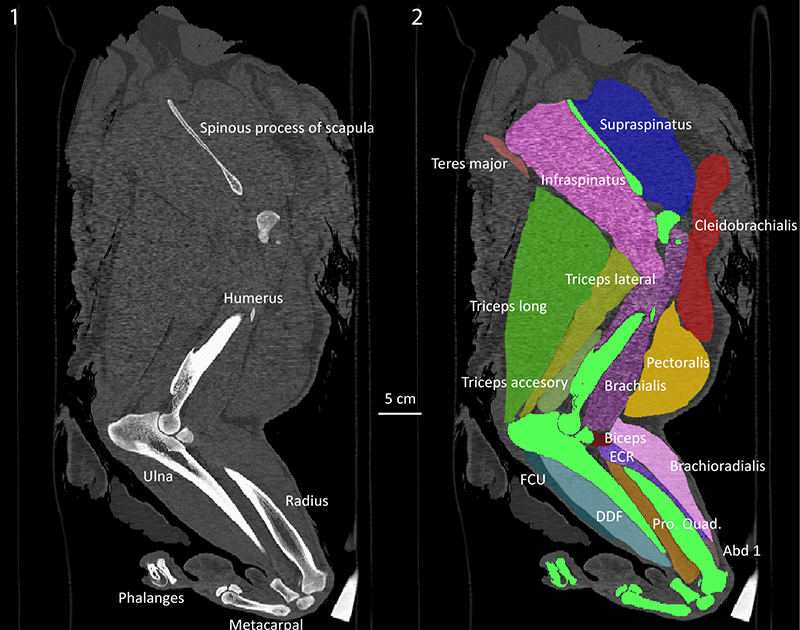
FIGURE 2. Skeletal reconstruction showing the original bones from Panthera atrox and those which have been copied from other vertebrae (red), or from P. leo persica (blue). 1, lateral; 2, dorsal; 3, anterior views. Scale bar is 50 cm.
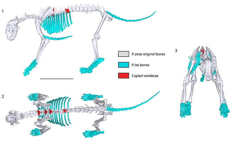
FIGURE 3. Muscled reconstruction of Panthera atrox showing the major muscle groups in lateral view. Abbreviations: FCU - m. flexor carpi ulnaris; ECU - m. extensor carpi ulnaris; ECR - m. extensor carpi radialis; EDL - m. extensor digitorum longus. Scale bar is 50 cm.
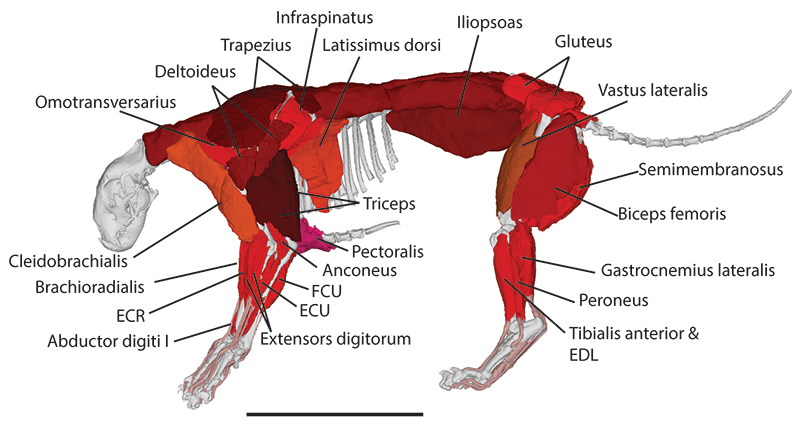
FIGURE 4. Convex hull model from the reconstructed Panthera atrox skeleton shown in Figure 2. 1, left lateral view; 2, dorsal view. Scale bar is 50 cm.
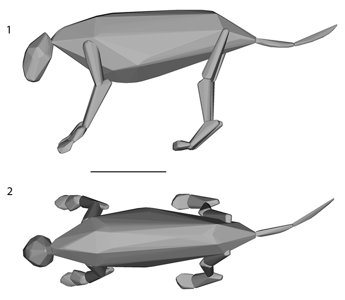
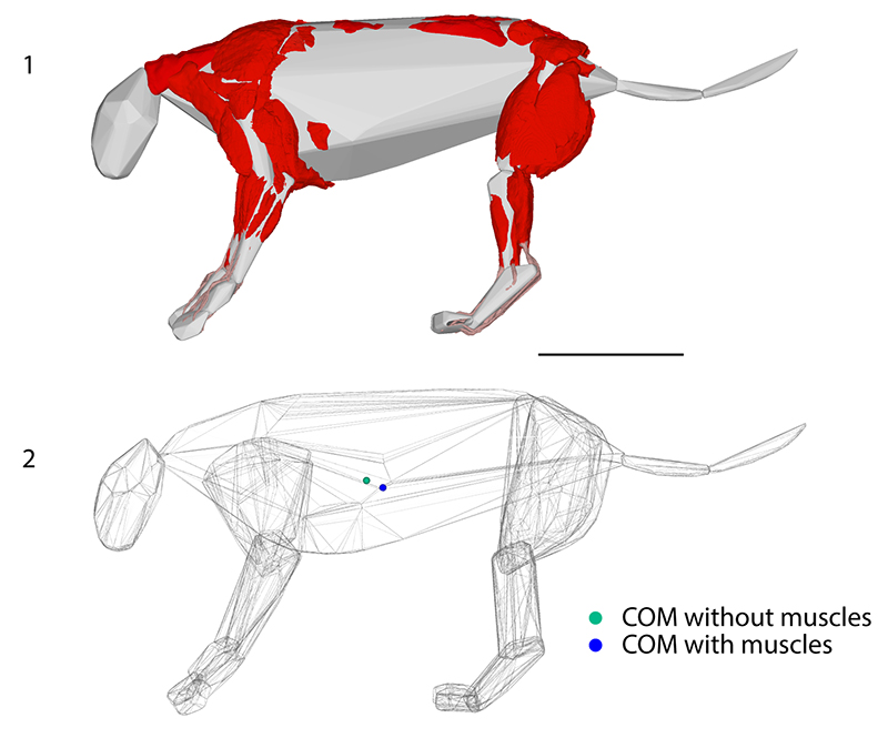
FIGURE 5. Panthera atrox reconstruction showing differences between simple convex hulls and more complex reconstructions. 1, Reconstructed muscles overlaid on the convex hull of just the bones. Any muscles that are visible extend beyond the range of the convex hull, thereby demonstrating the underestimation of size by convex hulls based solely on bones. 2, Reconstructions showing the posteroventral movement of the centre of mass (COM) between the bone convex hull and the muscled convex hull models of Panthera atrox. Scale bar is 50 cm.