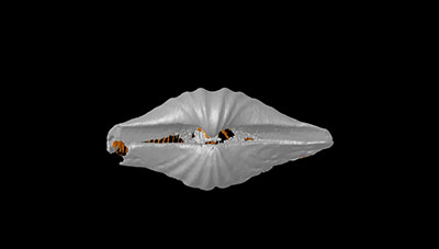
FIGURE 1. Thin section and cathodoluminescence (CL) photomicrographs: Timaniella harkeri (GSC26406) (1-4), Spiriferina sp.(BS-1) (5-8), Stenoscisma timorense (BS-2) (9-12), Cleiothyridina baracoodensis (ML32) (13-16) and Tylothyris transversa (TeP) (17-20). 1-2, transmitted light (TL) (1)and corresponding CL images (2) of nonluminescent shell and infilling sediment composed of fine sand grains. 3-4, TL (3) and CL (4) images of nonluminescent associated with partially luminescent shell in the cardinal area of dorsal valve. 5-8, TL (5,7) and CL (6, 8) images of nonluminescent associated with partially luminescent shell and luminescent infilling cement. 9-12, TL (9, 11) and CL (10, 12) images of luminescent shell with thin silicified layers and recrystallization along inner wall of brachiopod shell. 13, plane-polarized light (PPL) image of the original infilling sediment composed of calcareous sands. 14, PPL image showing the section of infilling cement. 15-16, TL (15) and CL (16) images of slightly luminescent shell. 17, PPL image of infilling comprising lime mud, calcite cement and relatively large, radially arranged, siliceous crystals. 18-19,, TL (18) and CL (19) images of nonluminescent associated with partially luminescent shell. 20, CL image showing the section of nonluminescent internal shell structure (spiralia) preserved within infilling (This structure is hardly recognized in the TL image). Abbreviations: bs, brachiopod shell; im, infilling material; sl, silicified layer; cc, calcite crystal; NL, nonluminescent; SL, slightly luminescent; L, luminescent.
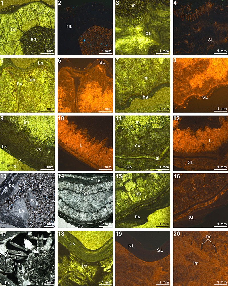
FIGURE 2. Thin section and cathodoluminescence (CL) photomicrographs: Indospirifer sp.(3229) (1-4), Cyrtospirifer whitneyi (CD) (5-8), Spiriferidae gen. sp. indet.(S1) (9-12), Spiriferella loveni (F8) (13-16), Meekella sangzhiensis (Q-1) (17-18) and Tyloplecta nanjingensis (Q-2) (19-20). 1, plane-polarized light (PPL) image of infilling micrite and shell fragments scattered. 2-3,transmitted light (TL) (2) and corresponding CL (3) images of luminescent shell. 4, CL image showing slightly luminescent shell. 5-8, TL (5, 7) and CL (6, 8) images of both luminescent shell and infilling sediment. 9-10, PPL images of brachiopod shell, infilling packstone and thin silicified layer along their boundary. 11-12, TL (11) and CL (12) images of nonluminescent shell. 13-16, TL (13, 15) and CL (14, 16) images showing a variety of shell luminescence and infilling sediment composed of calcitic skeletal grains (bryozoan in 15 and 16). 17-20, PPL (17, 19, 20) and TL (18) images of both silicified shells and infillings. Abbreviations: bs, brachiopod shell; im, infilling material; fs, fragmented shell; os, other shell material; sl, silicified layer; NL, nonluminescent; SL, slightly luminescent; L, luminescent.
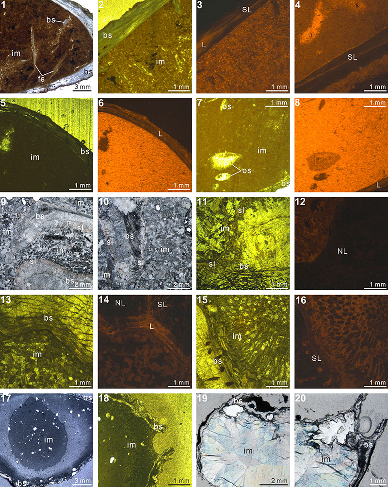
FIGURE 3. XMT result of Timaniella harkeri (GSC26406). 1-10, serial slices in the coronal plane (from posterior to anterior). 11-17, serial slices in the transverse plane (from ventral to dorsal). 18-22, serial slices in the sagittal plane (from lateral to middle). 23-25, lateral (23), ventral (24) and dorsal (25) views of the reconstructed 3-D model (external shell). 26, ventral view of the 3-D model in transparent mode. All the slice images were obtained under false-color lookup tables (Color 2 option in DataViewer). Abbreviations: tt, teeth; sp, spiralia; sk, socket.
FIGURE 4. XMT result of Stenoscisma timorense (BS-2). 1-10, serial slices in the coronal plane (from posterior to anterior). 11-17, serial slices in the transverse plane (from ventral to dorsal). 18-22, serial slices in the sagittal plane (from lateral to middle). 23-25, lateral (23), ventral (24) and dorsal (25) views of the reconstructed 3-D model (external shell). 26, posterior view of the coronally sectioned 3-D model. 27, ventral view of the 3-D model in transparent mode. All the slice images were obtained under false-color lookup tables (Color 1 option in DataViewer). Abbreviations: sd, spondylium; tt, teeth.
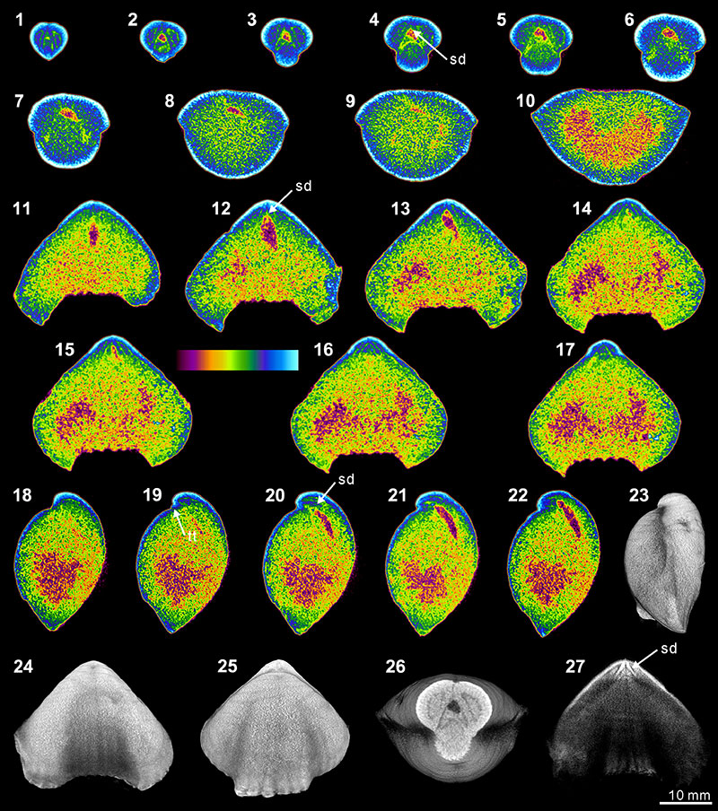
FIGURE 5. XMT result of Cleiothyridina baracoodensis (ML32). 1-11, serial slices in the coronal plane (from posterior to anterior). 12-21, serial slices in the transverse plane (from ventral to dorsal). 22-27, serial slices in the sagittal plane (from lateral to middle). 28-31, lateral (28), ventral (29), dorsal (30) and posterior (31) views of the reconstructed 3-D model (external shell). 32, ventral view of the 3-D model in transparent mode. All the slice images were obtained under false-color lookup tables (Color 2 option in DataViewer). Abbreviations: tt, teeth; sk, socket; cf, cardinal flanges.
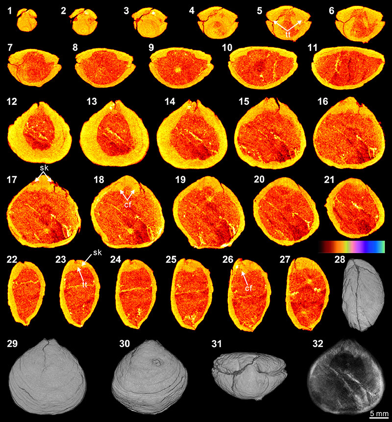
FIGURE 6. XMT result of Tylothyris transversa (TeP). 1-7, serial slices in the coronal plane (from posterior to anterior). 8-14, serial slices in the transverse plane (from ventral to dorsal). 15-19, serial slices in the sagittal plane (from lateral to middle). 20-22, lateral (20), ventral (21) and dorsal (22) views of the reconstructed 3-D model (external shell). 23, ventral view of the 3-D model in transparent mode. All the slice images were obtained under false-color lookup tables (Color 1 option in DataViewer). Abbreviation: sp, spiralia.
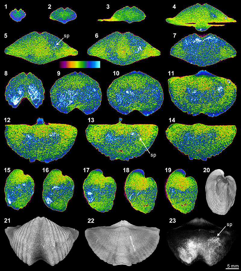
FIGURE 7. XMT result of Spiriferina sp. (BS-1). 1-7, serial slices in the coronal plane (from posterior to anterior). 8-14, serial slices in the transverse plane (from ventral to dorsal). 15-19, serial slices in the sagittal plane (from lateral to middle). 20-22, lateral (20), ventral (21) and dorsal (22) views of the reconstructed 3-D model (external shell). 23, ventral view of the 3-D model in transparent mode. All the slice images were obtained under false-color lookup tables (Color 1 option in DataViewer). Abbreviations: dp, dental plates; ms, median septum; sp, spiralia; tt, teeth; cr, crus; sk, socket.
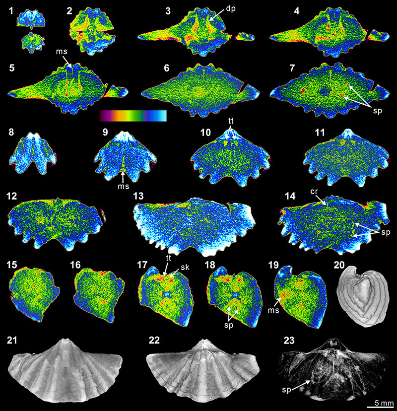
FIGURE 8. XMT result of Indospirifer sp. (3229). 1-8, serial slices in the coronal plane (from posterior to anterior). 9-15, serial slices in the transverse plane (from ventral to dorsal). 16-20, serial slices in the sagittal plane (from lateral to middle). 21-22, lateral (21) and ventral (22) views of the reconstructed 3-D model (external shell). 23-24, ventral (23) and ventroanterior (24) views of the 3-D model in transparent mode. All the slice images were obtained under false-color lookup tables (Color 1 option in DataViewer). Abbreviations: dp, dental plates; cp, cardinal process; hp, hinge plate; sp, spiralia.
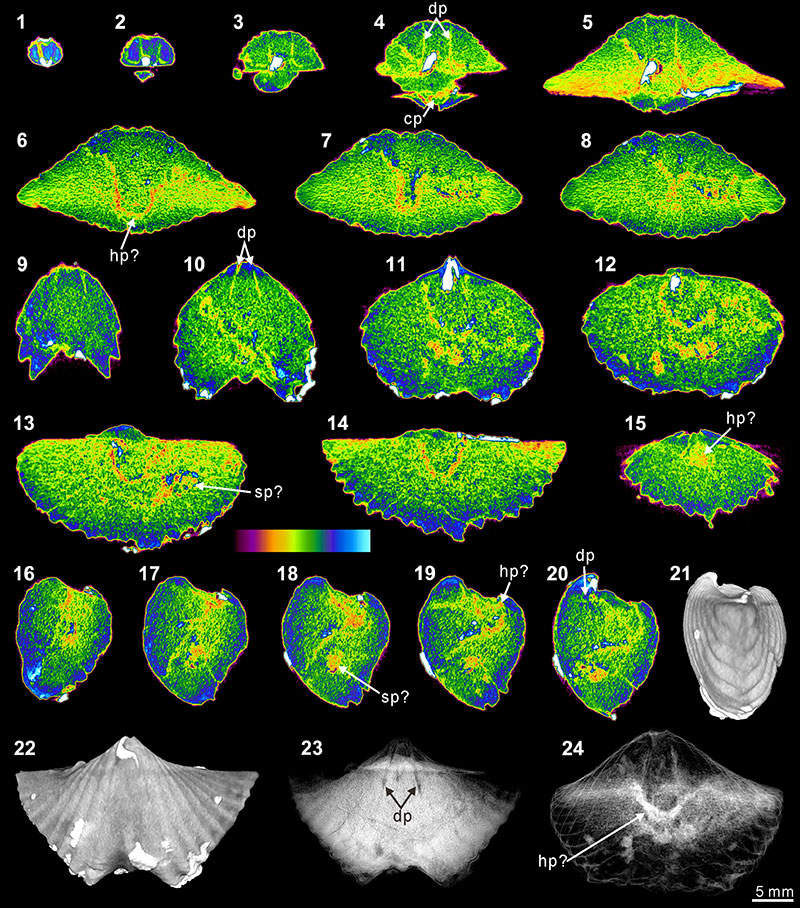
FIGURE 9. XMT result of Cyrtospirifer whitneyi (CD). 1-10, serial slices in the coronal plane (from posterior to anterior). 11-18, serial slices in the transverse plane (from ventral to dorsal). 19-24, serial slices in the sagittal plane (from lateral to middle). 25-28, lateral (25), ventral (26), dorsal (27) and posterior (28) views of the reconstructed 3-D model (external shell). 29, ventral view of the 3-D model in transparent mode. All the slice images were obtained under false-color lookup tables (Color 1 option in DataViewer). Abbreviations: dp, dental plates; tt, teeth.
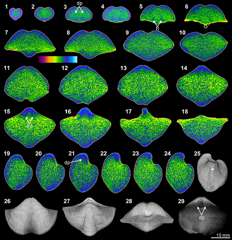
FIGURE 10. XMT result of Spiriferidae gen. sp. indet. (S1). 1-8, serial slices in the coronal plane (from posterior to anterior). 9-15, serial slices in the transverse plane (from ventral to dorsal). 16-21, serial slices in the sagittal plane (from lateral to middle). 22-24, lateral (22), ventral (23) and posterior (24) views of the reconstructed 3-D model (external shell). 25, ventral view of the 3-D model in transparent mode. All the slice images were obtained under false-color lookup tables (Color 1 option in DataViewer). Abbreviation: dp, dental plates.
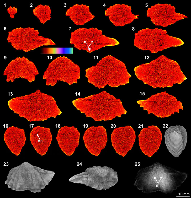
FIGURE 11. XMT result of Meekella sangzhiensis (Q-1). 1-7, serial slices in the coronal plane (from posterior to anterior). 8-15, serial slices in the transverse plane (from ventral to dorsal). 16-21, serial slices in the sagittal plane (from lateral to middle). 22-24, lateral (22), ventral (23) and dorsal (24) views of the reconstructed 3-D model (external shell). 25, ventral view of the 3-D model in transparent mode. All the slice images were obtained under false-color lookup tables (Color 1 option in DataViewer). Abbreviation: dp, dental plate.
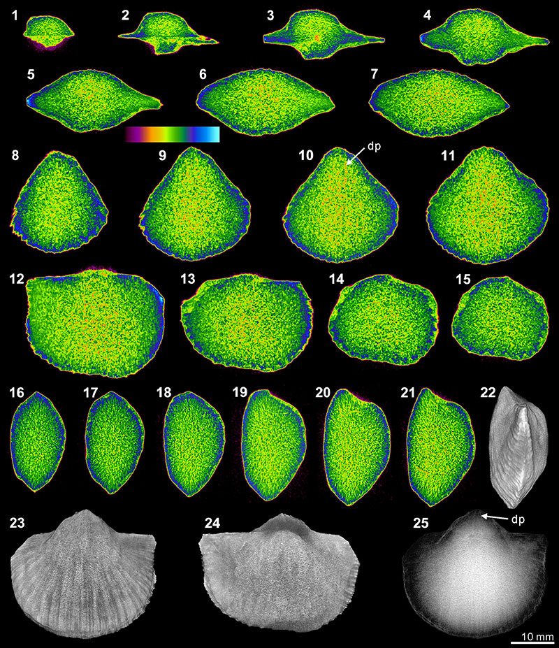
FIGURE 12. XMT result of Tyloplecta nankingensis (Q-2). 1-7, serial slices in the transverse plane (from dorsal to ventral). 8-14, serial slices in the coronal plane (from posterior to anterior). 15-18, serial slices in the sagittal plane (from lateral to middle). 19-21, lateral (19), ventral (20) and dorsal (21) views of the reconstructed 3-D model (external shell). 22, ventral view of the 3-D model in transparent mode. All the slice images were obtained under false-color lookup tables (Color 1 option in DataViewer). Abbreviations: cp, cardinal process; ap, adductor platform; ms, median septum; mc, muscle scar.
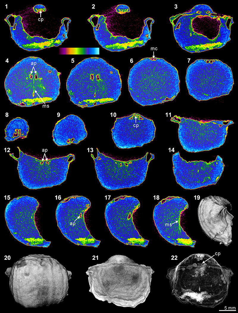
FIGURE 13. Three-dimensional reconstruction model of internal shell structures of Timaniella harkeri (GSC26406). 1-6, dorsal and anterior views of the whole shell interior through posteriorly continuous rotation. 7-12, dorsal and anterior views of shell interior without spiralia through posteriorly continuous rotation.
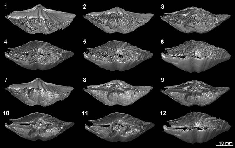
FIGURE 14. Three-dimensional rotational model (video) of Timaniella harkeri (GSC26406). Click on image to download or see video.