FIGURE 1. Cymastrobus irvingii gen. et sp. nov. NMVP 161998. 1, General view of the cone; cone axis at arrow. 2, Distal part of cone axis in transverse section. 3, Detail showing the stele (St) and five sporophyll traces at arrows. 4, Detail showing the wavy outline of the xylem ring, presumed location of the phloem (Ph), inner cortical cells (IC) and three sporophyll traces at arrows. 5, Detail showing the emission of a sporophyll trace from a groove of the primary xylem ring.
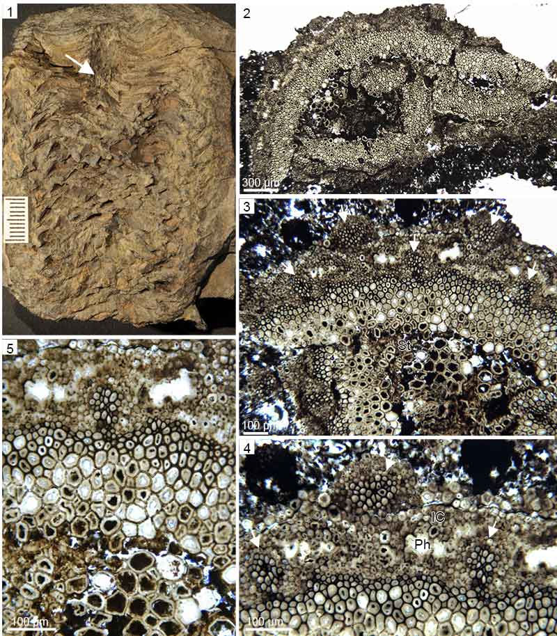
FIGURE 2. Cymastrobus irvingii gen. et sp. nov. NMVP 161998. Virtual sections, X-Ray synchrotron microtomography. 1, Cone in tangential section. 2, Cone in radial section; note the proximal position of the megasporangia. 3, Cone axis in transverse section. 4, Outer portion of the cone in tangential section showing four sporophyll-sporangium units in longitudinal row. 5-8, Inwards to outwards series of tangential sections through the cone showing the progressive changes in size of the sporophyll pedicels (P) and the sporangia (S).
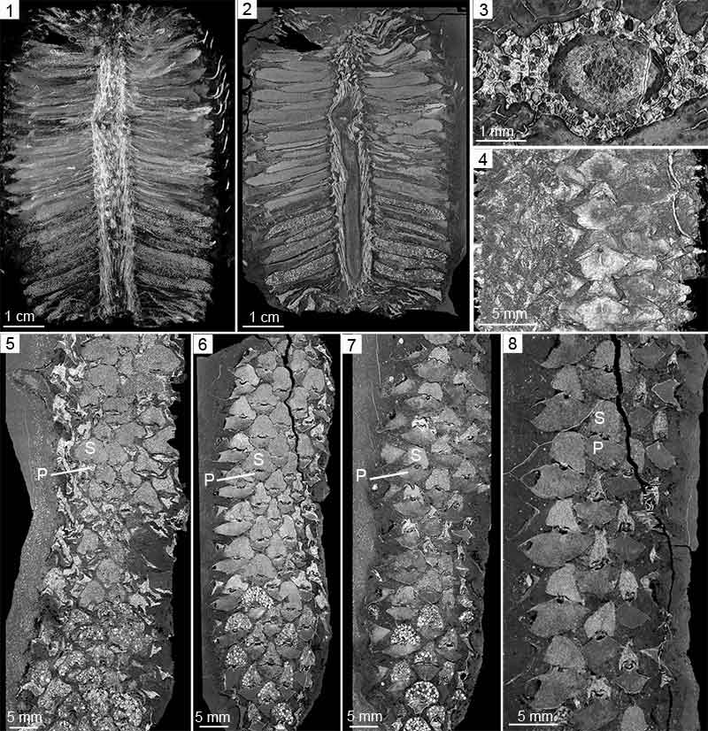
FIGURE 3. Cymastrobus irvingii gen. et sp. nov. NMVP 161998. Virtual sections, X-Ray synchrotron microtomography. 1-5, Proximal-distal series of longitudinal sections through a sporophyll-sporangium unit; note the heel (H) in 1, keel (K) in 2 and 3, microsporangium (mS) and longitudinal pad of tissue (P) in 4 and 5, vascular strand (VS) in 5.
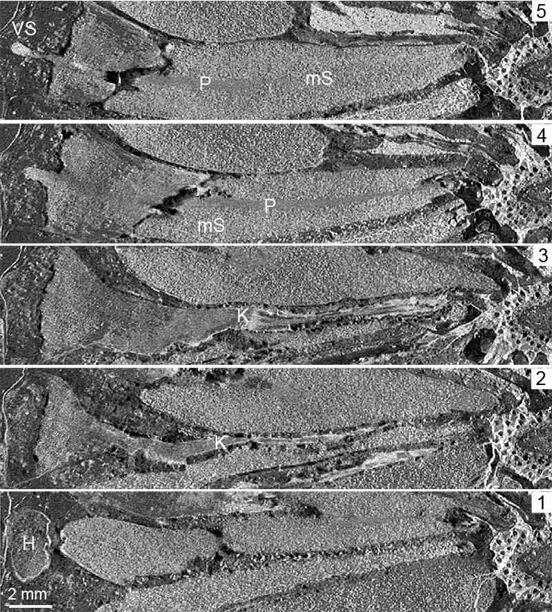
FIGURE 4. Cymastrobus irvingii gen. et sp. nov. NMVP 161998. Virtual reconstructions and volume rendering visualization of chosen anatomical units within the X-Ray synchrotron microtomography scan. 1-2, General view of the cone showing two reconstructed sporophyll-sporangium units, the proximal one producing megaspores, the distal one microspores. 3, Reconstructed sporophyll showing the enlarging pedicel and dissected lamina. 4-5, Two reconstructed sporophyll-microsporangium units in profile view. 6-7, Two reconstructed sporophyll-megasporangium units in profile view. VS: vascular strand.
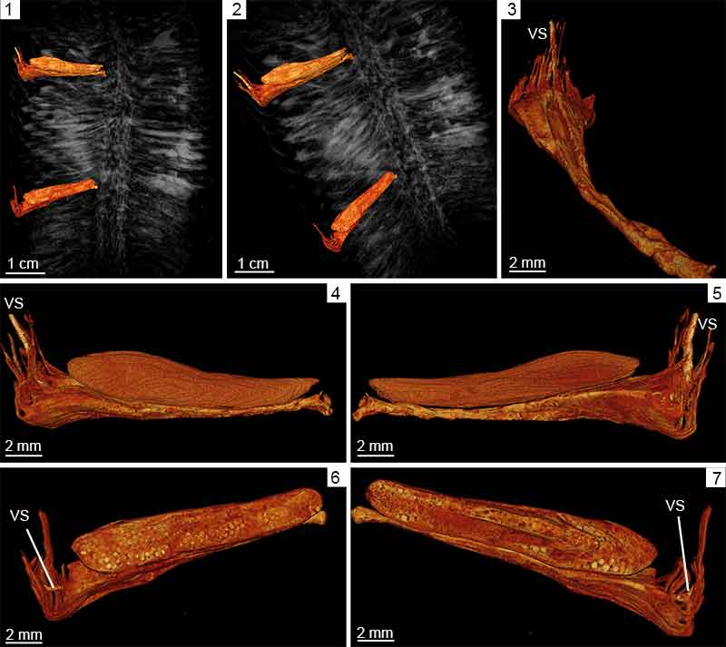
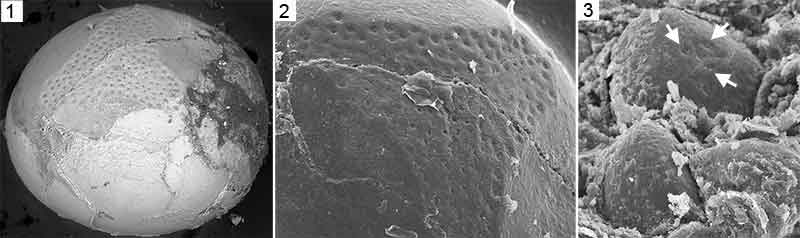
FIGURE 5. Cymastrobus irvingii gen. et sp. nov. NMVP 161998. 1, Cast of a megaspore central body showing numerous small circular pores arranged in several rows around the trilete mark. 2, Detail of previous view. 3, Casts of microspore central bodies; the largest one shows three pores between the rays of the trilete mark (arrows).