FIGURE 1. 1. Multiple views of Cryptoclidus eurymerus GLAHM V1799 left humerus (Top row) and; 2. normal humerus (GLAHM V1828) for comparison; 3. close up of the humerus showing tooth impacts (GLAHM V1799). White arrows indicate tubercle (t); black arrows indicate bite marks. Measurement bar is 5 cm.
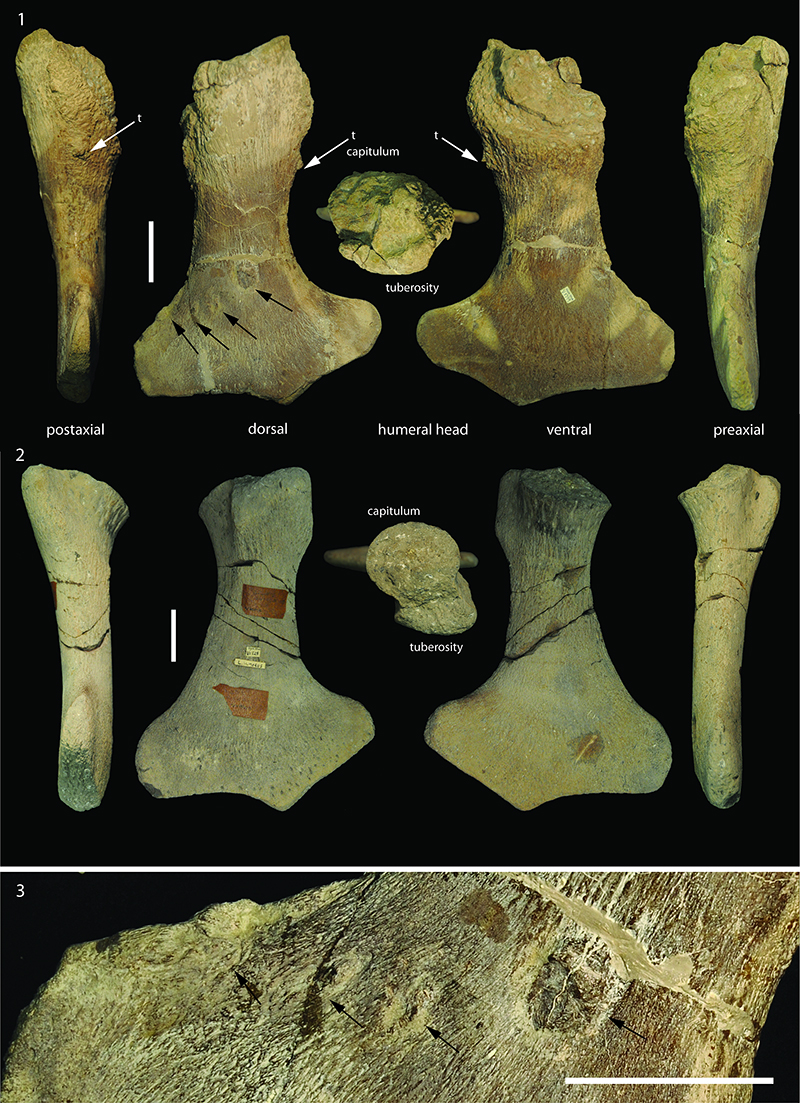
FIGURE 2. Table of pectoral girdle measurements and schematic of measurement sites. Juveniles (j) were recognized by the lack of prominent posterolateral processes and a curved posterior margin of the coracoids; (oa) denotes ‘old adult’ specimens where the scapulocoracoids are fused. * Difference in humeral fossa size extended by injury resulted in observed measurement variation. The mean of the angular measurements for the glenoid, coracoids, and scapula were used here (left and right measurements are included in the appendix).
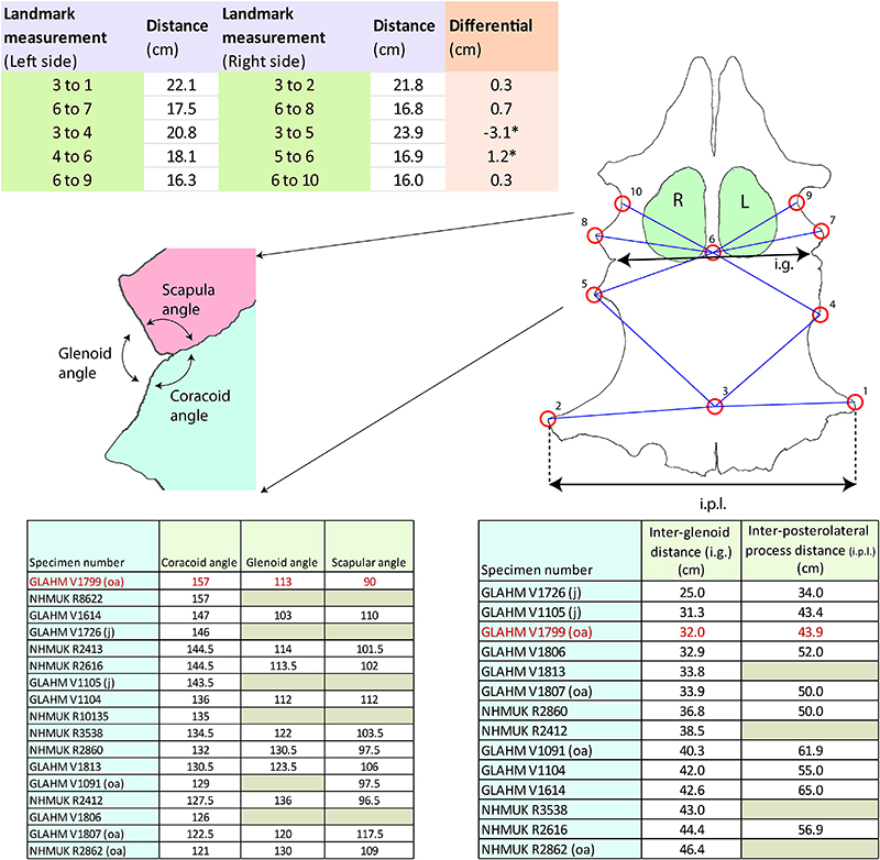
FIGURE 3. Results of the correlation principal component analysis using PAST ver. 2.17c (Hammer et al. 2001) showing that the juveniles generally morph from a larger coracoid angle towards a smaller angle in older adults (older adult (oa) n = 4 (red); adult (a) n = 11 (purple); juvenile (j) n = 3 (blue); and GLAHM V1799 plotted separately (green)).
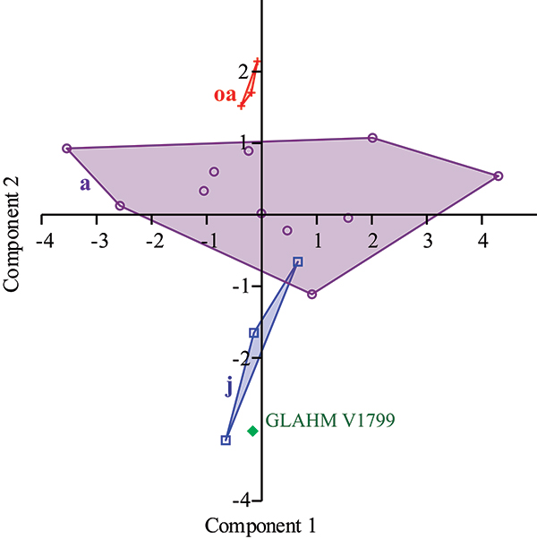
FIGURE 4. Ventral view of pectoral girdle of Cryptoclidus eurymerus GLAHM V1799. Measurement bar is 5 cm. Arrow indicates left side glenoid fossa pathology.
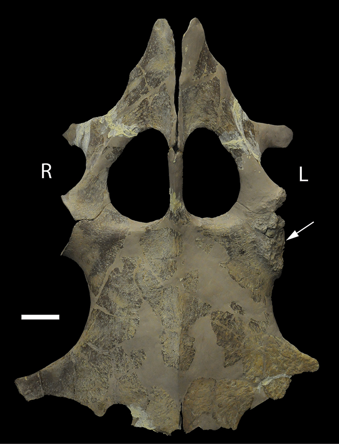
FIGURE 5. Cartoon of direction of attack on Cryptoclidus eurymerus specimen GLAHM V1799.
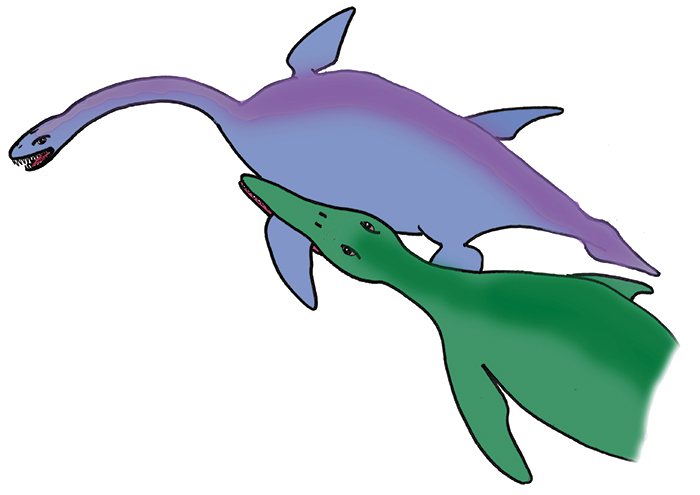
FIGURE 6. Dorsal view of left humerus and plastron, articulated as if the joint capsule had contracted. Measurement bar is 5 cm. Exostosis (arrow) would prevent joint movement.
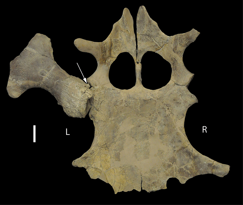
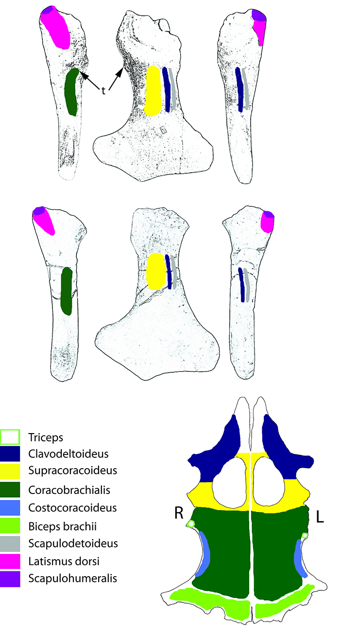
FIGURE 7. Plesiosaur muscle attachments to humerus and scapulocoracoid (Based on Araújo and Correia, 2015). Postaxial, ventral, and preaxial views of the affected left humerus GLAHM V1799 (top row) and the same of a healthy humerus (GLAHM V1828), as well as the ventral view of the pectoral girdle of GLAHM V1799 (bottom).