FIGURE 1. Geological setting. 1. Mexico and neighbouring countries (B for Belize, G for Guatemala, H for Honduras and S for El Salvador); area in the square is magnified in 2. 2. Main cities and roads of Sonora State, red square delimiting the area represented in 3. 3. Studied area. Indicated are the main roads (plain lines), trails (dashed lines) and cerros with Ediacaran/Cambrian succession outcrops, after Stewart et al. (1984), McMenamin (1987) and Sour-Tovar et al. (2007). 4. Geological map of Cerro Rajón reporting the distribution of basement rocks, Ediacaran and Cambrian formations, Mesozoic rocks and Quaternary alluvial deposits, after McMenamin (1984) and Stewart et al. (1984).
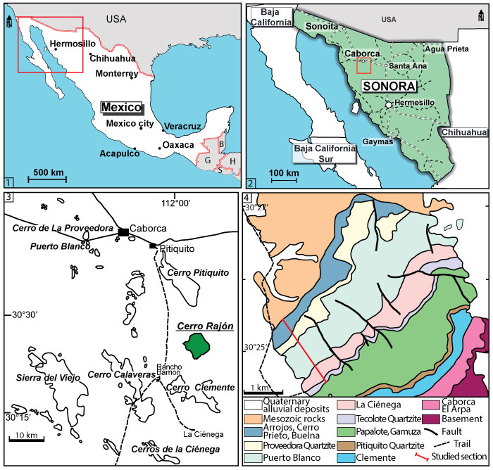
FIGURE 2. Stratigraphic section through the Puerto Blanco Formation at Cerro Rajón with the range of identified SSFs and other fossils. The transition between the underlying La Ciénega Formation and the PBF is not represented as well as the lowermost part of the PBF as no limestone beds suitable for SSF studies were identified and sampled. The upper limit of the PBF illustrated corresponds to the transition between the PBF and the overlying Proveedora Quartzite. The four identified microfaunal assemblages are indicated by Roman numerals. The characteristic microfossils are figured without scale. The characteristic microfossils are, for assemblage I: Xianfengella sp. and Allonnia tetrathallis; assemblage II: Mackinnonia corrugata and Archiasterella charma; assemblage III: Microdictyon multicavus, Parkula bounites and Rajonia ornata; assemblage IV: echinoderm ossicles).
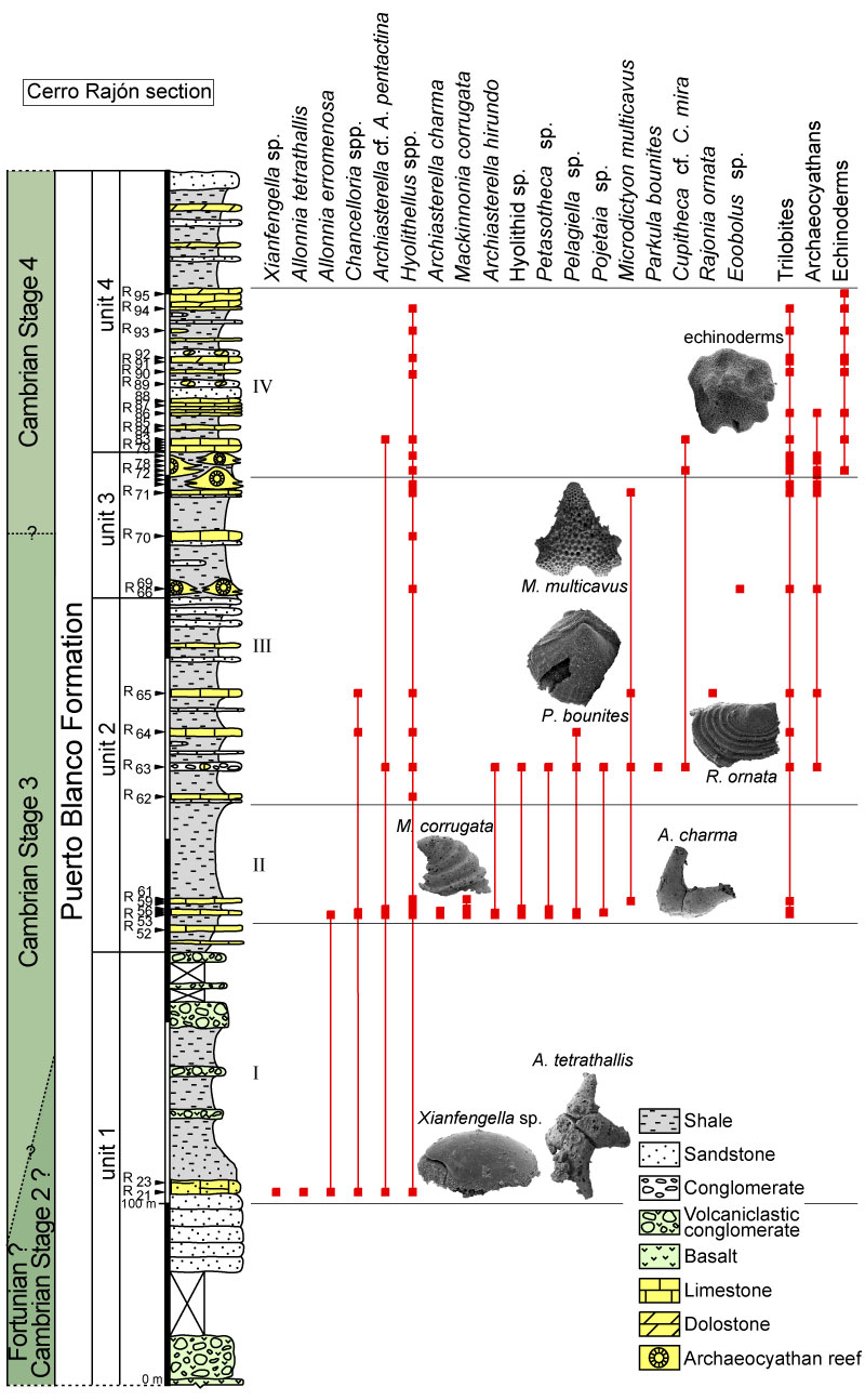
FIGURE 3. Range of globally distributed taxa recorded in the Puerto Blanco Formation. See Systematic Palaeontology (“other occurrences” of taxa) for references.
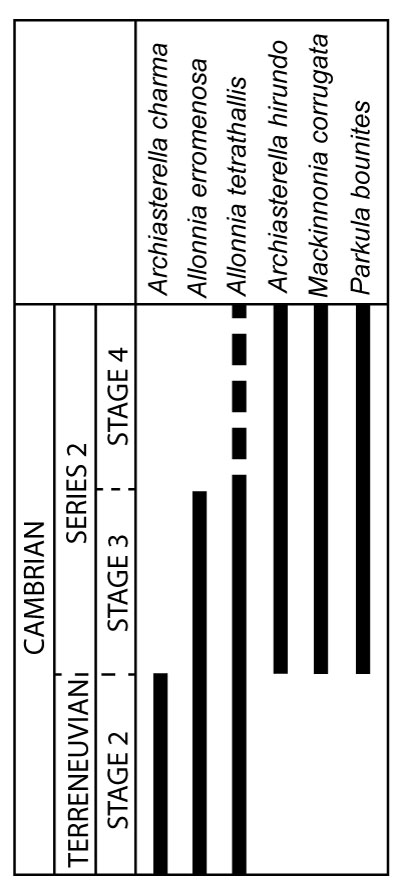
FIGURE 4. Distribution of formally identified taxa in the Puerto Blanco Formation at Cerro Rajón and comparison with Laurentian and global distribution. See Systematic Palaeontology (“other occurrences” of taxa) for references.
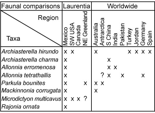
FIGURE 5. Internal moulds of sclerites with outer layer of Archiasterella hirundo Bengtson in Bengtson et al., 1990, from the Puerto Blanco Formation of Cerro Rajón, Sonora, Mexico. 1-5. Specimen USTL3170-3 from sample R54: 1. Adaxial view, area in the square is magnified in 3; 2. Abaxial view, area in the square is magnified in 4; 3. Detail of the basal plane showing the foramina and intersections of the rays articulatory facets; 4. Detail of broken tubercles of the outer layer forming circular perforations; 5. Lateral view showing the recurved lateral ray. 6-10, 17. Specimen USTL3175-4 from sample R63: 6. Adaxial view, area in the square is magnified in 9; 7. Abaxial view, area in the square is magnified in 17; 8. Lateral view showing the recurved lateral ray, area in the square is magnified in 10; 9. Detail of the outer layer near the foramen with broken inclined tubercles forming elongated perforations; 10. Detail of a transversally broken ray shows the gap between the outer phosphatic layer and the phosphatic internal mould of the lumen; 11-16. Specimen USTL3177-2 from sample R63: 11. Lateral view, area in the square is magnified in 16; 12. Adaxial view; 13. Abaxial view, area in the square is magnified in 15; 14. Detail of a transversally broken ray showing the gap between the outer phosphatic layer and the phosphatic internal mould of the lumen, note that a thin phosphatic crust is present on the outer surface of the internal mould; 15. Detail of the abaxial surface covered with broken tubercles that form perforations through the outer layer; 16. Detail of aligned tubercles along the suture between two adjacent rays; 17. Detail of the granular outer layer and preserved to partially broken tubercles at rays suture. Scale bars are: 17, 20 µm; 4, 9, 10, 16, 50 µm; 3, 14-15, 100 µm; 1-2, 5-8, 11-13, 200 µm.
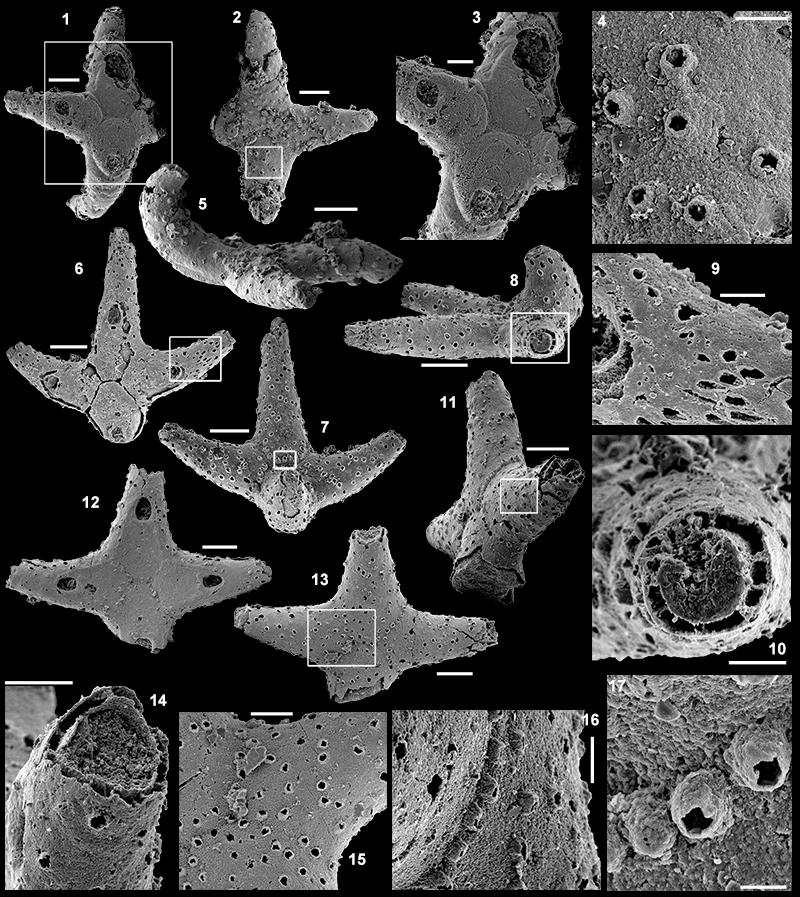
FIGURE 6. Articulated but broken internal moulds of sclerites of Archiasterella charma Moore, Li and Porter, 2014, from the Puerto Blanco Formation of Cerro Rajón, Sonora, Mexico. 1-3, 5. Specimen USTL3166-4 from sample R53: 1. Adaxial view showing the almost straight angle between the horizontal rays, area in the square is magnified in 3; 2, 5. Lateral views showing the curvature of the principal ray; 3. Detail of the basal plane showing the swollen rims of the foramina. 4. Disarticulated principal ray USTL3189-6 from sample R53 in lateral view showing the curvature and the swollen rim of the foramen. 6, 8. Specimen USTL3172-9 from sample R55: 6. Adaxial view; 8. Lateral view showing a relatively well-perserved (long) horizontal ray and the principal ray broken at the edge of the basal facet. 7, 9-10. Specimen USTL3168-4 from sample R54: 7. Lateral view from the principal ray; 9. Adaxial view showing the acute angle between the horizontal rays and the irregular junction between adjoining rays; 10. Lateral view showing the recurved principal ray partially covered by the external coating. 11, 14. Specimen USTL3167-7 from sample R53: 11. Adaxial view; 14. Lateral view. 12. Basal view of disarticulated principal ray USTL3167-3 from sample R53 showing the swollen rim of the foramen. 13. Adaxial view of one disarticulated horizontal ray USTL3173-4 from sample R55. 15. Lateral view of specimen USTL3169-6 R54b A_018 from sample R54b. 16-17. Specimen USTL3164-3 preserved with external coating from sample R53a: 16. Adaxial view; 17. Lateral view showing horizontal rays diverging from the basal plane. 18-19. Specimen USTL3168-6 preserved with external coating from sample R54: 16. Adaxial view; 17. Lateral view. Scale bars are: 3, 12, 100 µm; 1-2, 4-11, 13-19, 200 µm.
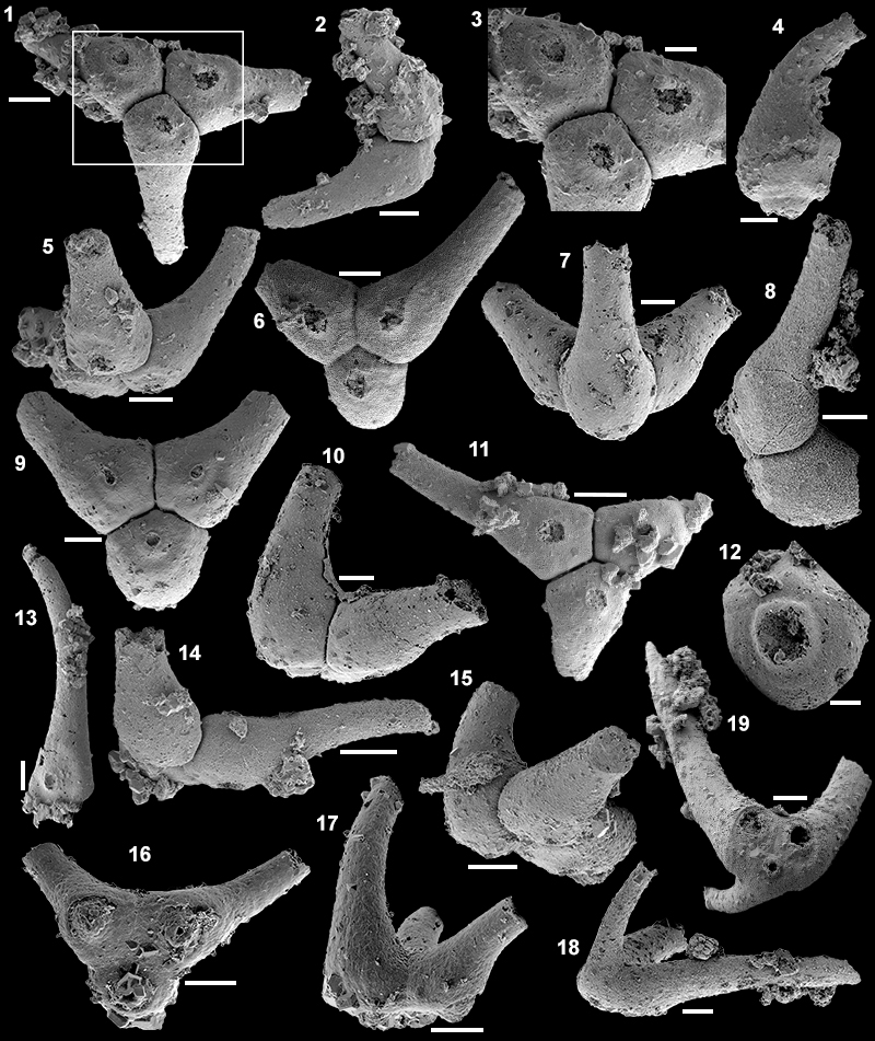
FIGURE 7. Internal moulds with partial external coating of sclerites of Archiasterella cf. A. pentactina Sdzuy, 1969 from the Puerto Blanco Formation of Cerro Rajón, Sonora, Mexico. 1. Specimen USTL3164-6 from sample R53a’: Adaxial view. 2-4. Specimen USTL3166-10 from sample R53: 2. Adaxial view, area in the square is magnified in 4; 3. Lateral view; 4. Detail of the basal plane with the circular and large foramina. 5-7, 10-11. Specimen USTL3171-5 from sample R54: 5. Adaxial view; 6. Abapical view showing the circular cross-section of the protruding broken principal ray; 7. Abaxial view; 10-11. Lateral views. 8, 12-13. Specimen USTL3175-13 from sample R63: 8. Lateral view; 12. Adaxial view showing the small perforations of the external coating corresponding to the foramina; 13. Abaxial view. 9, 14-15. Specimen USTL3167-4 from sample R53: 9. Abapical view; 14. Adaxial view, area in the square is magnified in 15; 15. Detail of the basal surface with elongated foramina surrounded by a swollen ring. Scale bars are: 4-7, 10-11, 15, 100 µm; 1-3, 8-9, 12-14, 200 µm.
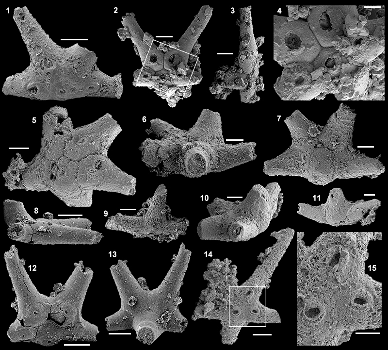
FIGURE 8. Articulated but broken internal moulds of sclerites of Allonnia erromenosa Jiang, in Luo et al., 1982 from the Puerto Blanco Formation of Cerro Rajón, Sonora, Mexico. 1-4. Specimen USTL3162-6 from sample R21: 1-2. Lateral view showing all the lateral rays recurved away from the basal plane; 3. Abaxial view; 4. Adaxial view showing the circular and wide foramina located on the basal edge of the rays. 5-7. Specimen USTL3162-12 from sample R21: 5. Adaxial view showing the foramina with raised rim surrounded by shallow depressions; 6-7. Lateral views. 8-10. Specimen USTL3163-11 from sample R21: 8, 10. Lateral views; 9. Adaxial view showing the circular and wide basally displaced foramina with raised rim and surrounding depression. Scale bars are all 200 µm.
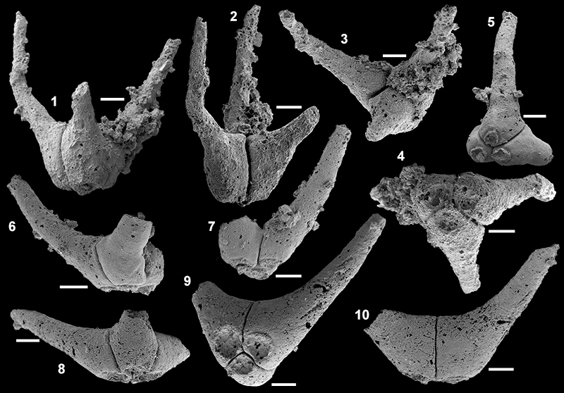
FIGURE 9. Articulated but broken internal moulds of sclerites of Allonnia tetrathallis from the Puerto Blanco Formation of Cerro Rajón, Sonora, Mexico. 1-3, 5. Specimen USTL3163-8 from sample R21 (4+0 form): 1. Adaxial view; 2-3, 5. Lateral views. 4. Specimen USTL3163-3 from sample R21 (4+0 form) in adaxial view. 6, 9, 12. Specimen USTL3163-10 from sample R21 (5+0 form): 6. Lateral view; 9. Adaxial view; 12. Abaxial view. 7-8, 10-11. Specimen USTL3162-4 from sample R21 (6+0 form): 7. Adaxial view; 8. Abaxial view; 10, 11. Lateral views. Scale bars are all 200 µm.
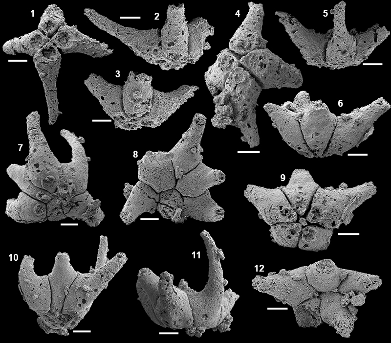
FIGURE 10. Internal moulds with external coatings of Chancelloria spp. from the Puerto Blanco Formation of Cerro Rajón, Sonora, Mexico. 1-6. Specimen USTL3189-4 from sample R53 (7+1 form): 1. Adaxial view, area in the square is magnified in 6; 2. Abaxial view showing the massive central ray; 3. Lateral view, area in the square is magnified in 4; 4. Detail of the curved and massive central ray with porous external coating; 5. Lateral view; 6. Detail of the slit-like lateral foramen running through the external coating. 7-9. Specimen USTL3168-5 from sample R54 (7+1 form): 7. Abaxial view; 8. Adaxial view; 9. Lateral view. 10, 13, 14. Specimen USTL3168-10 from sample R54 (8+1 form): 10. Adaxial view; 13. Abaxial view; 14. Lateral view. 11-12, 15-17. Specimen USTL3169-3 from sample R54 (7+1 form): 11. Adaxial view, area in the square is magnified in 15; 12. Lateral view showing the massive, recurved central ray; 15. Detail of the cross-section through a broken ray showing the external coating, the internal mould with the phosphatic internal layer and the void corresponding to dissolved shell material; 16. Abaxial view; 17. Lateral view. 18. Specimen USTL3170-2 from sample R54 (8+1 form). 19-20. Specimen USTL3170-6 from sample R54 (8+1 form): 19. Lateral view with long, slender lateral rays aligned with the basal plane; 20. Adaxial view. 21-22, 25-27. Specimen USTL3178-4 from sample R64 (5+1 form): 21. Adaxial view; 22. Abaxial view; 25. Detail of the basal plane; 26, 27. Lateral views. 23-24, 27-29. Specimen USTL3178-1 from sample R64 (5+1 form): 23, 28, 29. Lateral views; 24. Adaxial view. Scale bars are: 15, 20 µm; 4, 6, 50 µm; 1-3, 5, 7-14, 16-18, 25, 200 µm; 19-24, 26-29, 500 µm.
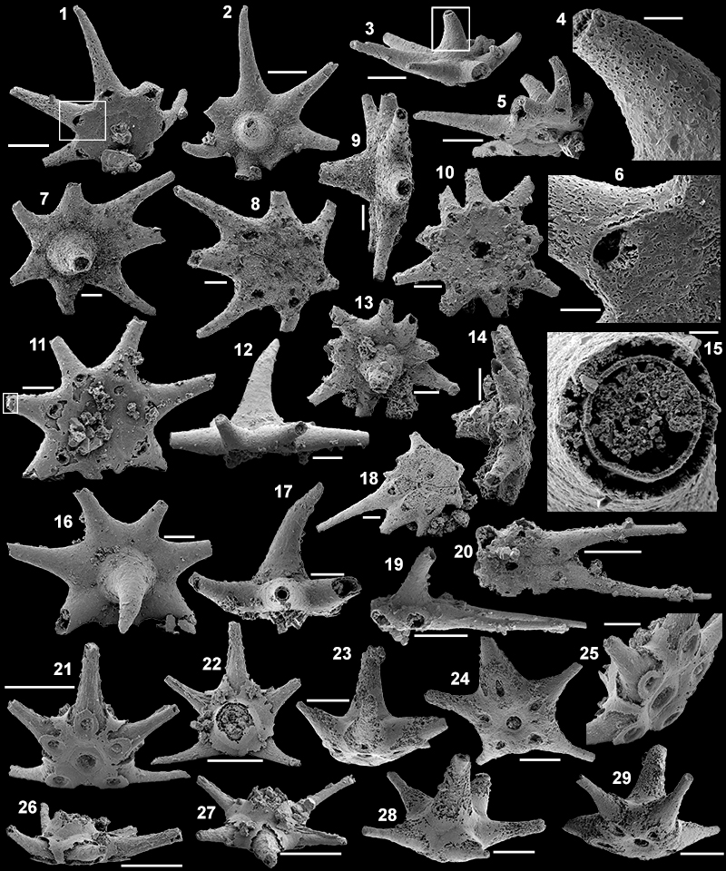
FIGURE 11. Incomplete tubes of Hyolithellus spp. from the Puerto Blanco Formation of Cerro Rajón, Sonora, Mexico. 1-2. Specimen USTL3189-2 from sample R53: 1. Lateral view; 2. Detail of the large aperture. 3. Specimen USTL3182-4 from sample R66 in lateral view. 4. Specimen USTL3169-8 from sample R54 in lateral view. 5-6, 9. Specimen USTL3168-9 from sample R54: 5. Lateral view, area in the square is magnified in 9; 6. Narrow aperture; 9. Detail of the external surface ornamentation. 7-8. Specimen USTL3181-5 from sample R71: 7. Lateral view; 8. Apertural view. 10-11. Specimen USTL3183-5 from sample R83: 10. Lateral view; 11. Apertural view. 12. Aperture of specimen USTL3187-3 from sample R92. 13. Aperture of specimen USTL3187-6 from sample R92. 14-16. Specimen USTL3184-2 from sample R83: 14. Lateral view, area in the lower square is magnified in 15 and area in the upper square is magnified in 16; 15. Detail of the laminar wall structure; 16. Detail of the external surface ornamentation. 17, 22. Specimen USTL3185-2 from sample R92: 17. Lateral view, area in the square is magnified in 22; 22. Detail of the irregular transverse ribs. 18-19. Specimen USTL3186-14 from sample R92: 18. Lateral view, area in the square is magnified in 19; 19. Detail of the external surface ornamentation. 20-21. Specimen USTL3186-2 from sample R92: 20. Lateral view, area in the square is magnified in 21; 21. Detail of the external surface ornamentation et exfoliation. 23. Lateral view of Specimen USTL3188-5 from sample R93. Scale bars are: 6, 11, 13, 15, 22, 50 µm; 2, 8-9, 12, 16, 19, 21, 100 µm; 10, 17-18, 20, 23, 200 µm; 1, 4-5, 7, 14, 500 µm; 3, 1 mm.
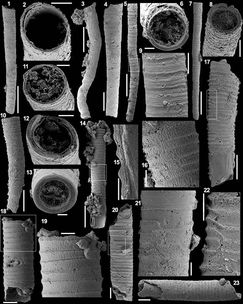
FIGURE 12. Incomplete conchs of Cupitheca cf. C. mira from the Puerto Blanco Formation of Cerro Rajón, Sonora, Mexico. 1-5, 8-10. Specimen USTL3176-9 from sample R63: 1. Lateral view, area in the square is magnified in 4; 2. Lateral view, area in the square is magnified in 5; 3. Adapical view, area in the square is magnified in 10; 4. Detail of the septum; 5. Detail of the flat and angular closed termination; 8. Abapical view, area in the square is magnified in 9; 9. Detail of the filling of a chamber; 10. Detail of the smooth septum. 6-7, 11-14. Specimen USTL3176-3 from sample R63: 6. Lateral view, area in the square is magnified in 14; 7. Lateral view; 11. Abapical view; 12, 13. Adapical view showing the hemispherical embryonic shell; 14. Detail of the relation between the septum and the lateral wall. Scale bars are: 14, 50 µm; 4-5, 9-10, 100 µm; 1-3, 6-8, 11-13, 200 µm.
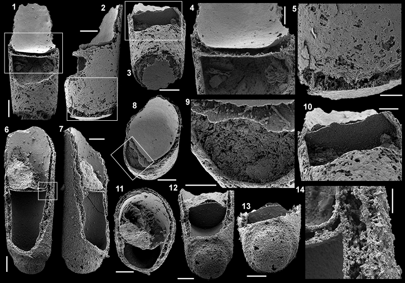
FIGURE 13. Reconstruction of a complete conch of Cupitheca cf. C. mira with delimited parts corresponding to the various parts that were preserved. The dashed lines represent material which was not preserved in the Mexican material, but which are inferred from the data of Bengtson et al. (1990).
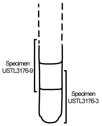
FIGURE 14. Conchs and opercula of Petasotheca sp. from the Puerto Blanco Formation of Cerro Rajón, Sonora, Mexico. 1-3. Specimen USTL3174-1 from sample R56: 1. Lateral view of the conch; 2. Apertural view; 3. Apical view. 4-6. Specimen USTL3175-7 from sample R63: 4. Lateral view of the conch; 5. Apertural view; 6. Apical view. 7. Specimen USTL3175-10 from sample R63, lateral view of the conch. 8. Specimen USTL3166-5 from sample R53, lateral view of the conch. 9-10. Specimen USTL3177-13 from sample R63: 9. External surface; 10. Lateral view of internal surface. 11-12. Specimen USTL3174-2 from sample R56: 11. Internal surface; 10. Lateral view of internal surface showing the organization of the raised concentric ridge and the clavicle-like structures. 13-15. Specimen USTL3174-5 from sample R56: 13. Internal surface; 14. External surface with fine concentric growth lines; 15. Lateral view. 16-19. Specimen USTL3177-5 from sample R63: 16. Lateral view, area in the square is magnified in 19; 17. Internal surface; 18. External surface with rounded subcentral apex and fine concentric growth lines; 19. Detail of the polygonal imprints on the internal concentric ridge. Scale bars are: 19, 20 μm; 2-3, 9-18, 100 μm; 1, 4-7, 200 μm; 8, 500 μm.
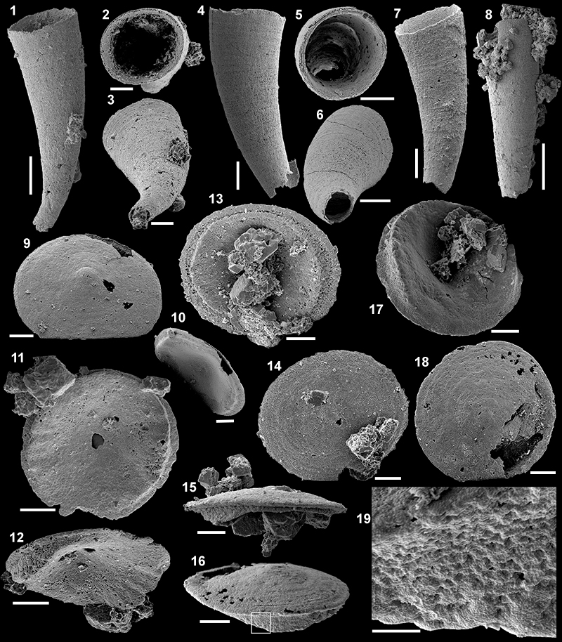
FIGURE 15. Conchs of hyolithid sp. and opercula of Parkula bounites Bengtson in Bengtson et al., 1990 from the Puerto Blanco Formation of Cerro Rajón, Sonora, Mexico. 1-3. Specimen USTL3169-5 of hyolithid sp. from sample R54: 1. Lateral view; 2. Dorsal view; 3. Apertural view. 4-6. Specimen USTL3169-1 of hyolithid sp. from sample R54: 4. Dorsal view; 5. Lateral view; 6. Apertural view. 7-12. Specimen USTL3175-9 of Parkula bounites Bengtson in Bengtson et al., 1990, from sample R63: 7. External view; 8. Internal view, area in the square is magnified in 11; 9. Lateral view, area in the square is magnified in 12; 10. Dorsal view; 11-12. Details of the cardinal processes. 13-20. Specimen USTL3176-1 of Parkula bounites Bengtson in Bengtson et al., 1990 from sample R63: 7. External view, area in the square is magnified in 19; 14. Dorsal view; 15. Lateral internal view, area in the square is magnified in 20; 16, 20. Details of the cardinal processes; 17. Lateral view; 18. Internal view, area in the square is magnified in 16; 19. Detail of apical area. Scale bars are: 11-12, 16, 19-20, 100 µm; 7-10, 13-15, 17-18, 200 µm; 1-6, 500 µm.
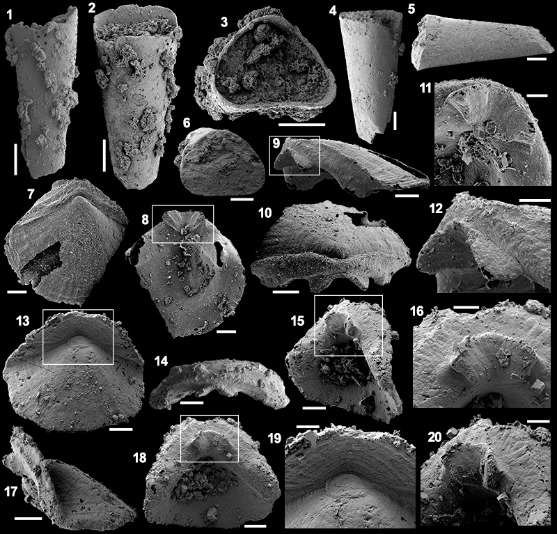
FIGURE 16. Internal moulds of Mackinnonia corrugata Runnegar in Bengtson et al., 1990 from the Puerto Blanco Formation of Cerro Rajón, Sonora, Mexico. 1-5. Specimen USTL3189-5 from sample R53: 1. Lateral view; 2. Apertural view; 3. Posterior view; 4. Anterior view, area in the square is magnified in 5; 5. Detail of the anterior field showing the polygonal imprints restricted to the summit of the comarginal corrugations. 6-9. Specimen USTL3189-3 from sample R53: 6. Upper view; 7. Lateral view, area in the square is magnified in 9; 8. Posterior view; 9. Detail of the polygonal imprints lining the apertural margin. 10-14. Specimen USTL3165-1 from sample R53: 10. Antero-lateral view, area in the square is magnified in 13; 11. Postero-lateral view, area in the square is magnified in 14; 12. Posterior view; 13. Detail of the high, sharp posterior part of the comarginal corrugations; 14. Detail of the polygonal imprints lining the apertural margin. 15-16. Specimen USTL3166-1 from sample R53: 15. Lateral view; 16. Posterior view. 17. Specimen USTL3172-8 from sample R55 in apertural view. 18. Specimen USTL3166-8 from sample R53 in lateral view. Scale bars are: 14, 20 μm; 5, 9, 13, 100 μm; 1-4, 6-8, 10-12, 15-18, 200 μm.
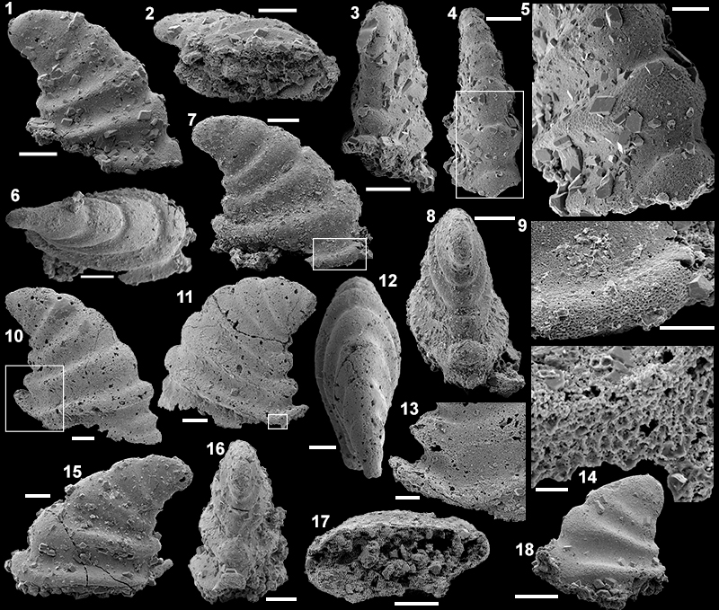
FIGURE 17. Internal moulds and coatings of Xianfengella sp. from the Puerto Blanco Formation of Cerro Rajón, Sonora, Mexico. 1-4, 8. Specimen USTL3163-6 from sample R21: 1. Upper view; 2. Lateral view; 3. Posterior view, area in the square is magnified in 8; 4. Anterior view; 8. Detail of the subapical notch in the internal mould. 5-7. Specimen USTL3162-5 from sample R21: 5. Lateral view, area in the square is magnified in 7; 6. Subapical view; 7. Detail of the subapical notch and wedge. 9-12, 17. Specimen USTL3163-12 from sample R21: 9. Upper view; 10. Lateral view, area in the square is magnified in 17; 11. Posterior view, area in the square is magnified in 12; 12, 17. Details of the apical region. 13, 15. Specimen USTL3162-10 from sample R21: 13. Lateral view; 15. Posterior view. 14, 16. Specimen USTL3162-3 from sample R21: 14. Upper lateral view, arrow pointing polygonal imprints on the internal mould; 16. Posterior view, arrow pointing polygonal imprints on the internal mould. Scale bars are: 7-8, 12, 17, 50 µm; 1-6, 9-11, 13-16 100 µm.
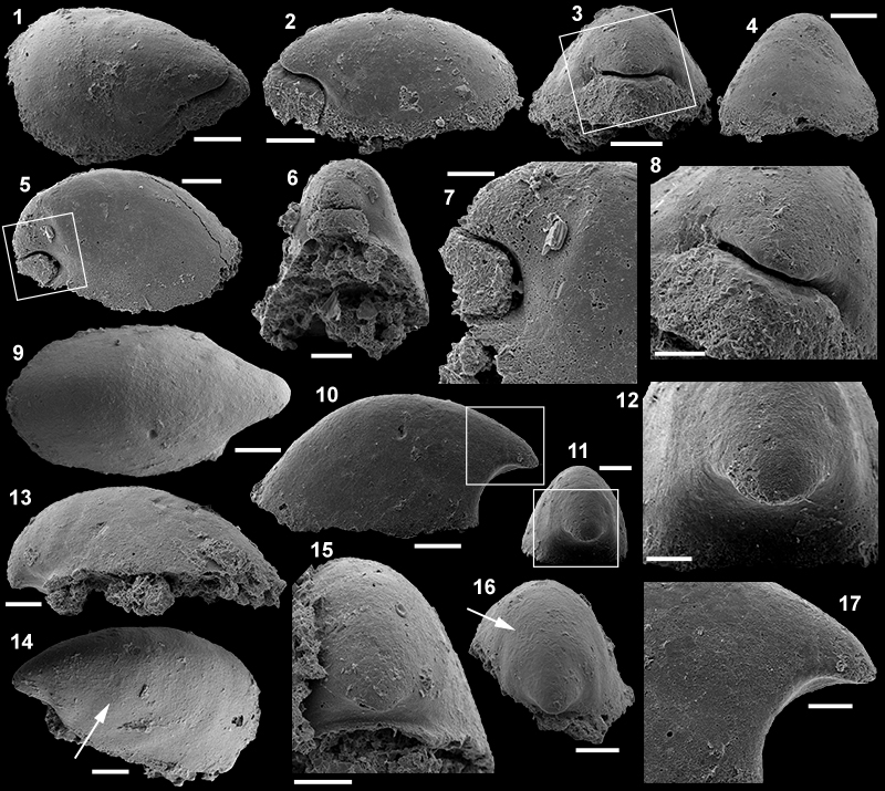
FIGURE 18. Pelagiella sp. from the Puerto Blanco Formation of Cerro Rajón, Sonora, Mexico. 1-7. Specimen USTL3168-2 (shell replacement) from sample R54: 1. Oblique apertural view, area in the square is magnified in 7; 2. Apertural view, area in the square is magnified in 4; 3. Abapical view; 4. Detail of the subumbilical furrow and of the radial shell striations; 5. Apertural view showing the triangular apertural cross-section; 6. Abapical view with radial striations visible; 7. Detail of the apex and umbilicus. 8-13. Specimen USTL3175-1 (internal coating) from sample R63: 8. Oblique apical view; 9. Apical view, area in the square is magnified in 12; 10. Abapertural view; 11. Apertural view, area in the square is magnified in 13; 12. Detail of the surface of the internal coating with comarginal striations; 13. Detail of the bulbous apex. 14-15. Specimen USTL3177-6 (internal mould and coating) from sample R63: 14. Apical view; 15. Apertural view. 16-17. Specimen USTL3175-6 (internal mould and coating) from sample R63: 16. Apical view; 17. Apertural view. 18-19, 24. Specimen USTL3177-10 (internal mould and coating) from sample R63: 18. Apical view; 19. Apertural view; 24. Lateral view. 20-23. Specimen USTL3175-3 (internal mould and coating) from sample R63: 20. Detail of the internal coating with comarginal striations; 21. Apical view, area in the square is magnified in 20; 22. Apertural view; 23. Lateral view. 25-27. Specimen USTL3177-8 (internal mould and coating, partial external coating) from sample R63: 25. Abapical view, area in the square is magnified in 24; 24. Detail of the external coating and internal mould and coating; 27. Apertural view. Scale bars are: 13, 20, 26, 50 µm; 4, 7, 12, 14-15, 100 µm; 1-3, 5-6, 8-11, 16-19, 21-25, 27, 200 µm.
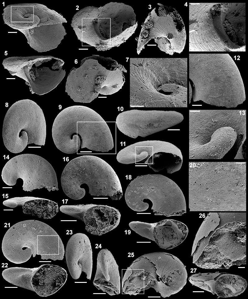
FIGURE 19. Pojetaia sp. from the Puerto Blanco Formation of Cerro Rajón, Sonora, Mexico. 1-4. Specimen USTL3171-4 (internal mould) from sample R54: 1. Right lateral view; 2. Anterior view; 3. Dorsal view, area in the square is magnified in 4; 4. Detail of the hinge showing the two teeth and associated sockets (one on each valve). 5-10, 12-13. Specimen USTL3175-2 (coating of disarticulated left valve) from sample R63: 5. Internal view, note the prosogyre umbo; 6. External view with the fine, dense comarginal, growth lines, area in the upper left square is magnified in 9 and area in the lower right square is magnified in 13; 7. Posterior view, area in the square is magnified in 12; 8. Anterior view; 9. Detail of the growth lines and the radial ribs of the anterior area; 10. Dorsal view; 12, 13. Details of the growth lines. 11, 17. Specimen USTL3176-7 from sample R63 (coating): 11. Internal view, area in the square is magnified in 17; 17. Detail of the hinge with the tooth. 14-16, 18. Specimen USTL3176-5 from sample R63 (coating): 14. Dorsal view; 15. Posterior view; 16. External view with external coating preserved in the anterior area (commarginal growth lines and radial ribs) and internal coating in the posterior area with branching microbial filaments, area in the square is magnified in 18; 18. Details of the microbial filaments. Scale bars are: 4, 9, 12-13, 17-18, 100 µm; 5-8, 10-11, 14-16, 200 µm; 1-3, 500 µm.
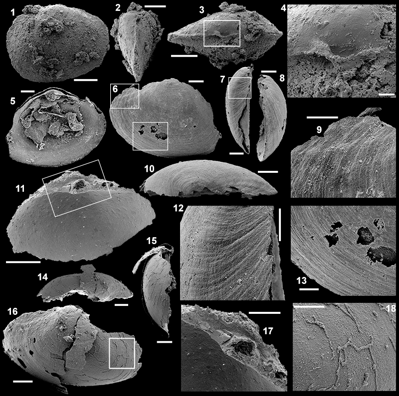
FIGURE 20. Valve of Bradoriid sp. from the Puerto Blanco Formation of Cerro Rajón, Sonora, Mexico. 1-9. Specimen USTL3182-4 from sample R66: 1. External view, area in the right square is magnified in 3, larea in the ower left square is magnified in 4 and area in the upper left square is magnified in 8; 2. Dorso-lateral view; 3-4. Details of the rim; 5. Ventral view; 6. Dorsal view, area in the square is magnified in 9; 7. Lateral view; 8-9. Details of the pustulose ornamentation. Scale bars are: 3, 8-9, 50 µm; 4, 100 µm; 2, 7, 200 µm; 1, 5-6, 500 µm.
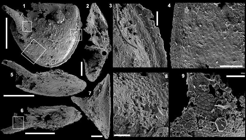
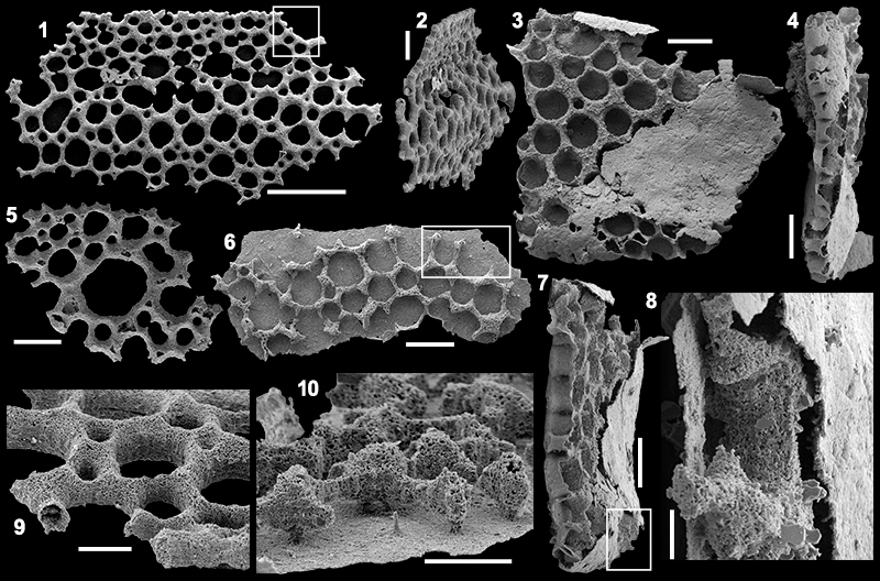
FIGURE 21. Microdictyon multicavus McMenamin, 1984, from the Puerto Blanco Formation of Cerro Rajón, Sonora, Mexico. 1-9. Specimen USTL3180-2 from sample R69: 1. Oblique view, area in the square is magnified in 8; 2. Lateral view, upper lower area in the square is magnified in 3 and lower area in the square is magnified in 4; 3. Detail of the node extremities from the edge to the centre of the sclerite (flat, flat cap, cap with subcentral apex); 4, 5. Detail of the caped-nodes with a subcentral apex; 6. External view, area in the square is magnified in 9; 7. Lateral view; 8. Detail of the peripheral girdle; 9. Detail of the nodes with a flat cap. 10-13, 15-16. Specimen USTL3179-2 from sample R65: 10. External view, area in the square is magnified in 13; 11. Lateral view, area in the square is magnified in 12; 12, 13. Detail of a caped-node with a subcentral apex; 15. Internal view, area in the square is magnified in 16; 16. Detail of the basal termination of a hole/depression. 14, 17-19. Specimen USTL3177-3 from sample R63: 14. External view, area in the square is magnified in 18; 17. Oblique view; 18. Detail of the peripheral girdle; 19. Detail of the holes and surrounding nodes. 20. Lateral view of specimen USTL3176-4 from sample R63. Scale bars are: 9, 13, 19, 20 µm; 4-5, 12, 18, 50 µm; 3, 8, 14, 16-17, 100 µm; 10-11, 15, 200 µm; 1-2, 6-7, 500 µm.
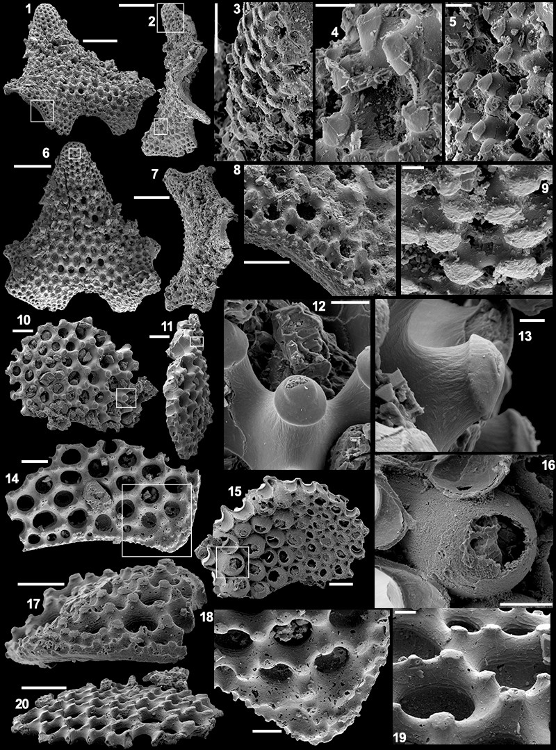
FIGURE 22. Brachiopods from the Puerto Blanco Formation of Cerro Rajón, Sonora, Mexico. 1-14. Rajonia ornata. 1-7. Specimen USTL3179-1 from sample R65: 1. External view, area in the lower square is magnified in 3 and area in the upper square is magnified in 4; 2. Lateral view; 3. Detail of the shell ornamentation; 4. Detail of the larval shell; 5. Internal view; 6. Subapical view, area in the square is magnified in 7; 7. Detail of the articulation. 8, 11. Specimen USTL3179-5 from sample R65: 8. External view; 11. Detail of the concentric fila. 9, 10, 12. Specimen USTL3179-3 from sample R65: 12. External view, area in the upper square is magnified in 9, area in the lower square is magnified in 10; 9-10. Details of the shell structure. 13-16. Specimen USTL3182-3 of a ventral valve of Eoobolus sp. from sample R66. Scale bars are: 10, 20 µm; 4, 11, 50 µm; 7, 9, 15, 100 µm; 1-3, 5-6, 8, 200 µm; 12-14, 16, 500 µm.
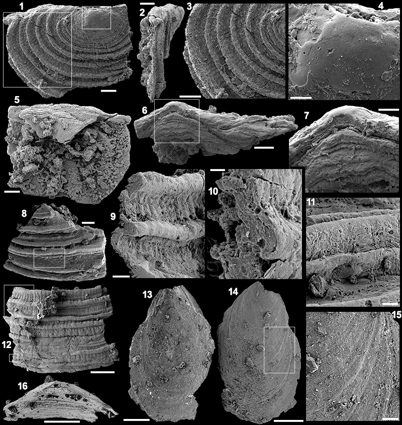
FIGURE 23. Indeterminate fossil from the Puerto Blanco Formation of Cerro Rajón, Sonora, Mexico. 1-2, 9. Specimen USTL3174-12. 3-4, 7-8. Specimen USTL3176-11. 5. Specimen USTL3174-13. 6, 10. Specimen USTL3174-11. Scale bars are: 8, 50 µm; 9, 10, 100 µm; 2-7 200 µm; 1, 500 µm.