

SUPPLEMENTARY FIGURE 1. Depiction of landmark placement. Reconstruction courtesy of Stephen Pates.
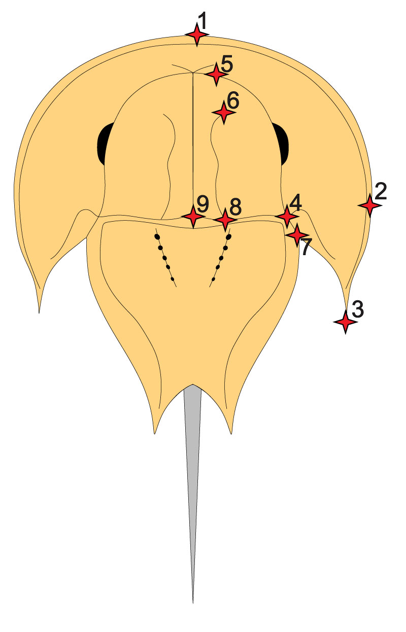
SUPPLEMENTARY TABLE 1. Table of landmarks used for the geometric morphometric analysis. When the right cephalothoracic side was not preserved, the left side of the cephalothorax was digitised and the data were reflected.
| Landmark number | Description |
| Number 1 | Most anterior section of cephalothorax |
| Number 2 | Most lateral extent of right cephalothoracic edge |
| Number 3 | Most distal point of genal spine |
| Number 4 | Most posterior point of right ophthalmic ridge |
| Number 5 | Most anterior point of right ophthalmic ridge |
| Number 6 | Most anterior point of right interophthalmic ridge |
| Number 7 | Most right lateral extent of cephalothoracic-thoracetronic hinge |
| Number 8 | Most posterior point of right interophthalmic ridge |
| Number 9 | Most posterior point of cephalothoracic midline |
SUPPLEMENTARY INFORMATION 1. The TPS file of analysed specimens (all supplementary information files available in zipped file).
SUPPLEMENTARY INFORMATION 2. CSV file of PCA results, family and generic assignment of specimens (all supplementary information files available in zipped file).
SUPPLEMENTARY INFORMATION 3. NEXUS file of the analysed phylogenetic matrix. Originally presented in Lamsdell (2016) (all supplementary information files available in zipped file).
