FIGURE 1. Location and geologic setting of the Arlington Archosaur Site (AAS marked with star). 1, Stratigraphic column and selected ages of Cretaceous rock units in north-central Texas. Terrestrial deposits are marked by stippled regions. Modified from Winkler et al. (1989) and Jacobs et al. (2013); 2, Generalized map of geologic units in the Fort Worth Basin; 3, Composite stratigraphic section of AAS (modified after Noto, 2015); 4, Inset map showing approximate location of the AAS in relation to the southern WIS (modified after Barnes et al., 1972 and Strganac, 2015). Approximate WIS shorelines during the Cenomanian are dotted (after Blakey, 2014).
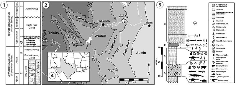
FIGURE 2. Carapace of “Trinitichelys” maini, sp. nov. DMNH 2013-07-0784, co-ossified partial carapace, with the anterior portion in 1-2, dorsal view, the associated co-ossified posterior carapace in 4-5 dorsal, and 5-6, ventral views. 3(A-H): DMNH 2013-07-0499, an isolated p1 in 3A-B dorsal, and 3C-D ventral views. Red line indicates possible tooth mark. DMNH 2013-07-1711, an isolated nu in 3E-F dorsal, and 3G-H, ventral views. Note scale bar for 3A-H is 10 mm, half the size of scale bar for DMNH 2013-07-0784.
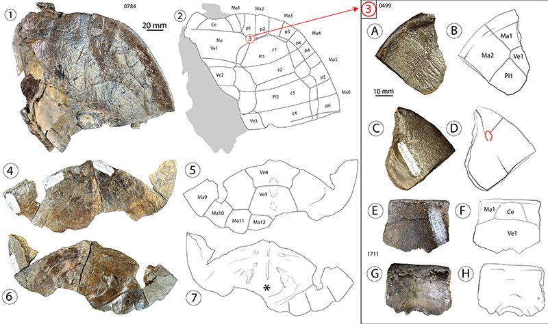
FIGURE 3. “Trinitichelys” maini sp. nov. plastral elements; red lines indicate possible tooth marks. DMNH 2013-07-0712, type specimen for “T.” maini sp. nov., an anterior plastral lobe, in 1-2, ventral, and 3-4, dorsal views. Please note midline contact between extragular scutes. DMNH 2013-07-0696 and -0704, left co-ossified bridge series, in 5-6, ventrolateral, and 7-8, dorsal views. DMNH 2013-07-0783, right co-ossified bridge series in 9-10, ventrolateral, and 11-12, dorsal views. DMNH 2013-07-1703, isolated mesoplastron in 13-14, dorsal, 15-16, ventral, and 17, posterior views. DMNH 2013-07-1708, isolated xiphiplastron from likely juvenile in 18-19, ventral, and 20-21, dorsal views. Red outlines in line drawings indicate edges of taphonomic traces.
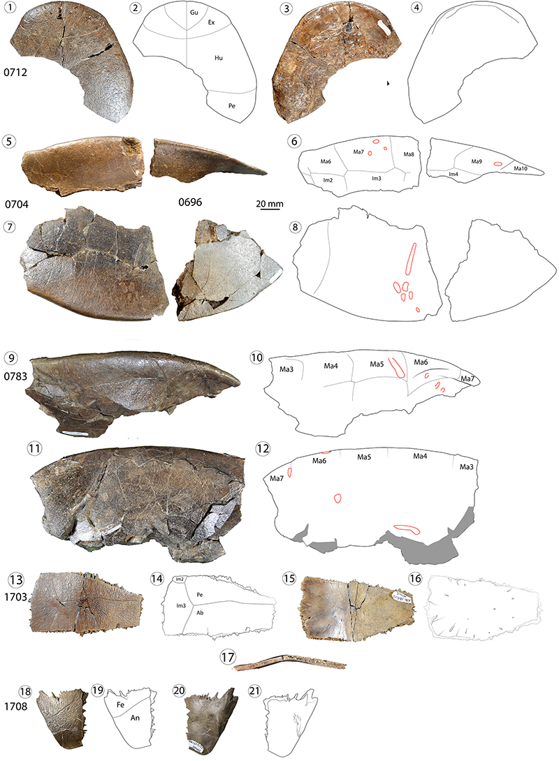
FIGURE 4. Isolated carapace elements of Naomichelys sp. DMNH 2013-07-1715, nu, in 1-2, dorsal, and 3-4, ventral views. DMNH 2013-07-0530, left c1, in 5-6, dorsal, and 7-8, ventral views; DMNH 2013-07-0543, partial left cf. c3 in 9-10, dorsal, and 11-12, ventral views. DMNH 2013-07-0559, left c7, in 13-14, dorsal, and 15-16, ventral views. DMNH 2013-07-0706, cf. n4, in 17-18, dorsal, and 19-20, ventral views. DMNH 2013-07-0770, a medial right partial n7/8 in 21-22, dorsal, and 23-24, ventral views. DMNH 2013-07-0570, pygal, in 25-26, dorsal, and 27-28, ventral views. Red outlines in line drawings indicate edges of taphonomic traces.
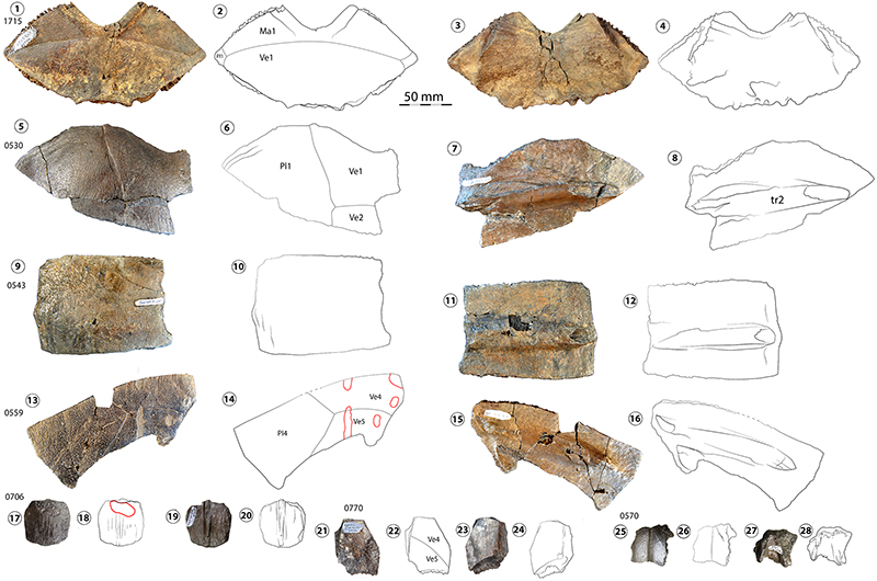
FIGURE 5. Plastral elements of Naomichelys sp. Red arrows indicate ventral knob-like processes. DMNH 2013-07-0711, isolated right hyoplastron and Inframarginal scute 2 in 1-2, ventral, and 3-4, dorsal views. DMNH 2013-07-0511, a left hyoplastron, in 5-6, ventral, and 7-8, dorsal views. DMNH 2013-07-1717, isolated right xiphiplastron in 9-10, ventral, and 11-12, dorsal views. DMNH 2013-07-0603, partial mesoplastron in 13-14, ventral, and 15-16, dorsal views. Red outlines in line drawings indicate edges of taphonomic traces.
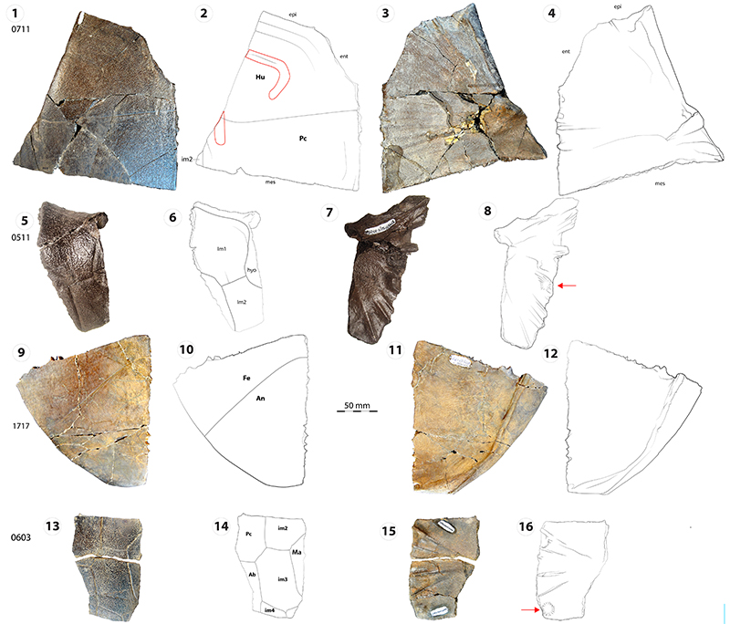
FIGURE 6. Various conditions of shell bone preservation in Naomichelys sp., with: 1, raised intact tubercles, 2, a tubercle broken in half along a bone edge, and 3, external bone surface with pattern of missing tubercles, by far the most common state in the taxon at the AAS. Please note the matrix adhered to the surface, the concentric layers of bulging parallel-fibred bone, and the continuance of the tubercle into deeper layers of the external cortex.
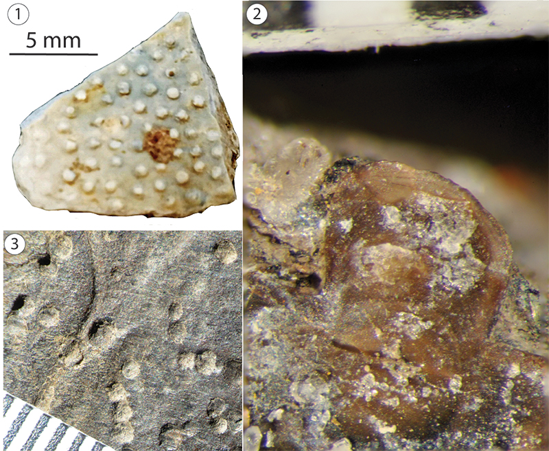
FIGURE 7. Trionychidae indet. DMNH 2013-07-1304, isolated left nu, in 1-2, dorsal, and 3-4, ventral views. Dashed lines indicate free margins and arrows indicate possible suprascapular fontanelles. DMNH 2013-07-1405, an indeterminate bothremydid (cf. Algorachelus) c4 in 5-6, dorsal and 7-8, ventral views. Gray shading indicates damaged surface from likely axillary bridge connection. Please note that the specimens have a 10 mm scale.
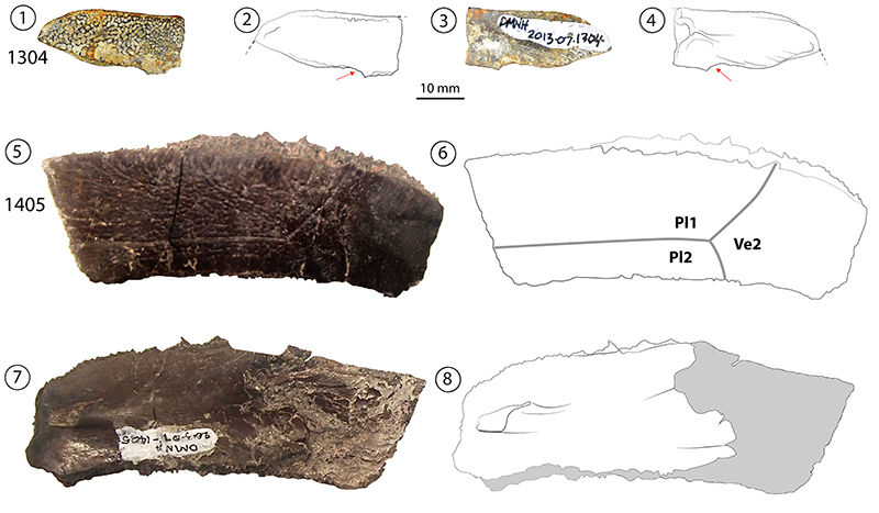
FIGURE 8. Proposed phylogenetic position of “T.” maini sp. nov. within Paracryptodira from the phylogenetic analysis performed using characters from Joyce et al. (2016), with Kayentachelys aprix as the outgroup.