FIGURE 1. Stratigraphical distribution of coral genera that became extinct before the base of the Hauterivian. Orange lines indicate genera that range was extended by the present fauna.
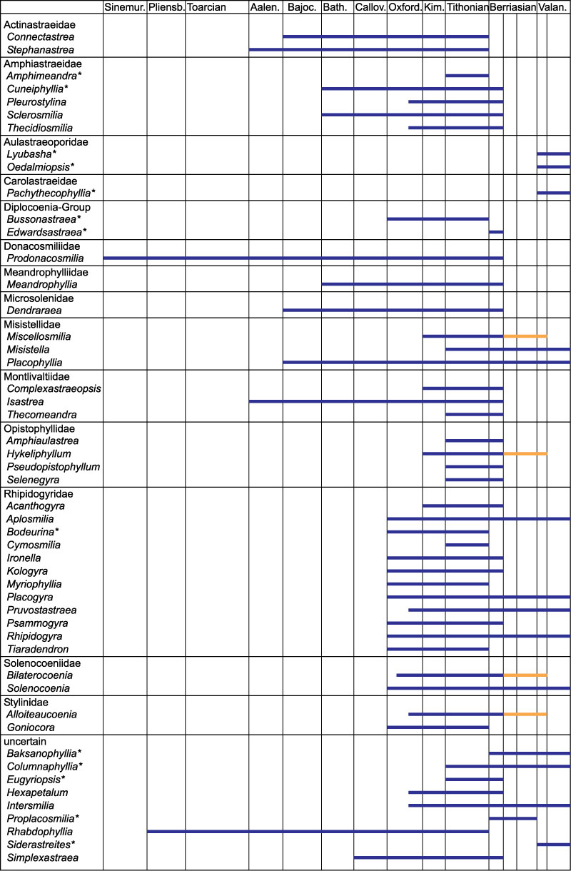
FIGURE 2. Geographical and geological location of the Puerto Llano section. 1. Location of the Betic Cordillera in the South of Spain. 2. Palaeogeographical reconstruction of the South Iberian Palaeomargin in Late Kimmeridgian according to Vera (2001). 3. Geological sketch of the Betic Cordillera with location of the Prebetic zone shown in 4. 4. Geological sketch of the Prebetic in the ended of the Sierra de Cazorla (External Prebetic) and Sierra de Segura (Internal Prebetic) with location of the Puerto Llano section.
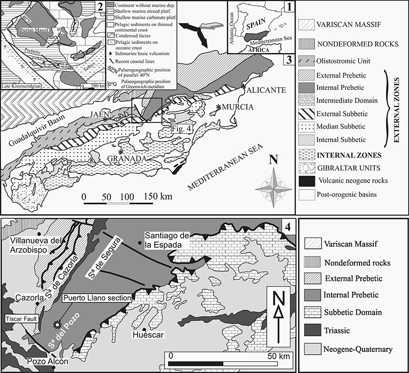
FIGURE 3. Stratigraphic section of the Sierra del Pozo Formation (Tithonian-Valanginian). 1. Detailed geological sketch of the area where the formation crops out. 2. General stratigraphic section of the Sierra del Pozo Formation and detailed section of the upper member, where corals are present.
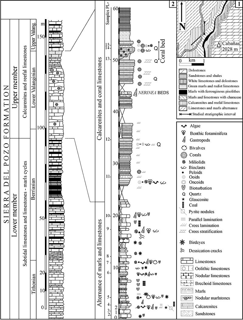
FIGURE 4. Detailed stratigraphic section of the calcarenites and coral limestones (upper member of the Sierra del Pozo Formation) with the position of the samples and their microfacies features.
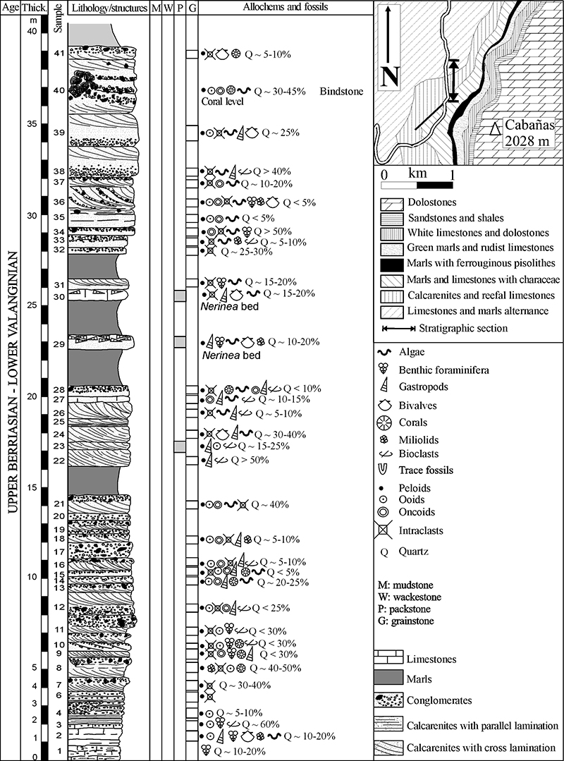
FIGURE 5. ? Actinastrea sp., MGB 83276. 1: transversal thin section. 2: transversal thin section, detail. 3: longitudinal thin section. Scale 1 mm.

FIGURE 6. Floriastrea iberica sp. nov., Löser. 1: MGB 83251, transversal thin section. 2: MGB 83251, transversal thin section, detail. 3: MGB 83314, transversal thin section, larger view. 4: MGB 83251, longitudinal thin section. Scale 1 mm.

FIGURE 7. Stelidioseris melkarthi Felix, 1909, MGB 83295. 1: transversal thin section. 2: transversal thin section, detail. 3: longitudinal thin section. Scale 1 mm.
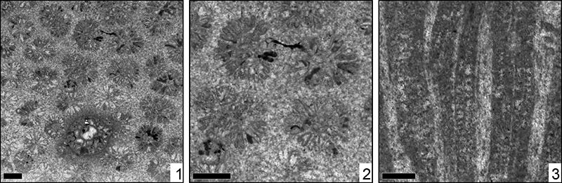
FIGURE 8. Stelidioseris sp., MGB 83270. 1: transversal thin section. 2: transversal thin section, detail. Scale 1 mm.
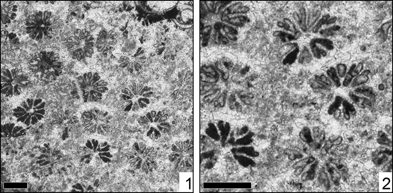
FIGURE 9. Stelidioseris ? sp., MGB 83366. 1: transversal thin section. 2: transversal thin section, detail. 3: longitudinal thin section. Scale 1 mm.
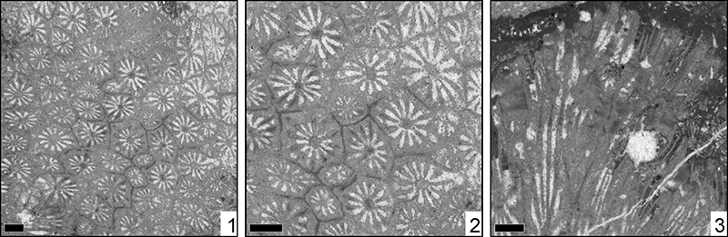
FIGURE 10. Amphiastrea basaltiformis Etallon, 1859, MGB 83262. 1: transversal thin section. 2: transversal thin section, detail. Scale 1 mm.
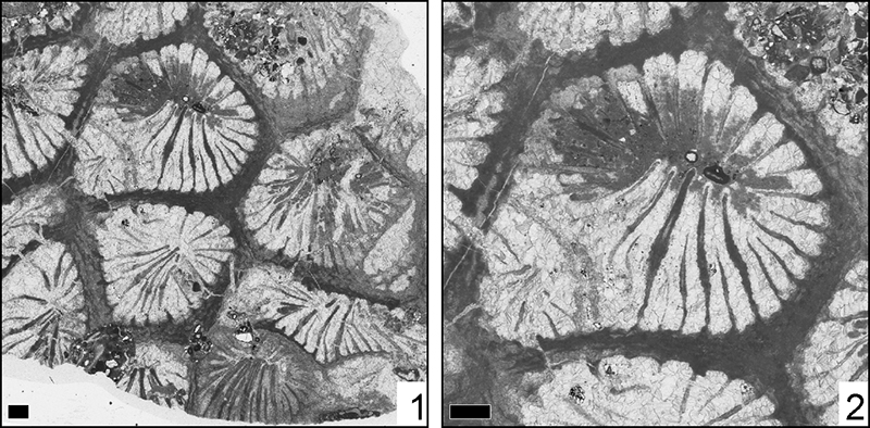
FIGURE 11. Amphiastrea cf. basaltiformis Etallon, 1859, MGB 83319. 1: transversal thin section. 2: transversal thin section, detail. 3: Oblique thin section. Scale 1 mm.
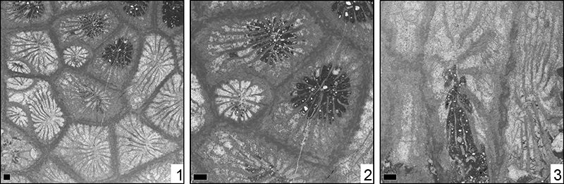
FIGURE 12. Amphiastrea cf. woodiae (Gregory, 1930), MGB 83274. 1: transversal thin section. 2: transversal thin section, detail. 3: longitudinal thin section. Scale 1 mm.
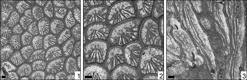
FIGURE 13. Amphiastrea sp., MGB 83249. 1: transversal thin section. 2: transversal thin section, detail. Scale 1 mm.
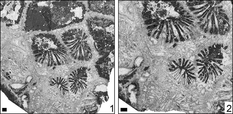
FIGURE 14. Hykeliphyllum sp., MGB 83261. 1: transversal thin section. 2: transversal thin section, detail. Scale 1 mm.
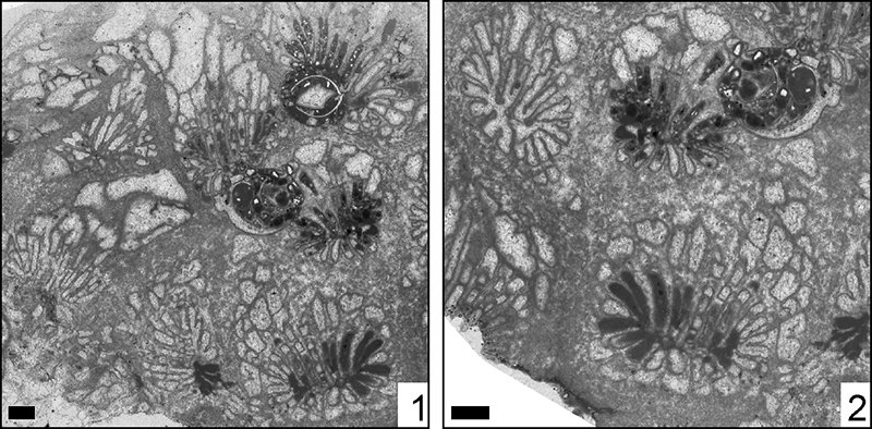
FIGURE 15. Astraeofungia diversisepta (Hackemesser, 1936), MGB 83350. 1: transversal thin section. 2: transversal thin section, detail. 3: longitudinal thin section. Scale 1 mm.
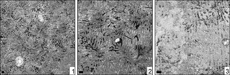
FIGURE 16. Astraeofungia tenochi Felix, 1891, MGB 83267. 1: transversal thin section. 2: transversal thin section, detail. 3: longitudinal thin section. Scale 1 mm.
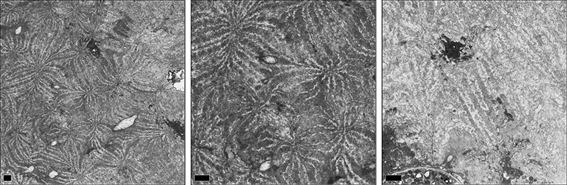
FIGURE 17. Latiastrea canavarii (Prever, 1909), MGB 83311. 1: transversal thin section. 2: transversal thin section, detail. 3: longitudinal thin section. Scale 1 mm.
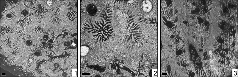
FIGURE 18. Latiastrea somaensis Eguchi, 1951, MGB 83244. 4: transversal thin section. 5: transversal thin section, detail. 6: longitudinal thin section. Scale 1 mm.
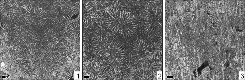
FIGURE 19. Latiastrea sp., MGB 83387. 1: transversal thin section. 2: transversal thin section, detail. Scale 1 mm.
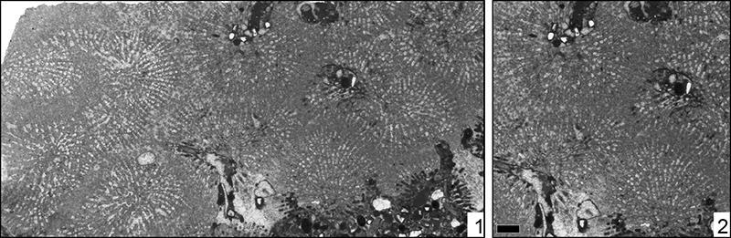
FIGURE 20. Latomeandra isseli (Prever, 1909), MGB 83232. 1: transversal thin section. 2: transversal thin section, detail. 3: longitudinal thin section. Scale 1 mm.
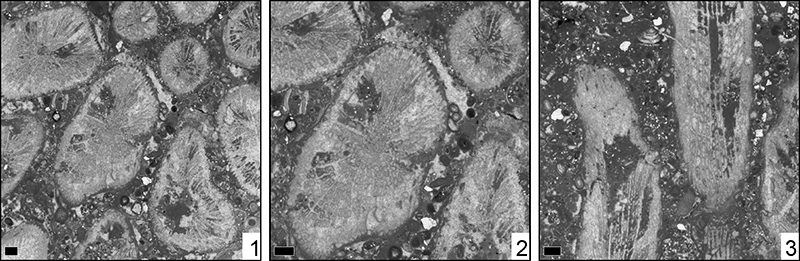
FIGURE 21. Ovalastrea caryophylloides Goldfuss, 1826, MGB 83236. 1: transversal thin section. 2: transversal thin section, detail. 3: longitudinal thin section. Scale 1 mm.
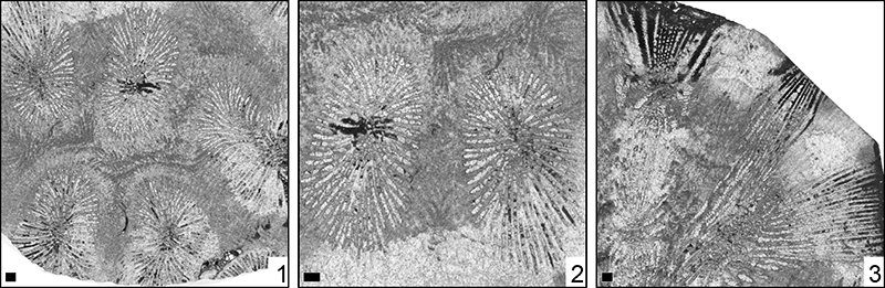
FIGURE 22. Periseris crassisepta Sikharulidze, 1985, MGB 83268. 1: transversal thin section. 2: transversal thin section, detail. 3: longitudinal thin section. Scale 1 mm.
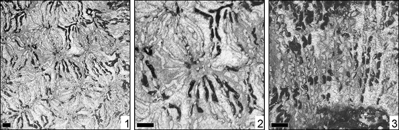
FIGURE 23. Periseris elegantula Orbigny, 1850, MGB 83277. 1: transversal thin section. 2: transversal thin section, detail. 3: longitudinal thin section. Scale 1 mm.
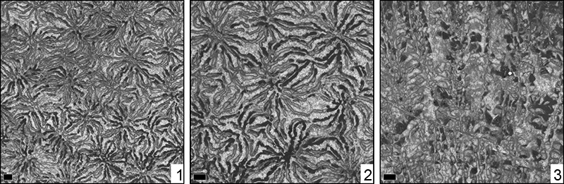
FIGURE 24. Periseris frondescens Orbigny, 1850, MGB 83323. 10: transversal thin section. 11: transversal thin section, detail. 12: longitudinal thin section. Scale 1 mm.
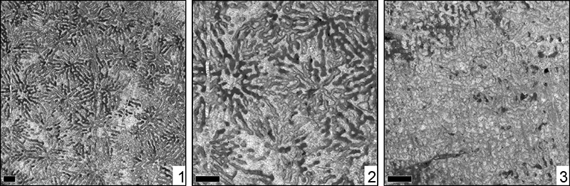
FIGURE 25. Placoseris poculum (Fromentel, 1857), MGB 83260. 1: transversal thin section. 2: transversal thin section, detail. Scale 1 mm.
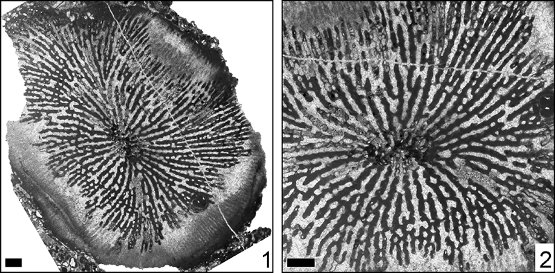
FIGURE 26. Thalamocaeniopsis explanata Reig Oriol, 1994, MGB 83275. 1: transversal thin section. 2: transversal thin section, detail. Scale 1 mm.
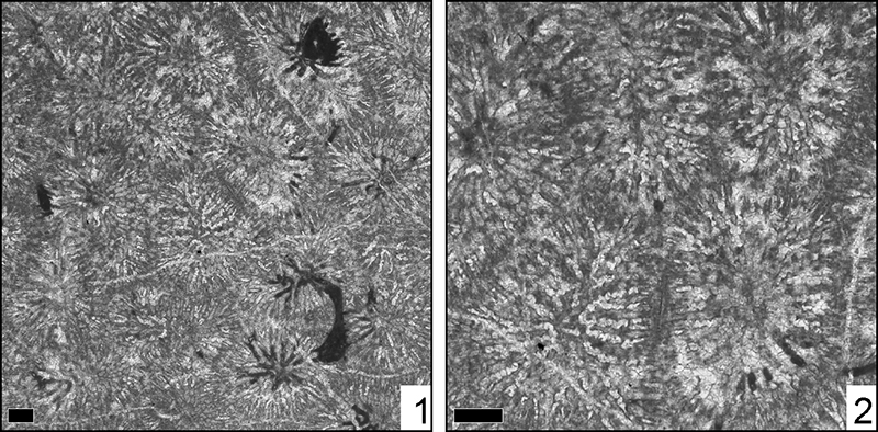
FIGURE 27. Eocomoseris sp. 1, MGB 83343. 1: transversal thin section. 2: transversal thin section, detail. 3: longitudinal thin section. Scale 1 mm.
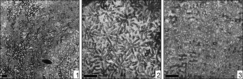
FIGURE 28. Eocomoseris sp. 2, MGB 83247. 1: transversal thin section. 2: transversal thin section, detail. 3: longitudinal thin section. Scale 1 mm.
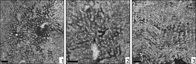
FIGURE 29. Eocomoseris sp. 3, MGB 83234. 1: transversal thin section. 2: transversal thin section, detail. 3: longitudinal thin section. Scale 1 mm.
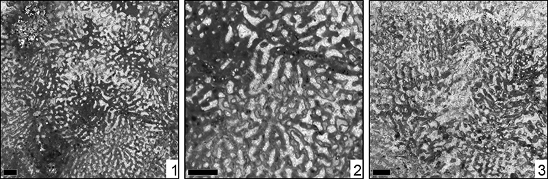
FIGURE 30. Eocomoseris sp. 4, MGB 83231. 1: transversal thin section. 2: transversal thin section, detail. 3: longitudinal thin section. Scale 1 mm.
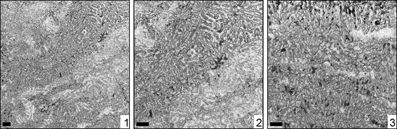
FIGURE 31. Meandraraea miyakoensis Eguchi, 1951, MGB 83226. 1: transversal thin section. 2: transversal thin section, detail. 3: longitudinal thin section. Scale 1 mm.
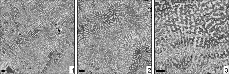
FIGURE 32. Microsolena interjecta Alloiteau, 1958, MGB 83338. 10: transversal thin section. 11: transversal thin section, detail. 12: ongitudinal thin section. Scale 1 mm.
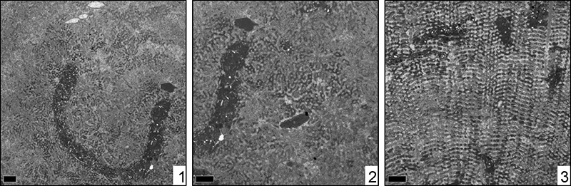
FIGURE 33. Microsolena sp. 1, MGB 83335. 1: transversal thin section. 2: transversal thin section, detail. 3: longitudinal thin section. Scale 1 mm.
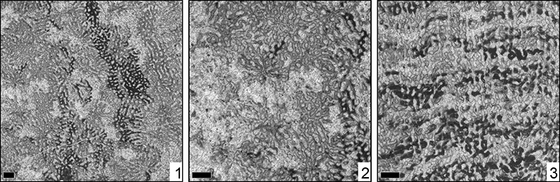
FIGURE 34. Microsolena sp. 2, MGB 83342. 1: transversal thin section. 2: transversal thin section, detail. 3: longitudinal thin section. Scale 1 mm.
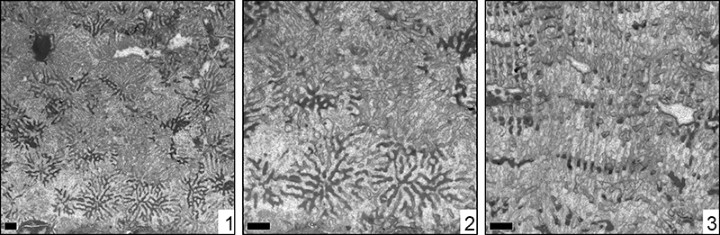
FIGURE 35. Bilaterocoenia sp., MGB 83281. 1: transversal thin section. 2: transversal thin section, detail. 3: longitudinal thin section. Scale 1 mm.
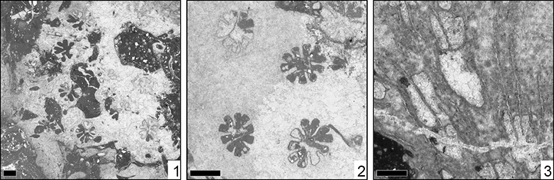
FIGURE 36. Confusaforma prima sp. nov. Löser, MGB 83346. 1: transversal thin section. 2: transversal thin section, detail. 3: longitudinal thin section. Scale 1 mm.
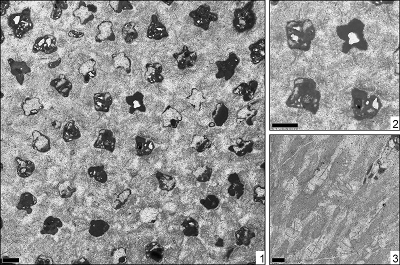
FIGURE 37. Cryptocoenia neocomiensis Orbigny, 1850, MGB 83310. 1: transversal thin section. 2: transversal thin section,detail. 3: longitudinal thin section. Scale 1 mm.
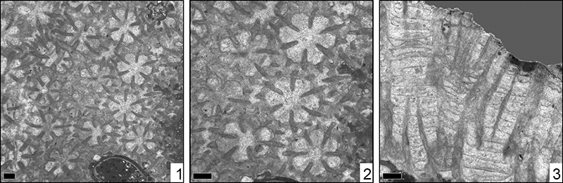
FIGURE 38. Solenocoenia gracilis Roniewicz, 1976, MGB 83379. 1: transversal thin section. 2: transversal thin section, detail. 3: longitudinal thin section. Scale 1 mm.
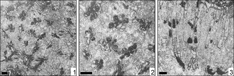
FIGURE 39. Comalia fasciculata Wells, 1932, MGB 83329. 1: transversal thin section. 2: transversal thin section, detail. 3: longitudinal thin section. Scale 1 mm.
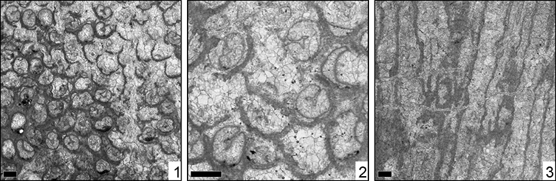
FIGURE 40. Holocoenia cf. micrantha Roemer, 1841, MGB 83284. 1: transversal thin section. 2: transversal thin section, detail. 3: longitudinal thin section. Scale 1 mm.
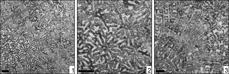
FIGURE 41. Miscellosmilia sp., MGB 83264. 1: transversal thin section. 2: transversal thin section, detail. Scale 1 mm.
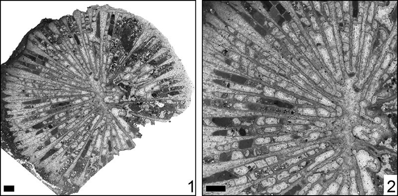
FIGURE 42. Clausastrea bolzei Alloiteau, 1960, MGB 83227. 1: transversal thin section. 2: transversal thin section, detail. 3: longitudinal thin section. Scale 1 mm.
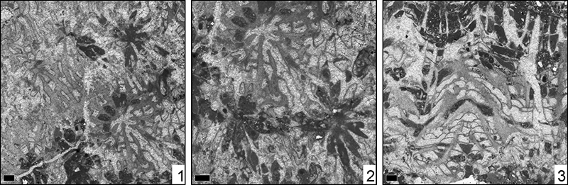
FIGURE 43. Dimorphocoenia multitabulata (Morycowa, 1971), MGB 83328. 1: transversal thin section. 2: transversal thin section, detail. 3: longitudinal thin section. Scale 1 mm.
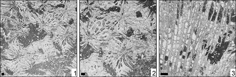
FIGURE 44. Eopreverastrea llanoensis gen.nov. sp. nov., Löser. Drawing after MGB 83265. Ms, main septum. Arrows point to apophysal septa. Scale 1 mm.
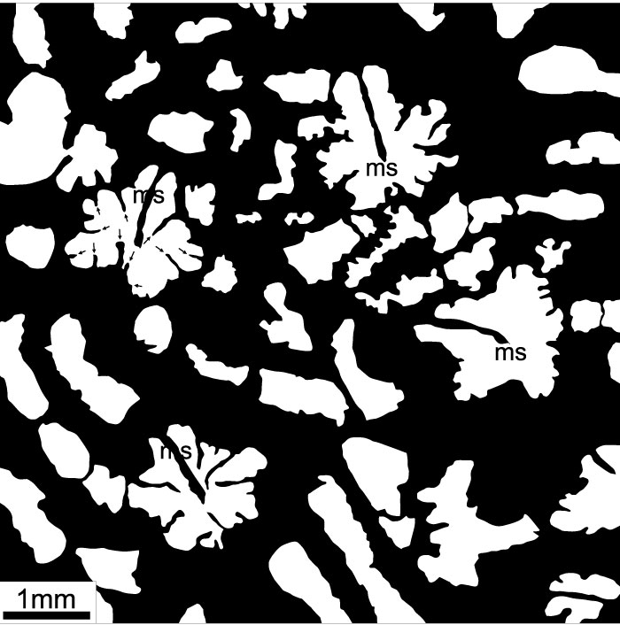
FIGURE 45. Eopreverastrea llanoensis gen.nov. sp. nov., Löser, MGB 83265. 1: transversal thin section. 2: transversal thin section, detail. 3: transversal thin section, detail. 4: longitudinal thin section. Scale 1 mm.
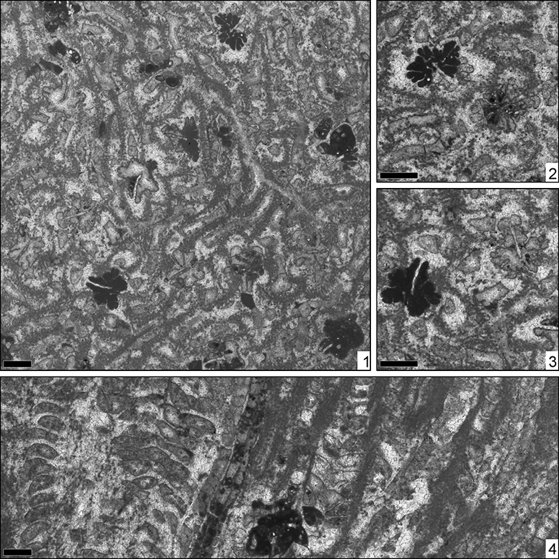
FIGURE 46. Placogyra cf. hykeli Eliášová, 1973, MGB 83345. 1: transversal thin section. 2: transversal thin section, detail. Scale 1 mm.
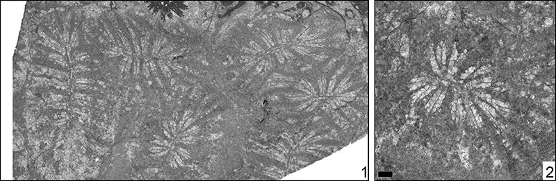
FIGURE 47. Rhipidogyra sp., MGB 83299. transversal thin section. Scale 1 mm.
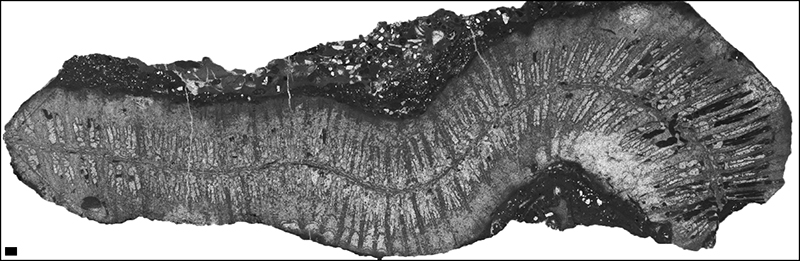
FIGURE 48. Alloiteaucoenia bernardina (Orbigny, 1850), MGB 83304. 1: transversal thin section. 2: transversal thin section, detail. 3: longitudinal thin section. Scale 1 mm.
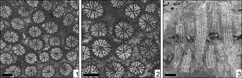
FIGURE 49. Stylina arborea Achiardi, 1880, MGB 83384. 1: transversal thin section. 2: transversal thin section, detail. 3: longitudinal thin section. Scale 1 mm.
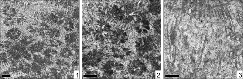
FIGURE 50. Stylina digitiformis Achiardi, 1880, MGB 83355. 1: transversal thin section. 2: transversal thin section, detail. 3: longitudinal thin section. Scale 1 mm.
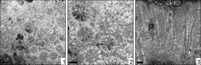
FIGURE 51. Stylina inflata Fromentel, 1856, MGB 83258. 1: transversal thin section. 2: transversal thin section, detail. 3: longitudinal thin section. Scale 1 mm.
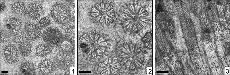
FIGURE 52. Stylina lamellosa Trautschold, 1886, MGB 83255. 1: transversal thin section. 2: transversal thin section, detail. 3: longitudinal thin section. Scale 1 mm.
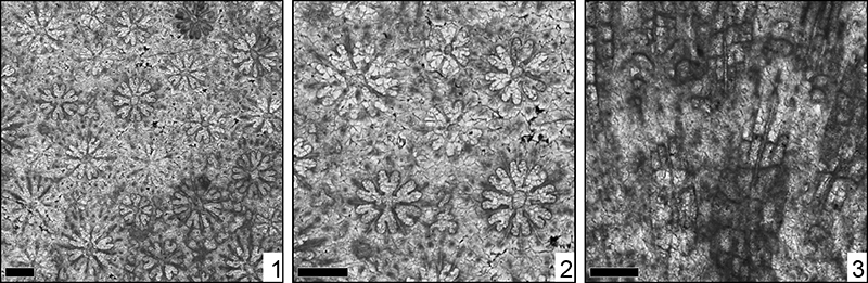
FIGURE 53. Stylina sp. 1, MGB 83306. 1: transversal thin section. 2: transversal thin section, detail. 3: longitudinal thin section. Scale 1 mm.
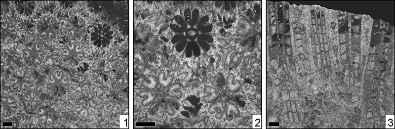
FIGURE 54. Stylina sp. 2, MGB 83320. 1: transversal thin section. 2: transversal thin section, detail. Scale 1 mm.
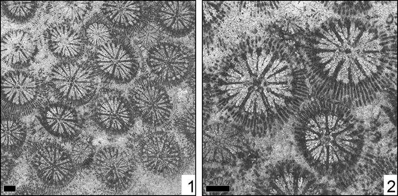
FIGURE 55. Stylina sp. 3, MGB 83248. 1: transversal thin section. 2: transversal thin section, detail. 3: longitudinal thin section. Scale 1 mm.
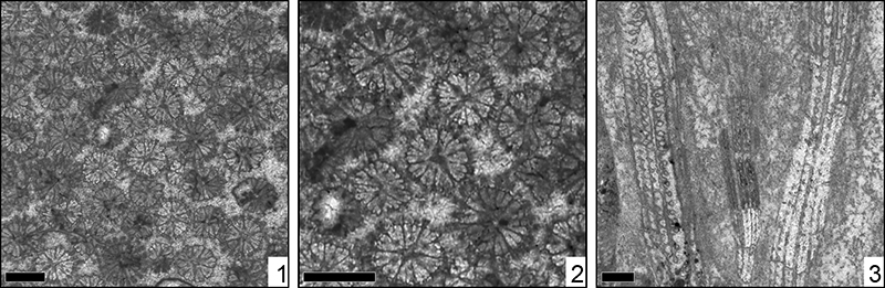
FIGURE 56. Stylina sp. 4, MGB 83389. 1: transversal thin section. 2: transversal thin section, detail. 3: longitudinal thin section. Scale 1 mm.
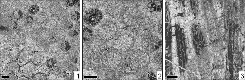
FIGURE 57. Distribution of coral species among superfamilies.
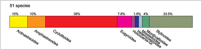
FIGURE 58. Distribution of species in localities outside of the study area. The thickness of bars corresponds to the number of localities where the species was found. Black bars correspond to species that occurred before and after the base of the Valanginian, light blue bars to species that occurred only in the Jurassic and Berriasian, and light green bars to species that are only known from the Valanginian on. The vertical dark green bar marks the age of the investigated coral fauna.
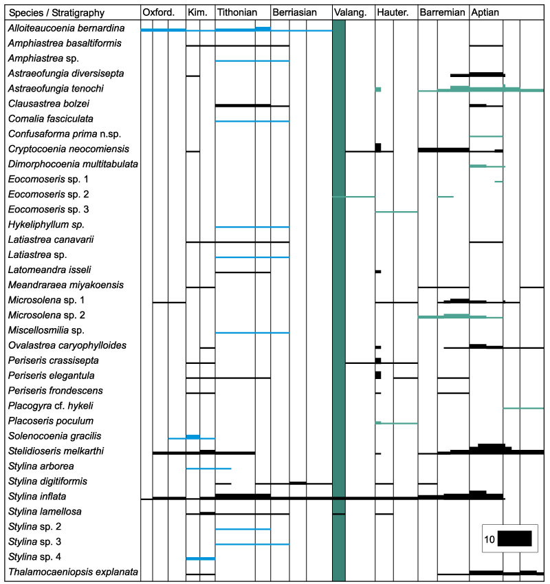
FIGURE 59. Summarised distribution of species in localities outside of the study area. The thickness of bars corresponds to the number of localities where the species was found. The vertical green bar marks the age of the investigated coral fauna.
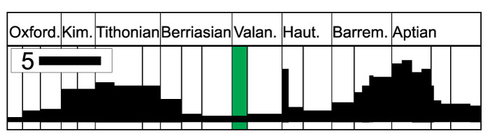
FIGURE 60. Correlation of the palaeo-provinces where species of the studied fauna occur. Only provinces with more than two species were included, and only the time period Oxfordian to Aptian was considered. The Correlation Ratio coefficient was applied, the graph is logarithmic. Abbreviations: Ju, Jurassic; Ox, Oxfordian; Ki, Kimmeridgian; Ti, Tithonian; C, Cretaceous; Be, Berriasian; Va, Valanginian; Ha, Hauterivian; Ba, Barremian; Ap, Aptian; Al, Albian. The number one indicate lower, the number two upper, in the Ju2-3 indicates Middle to Upper Jurassic. The numbers in brackets are the numbers of joint species. The study area is marked in bold letters.
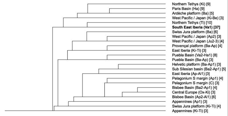
FIGURE 61. Critical stratigraphic ranges of the coral genera in the study area. Ranges after Löser (2016), but improved by newer data. Black lines indicate genera that occurred before and after the Early Valanginian, light blue lines genera that have their last occurrence in the outcrop area, light green lines genera that have the first occurrence in the outcrop area. The vertical green area shows the age of the studied fauna.
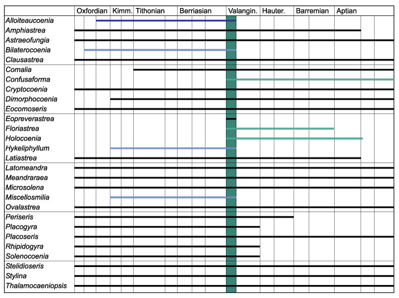
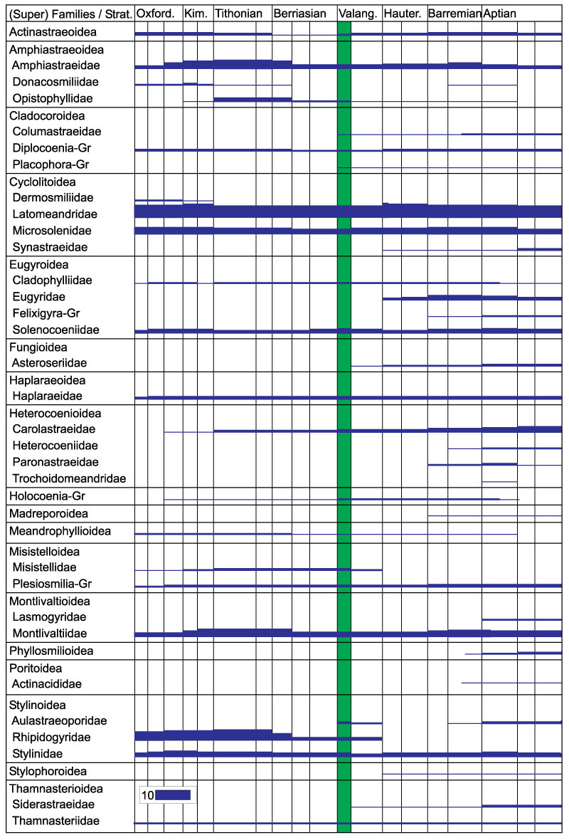
FIGURE 62. Critical stratigraphic ranges of coral families with a distribution in the time period Oxfordian to Aptian. Ranges after Löser (2016), but improved by newer data. The thickness of bars corresponds to the number of genera of the families. When a range is given for a superfamily, it collects only one family. The vertical green area shows the age of the studied fauna.