FIGURE 1. Measurements performed on the labyrinth and the cochlea. A, B and C the planes that the angles were measured between the respective canals A, anterior- lateral; B, anterior-posterior; C, lateral-posterior; D, the width and height of the canals and the overall length of the labyrinth; and E, width and height of the cochlea.
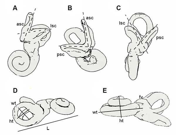
FIGURE 2. 3D reconstruction of the petrosals, samples (A-C) T2406 along with transparent views locating the orientation of the labyrinth in the bone, (D-F) T2506 and (G-I) T137. A, D, G ventrolateral view; B, E, H dorsolateral view; C, F, I dorsomedial view. See text for abbreviations.
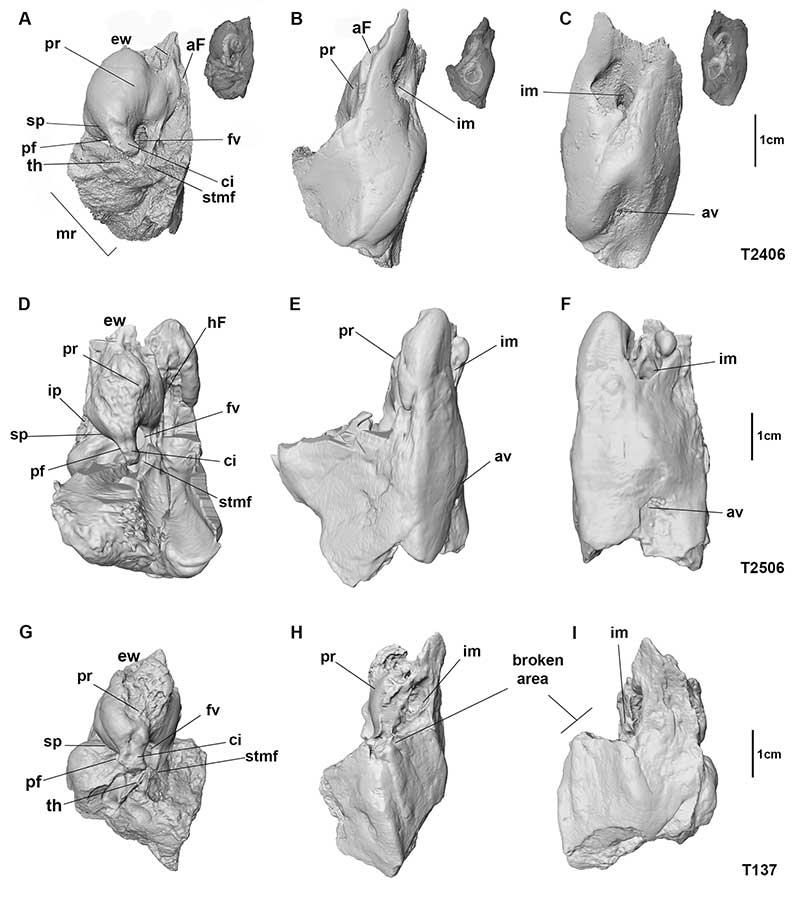
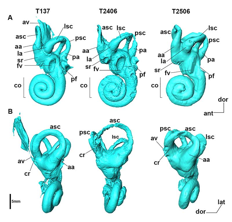
FIGURE 3. The bony labyrinth of the three samples of Palaeoloxodon tiliensis. A, cochlear ventral view; B, anterior view. See text for abbreviations.