FIGURE 1. Stratigraphic positions and locations of different Upper Jurassic plattenkalk localities of the Franconian and Swabian Alb. 1 Nusplingen, 2 Daiting, 3 Solnhofen, 4 Mörnsheim, 5 Eichstätt, 6 Schamhaupten, 7 Zandt, 8 Brunn. Stratigraphic positions after Schweigert (2007).
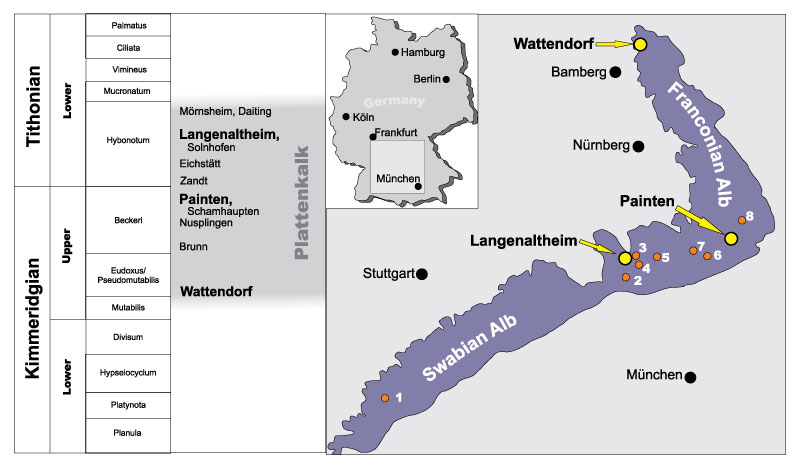
FIGURE 2. Metriorhynchidae indet., LF 3474, patch of trunk integument with displaced vertebrae and rib (uncertain position on body), same section photographed under visible light (above) and UV (below; UV A, B, and C, with orange filter and linear polarizing filter). Skin traits comprise soft-looking folds (left), sharp striae identified as fibers (right portion), and irregular skin scars (arrows pointing at three circular irregularities in the center, and curl-like structure in the right half; see discussion for interpretation). Scale bar for both parts equals 10 cm.
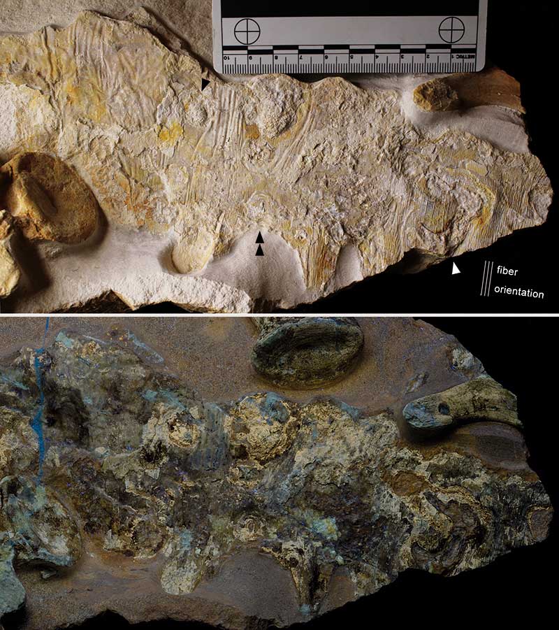
FIGURE 3. Metriorhynchidae indet., LF 3474, scattered patches of skin preservation in visible light, from partially disarticulated trunk. A: Fold systems with dominant and additional orientations. B: Sharp lines forming a net of cracks or distinctly flexed folds; displaced portion with unknown body orientation. C: Displaced patch as in A in a wider section, with at least one circular irregularity in the lower part (arrow). D: Soft folding of skin surface, exposed near to the knee. E: Potential bundles of muscle fibers (horizontal), with oblique crossing fold traces on the right (chest, anterior to pectoral girdle). F: Softly folded integument associated to the ischium (upper middle). All with mm scale.
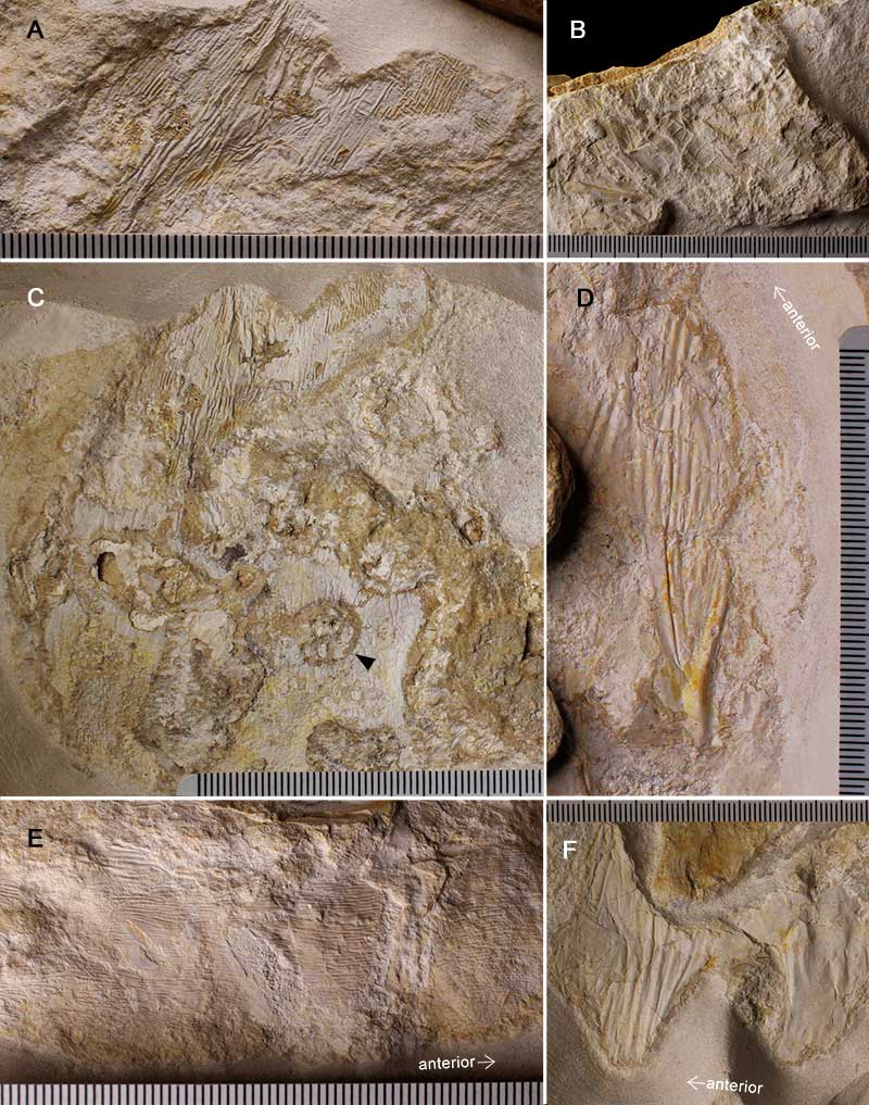
FIGURE 4. Metriorhynchidae indet., LF 3474, distal half of tail, photographed under UV A, B, and C, with orange filter and linear polarizing filter. Skin preservation mostly reacting as yellowish areas. Note the pronounced fluke. The edge of the base of the upper lobe is marked by a white line, as mapped from the below detail under visible light to demonstrate soft tissue distribution (arrow pointing to leading edge). Scale bar equals 20 cm; for inset in mm.
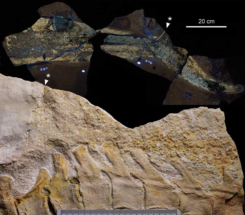
FIGURE 5. Dakosaurus sp., DMA-JP-2009/001, skin preservation on dorsal margin of trunk. A: Shoulder area, skin with transversal fibers and associated folding, visible light. Width of frame equals approximately 18.2 cm. B: Lumbar region, similar structures as in A, under UV. Width of frame equals approximately 20.5 cm.
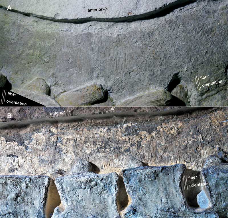
FIGURE 6. Dakosaurus sp., DMA-JP-2009/001, integumentary details of various regions of the body, all under visible light. A: Patch on proximal tail, with widely spaced striation right-angled to vertebral column. Width of frame equals approximately 6.7 cm. B: Crisscross folding in neck region. Width of frame equals approximately 3.7 cm. C: Patch on proximal tail with rare occurrence of longitudinal folding, right-angled to transversal fiber system (section of the middle left of A, under different lighting). Width of frame equals approximately 5.3 cm.
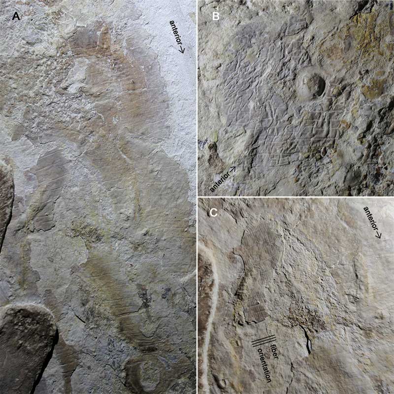
FIGURE 7. Dakosaurus sp., DMA-JP-2009/001, visible light photograph of dorsal region of anterior middle tail, with numerous dark irregularities on the skin. Width of frame equals approximately 12.1 cm.
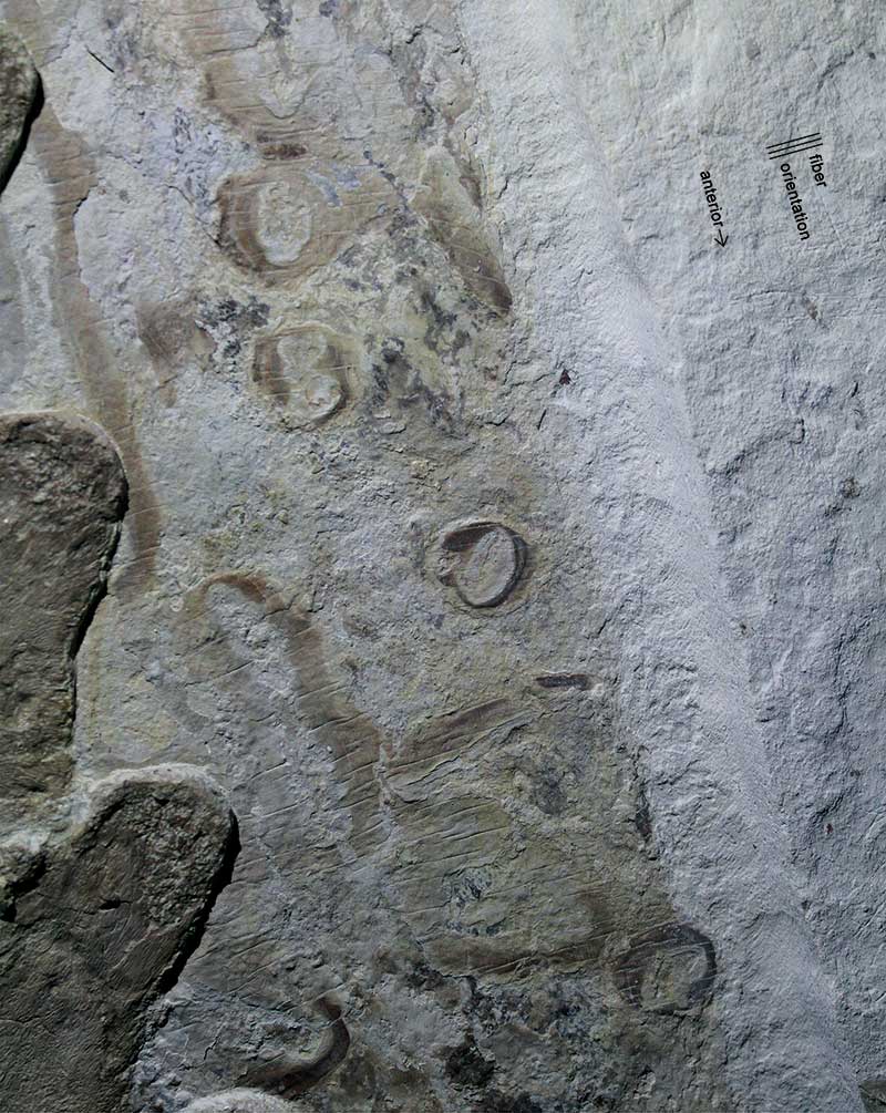
FIGURE 8. Dakosaurus sp., DMA-JP-2009/001, anatomically proximal continuation of Figure 7. On the lower left and right, the dark patches (white arrows) expose a skin layer that is overlain by the bright matter in most of the preserved skin. Width of frame equals approximately 8.2 cm.
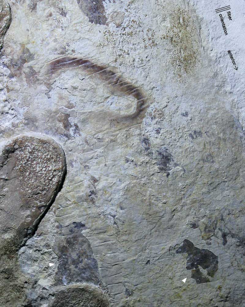
FIGURE 9. Dakosaurus sp., DMA-JP-2009/001, UV image of tail portion with the highest concentration of skin irregularities (arrows; see previous figures). Width of frame equals approximately 28.0 cm.
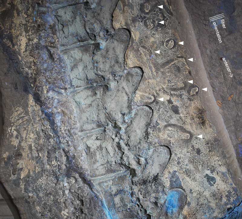
FIGURE 10. Dakosaurus sp., DMA-JP-2009/001, close-ups of skin irregularities, courtesy of Yanina Herrera, used with permission. A: Large oval scar, see also Figure 8 and Figure 9. Width of frame equals approximately 4.5 cm. B: Circular scar representing the most abundant type and size of skin irregularities in this specimen, adjacent to blurred, open irregularity, see also Figure 6A, Figure 7, and upper edge of Figure 9. Width of frame equals approximately 4.5 cm. C: Circular scar retaining the texture of uninterrupted skin, see also Figure 7 and Figure 9. Width of frame equals approximately 5.5 cm.
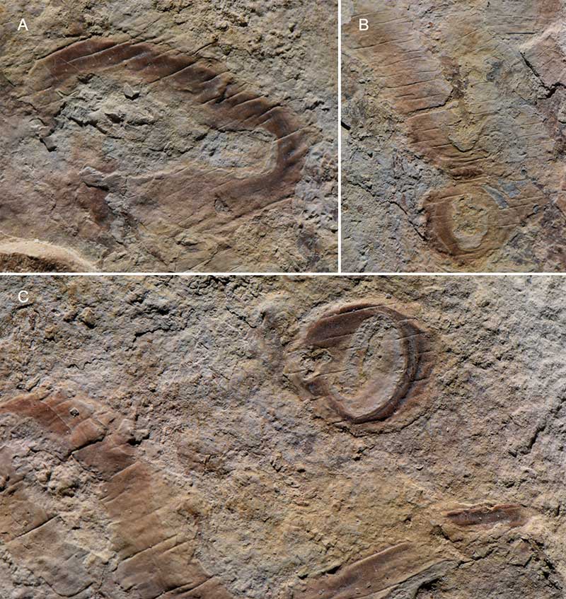
FIGURE 11. Metriorhynchidae indet., NKMB-P-Watt06/508, specimen under UV, with soft tissue preservation surrounding most of the skeleton. A separated piece containing the distal tail is not depicted here, since no soft tissue is exposed. Scale bar equals 20 cm.
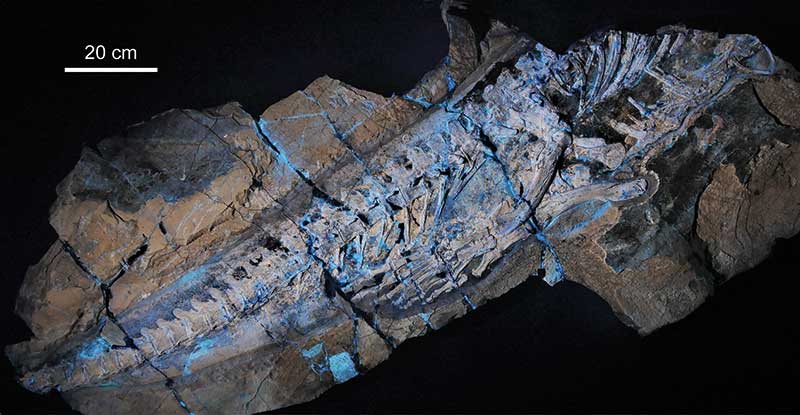
FIGURE 12. Metriorhynchidae indet., NKMB-P-Watt06/508, dorsal region of middle tail, with longitudinal and transversal skin folding, along with a nest of irregularities (grey knobs). Width of frame equals approximately 7.6 cm.
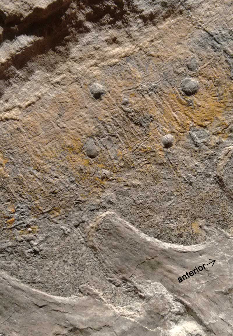
FIGURE 13. Metriorhynchidae indet., NKMB-P-Watt06/508, feet of both sides under UV. The caudal soft tissue ends slightly ventral to the hemapophyses. Minor patches of yellowish skin indication are uncertainly belonging to the webbing or metatarsal area of one of the feet (arrow). Width of frame equals approximately 35.5 cm.
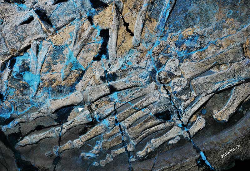
FIGURE 14. Metriorhynchidae indet., NKMB-P-Watt06/508, middle tail with soft tissue preservation. A: Visible light image with structural details of the skin, mostly folding, and knob-like irregularities (some indicated by arrows). Width of frame equals approximately 30.6 cm. B: UV image with darker bluish signals probably reflecting varnish. Width of frame equals approximately 33.9 cm.
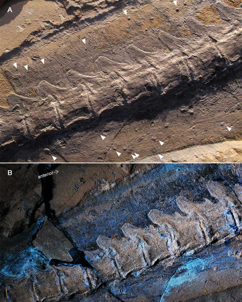
FIGURE 15. Metriorhynchidae indet., NKMB-P-Watt06/508, UV close-ups of skin surrounding the middle tail. A: Dorsal region with numerous knob-like irregularities. Width of frame equals approximately 10.1 cm. B: Ventral region with longitudinal folding (by skin substance preservation or imprints), and further irregularities. Width of frame equals approximately 18.0 cm.
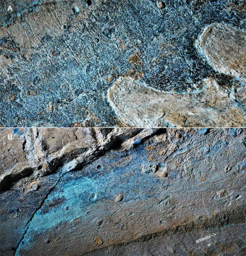
FIGURE 16. Cricosaurus albersdoerferi, BMMS-BK 1-2, soft tissue patch under UV, with yellowish areas indicating skin preservation. The posterior skull can be seen on the left, the scapulocoracoid on the lower right. Width of frame equals approximately 23.0 cm.
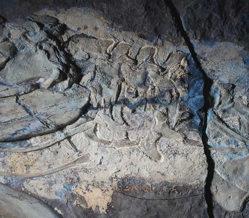
FIGURE 17. Cricosaurus albersdoerferi, BMMS-BK 1-2, soft tissue patch on the ventral chest, posterior to pectoral girdle. On the middle left (below terminus of rib), horizontal striation reflects musculature. The covering layer exhibits soft skin folds. Circular irregularities (some marked by arrows) are interpreted in the text. Width of frame equals approximately 6.5 cm.
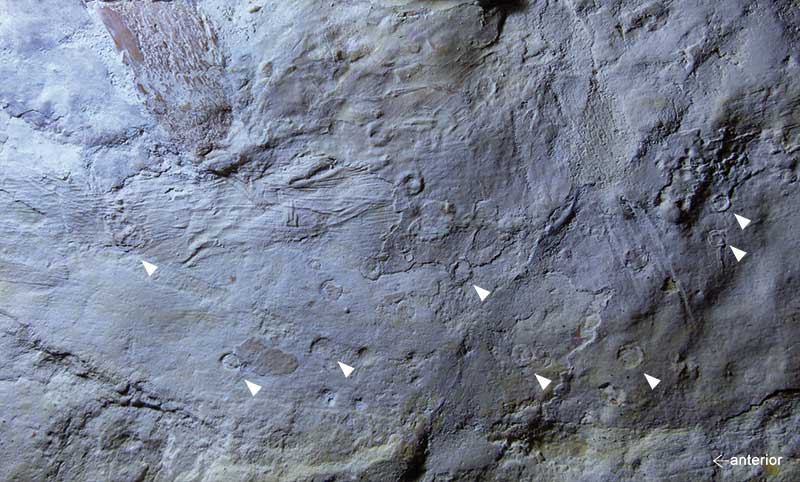
FIGURE 18. Cricosaurus albersdoerferi, BMMS-BK 1-2, details of skin preservation. A: Anterior part of dorsal edge of neck patch (Figure 16). From the center to the lower right longitudinal muscle fascia are exposed. In the upper part, sets of folds and possibly fibers overlap. Width of frame equals approximately 3.1 cm. B: Indistinct fragmentation of skin patch in posterior trunk, interpreted as potential taphonomic shrinking. Width of frame equals approximately 3.5 cm. C: Dorsal edge of neck patch (Figure 16) with curved striation (arrows indicate acute rhomboidal directions). Width of frame equals approximately 3.7 cm. D: Position little more posterior as in C. Additional to the dominant orientation of skin surface striation, another, acute-angled set crosses and is visible under a certain angle of light. Width of frame equals approximately 3.2 cm. E: Skin details associated to femur (lower right), with oblique striation and rhomboidal, superficially scale-like compaction pattern (lower left, indicated by arrows). Width of frame equals approximately 4.2 cm. F: Anterior hemapophysis with associated skin folds that radially spread from the bone terminus as an effect of load during compaction. A circular skin irregularity is seen above the bone (arrow). Width of frame equals approximately 5.4 cm.
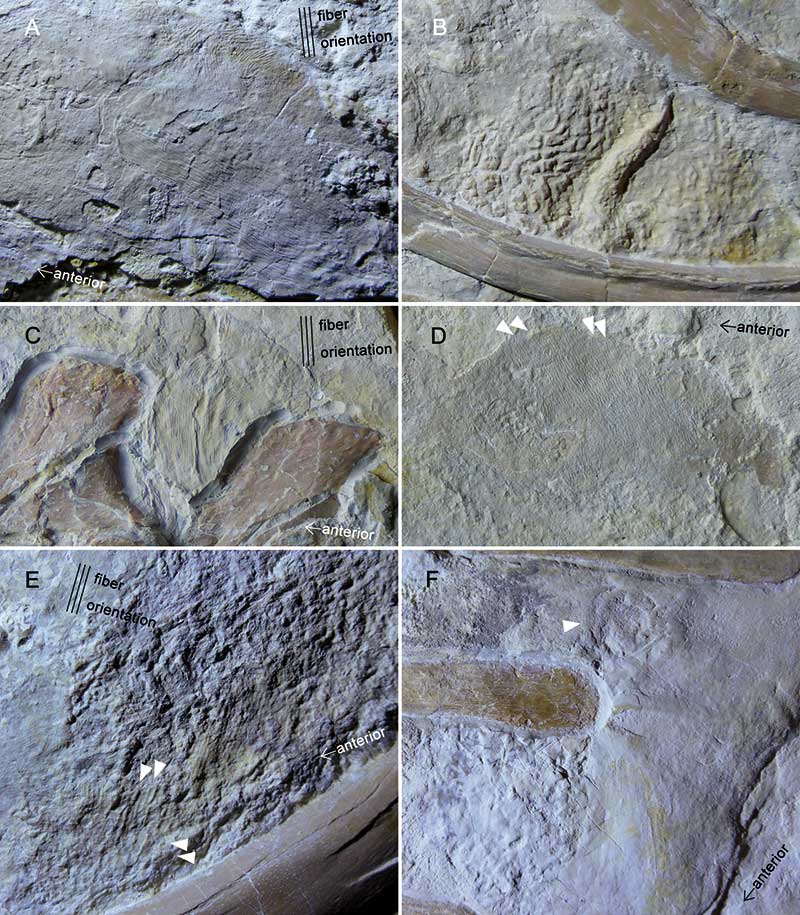
FIGURE 19. Cricosaurus bambergensis, NKMB-P-Watt14/274, skin preservation in dorsal neck, illustrating transversal fibers. Width of frame equals approximately 5.5 cm.
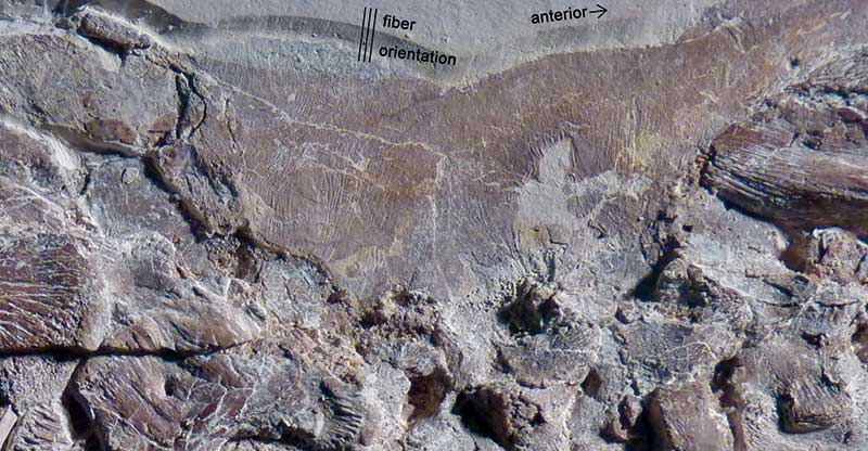
FIGURE 20. Cricosaurus bambergensis, NKMB-P-Watt14/274, body trunk portion with folded skin (upper red margin) embedding dorsal column. In the lower right corner, gut contents preserved a crustacean (arrow marking hinge between carapace and abdomen). Width of frame equals approximately 9 cm.
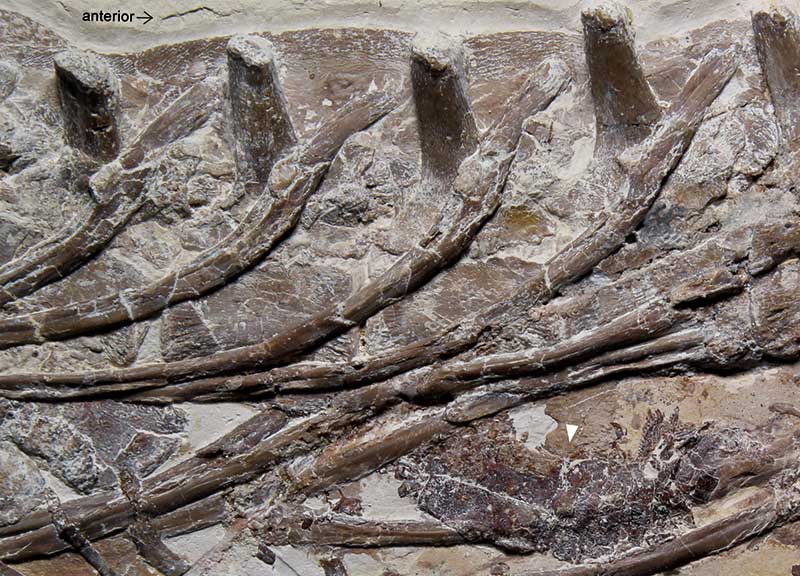
FIGURE 21. Cricosaurus bambergensis, NKMB-P-Watt14/274, proximal tail portion, column surrounded by skin patches with folds and fibers. The arrow points to a patch tentatively identified as musculature. Width of frame equals approximately 12 cm.
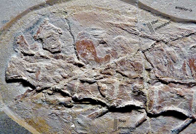
FIGURE 22. Cf. Rhacheosaurus gracilis, LF 2426, photographed under UV A, B, and C, with orange filter and linear polarizing filter. Yellowish areas represent skin preservation as well as bone. Cube length 1 cm; total width for frame equaling 57.8 cm.
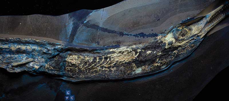
FIGURE 23. Rhacheosaurus gracilis, NHMUK R.3948. A: Skeleton with soft tissue outlines. B: Detail of tail fluke. The outline of the upper lobe is blurred and not clearly indicated by darker color. Courtesy of Yanina Herrera, used with permission. Scale bar in cm.
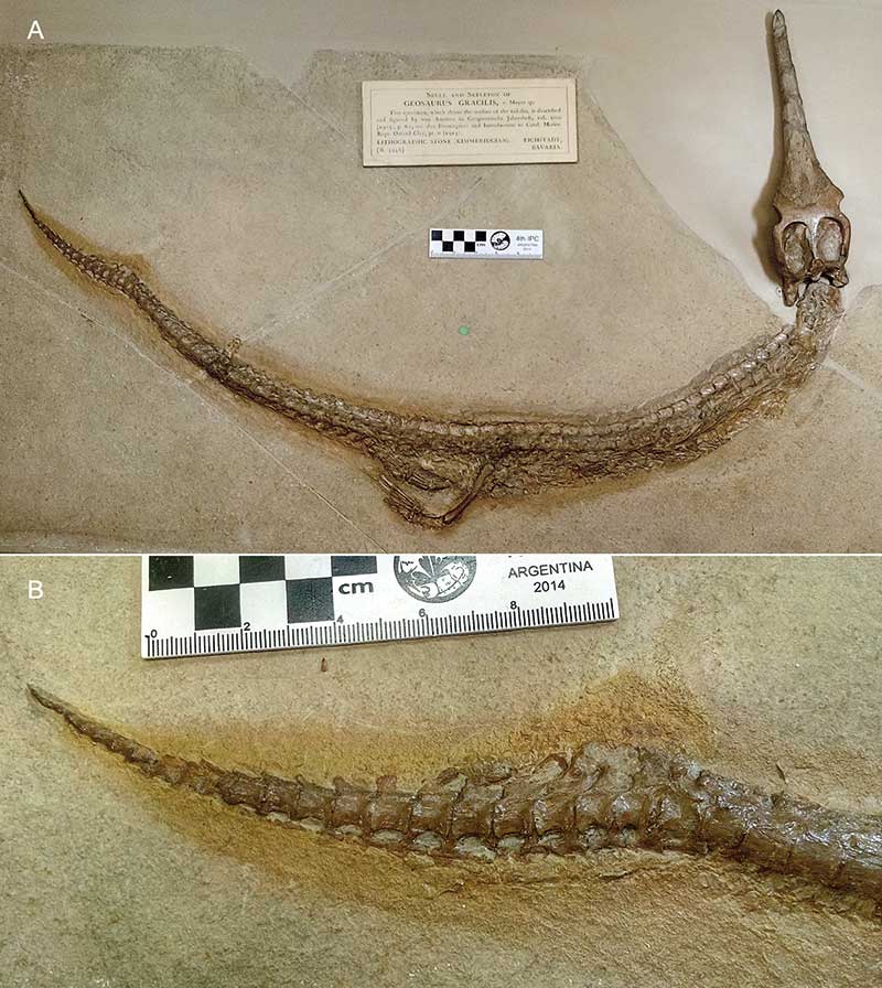
FIGURE 24. Schematic drawings of metriorhynchid tail flukes, distal caudal columns and integumentary contours. A: Rhacheosaurus NHMUK R.3948, historical reconstruction (Andrews, 1913, figure A). B: Same specimen, outline from photograph (Figure 23B), with potential outlines of the upper lobe evaluated from textures on the fossil in visible light. C: Metriorhynchidae indet., LF 3474 (new specimen, Figure 4), with possible outlines for the upper lobe inferred from preserved edges. D: Proposed outline in ontogenetic stages with less pronounced tail bending (generalized sketch comparable to unpublished specimen, see text for further information). All drawn to similar size.
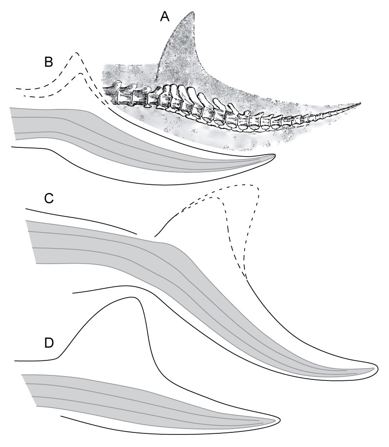
FIGURE 25. Teleosauroid SMNS 10985 (formerly assigned to Steneosaurus), right hind leg with soft tissue preservation. Courtesy of Erin Maxwell, used with permission. Scale bar equals 10 cm.
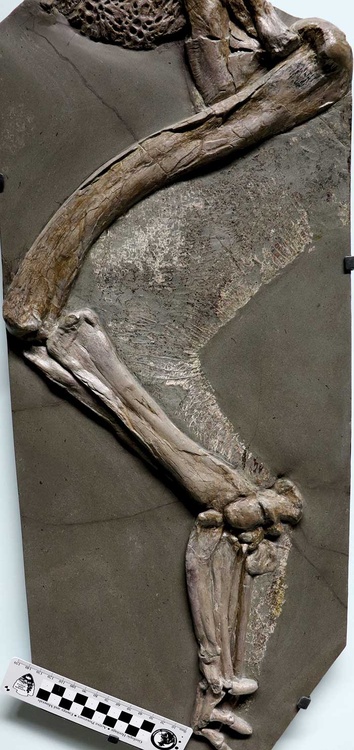
FIGURE 26. Teleosauroid SMNS 10985 (formerly assigned to Steneosaurus), detail of integument on rear side of hind leg, illustrating flexed joints and square to rhomboidal shaped scutes. Courtesy of Erin Maxwell, used with permission. Width of frame equals 16 cm.
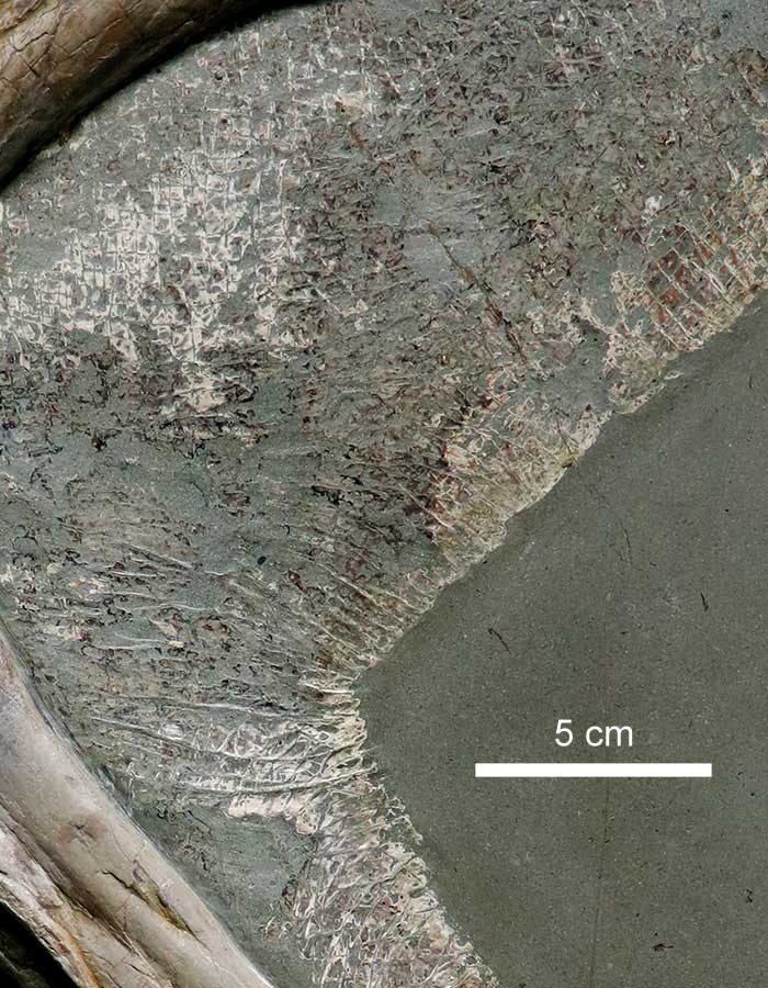
FIGURE 27. Aeolodon priscus, JME (uncatalogued, firmly installed in the permanent exhibition). A: Distal tail portion preserving enlarged rhomboidal scutes on dorsal edge (arrows mark dorsal points of transversal hinges); width of frame equals 15 cm. B: Skin preservation around the hollow of the knee (femur visible in left lower corner), with patches of distinct rhomboidal scutes (arrows mark example of hinge orientation); width of frame equals 23.5 cm. C: Webbing of foot with stretched polygonal scutes; width of frame equals 15.8 cm.
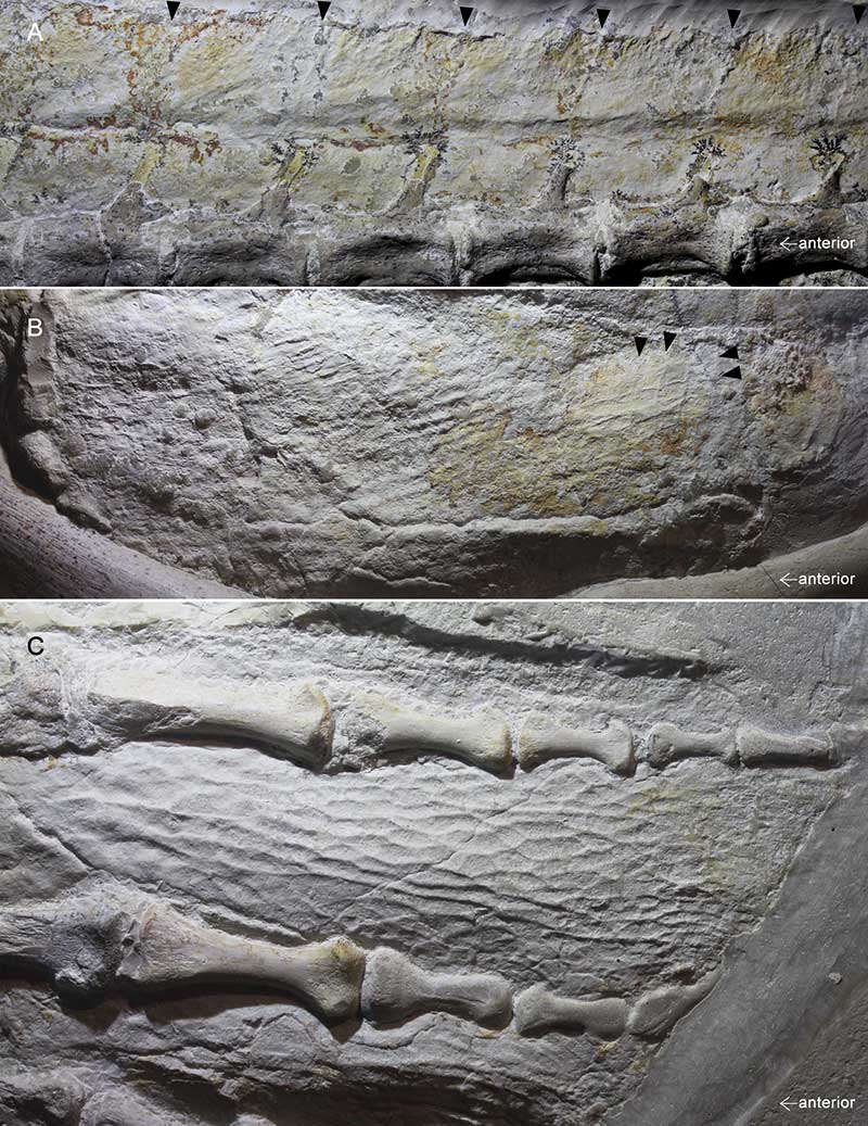
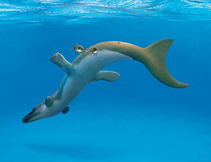
FIGURE 28. Life reconstruction of a Dakosaurus -like metriorhynchid, illustrating the skin surface, fluke outline, and potential epizoic parasites, according to new data.