FIGURE 1. The Aetosauroides scagliai specimen MCN-PV 2347 and the location of the study area. A, map of the Santa Maria Supersequence in the Rio Grande do Sul state, southern Brazil (modified from Jenisch et al., 2017). B, map of the São João do Polêsine area, with the Candelária Sequence exposures highlighted (brown), showing the location (arrow) of the Piche Site outcrop (modified from Müller et al., 2018). C, stratigraphic column of the Piche Site (modified from Jenisch et al., 2017), with position of MCN-PV 2347 indicated (arrow). D, reconstruction of MCN-PV 2347 with preserved elements in brown. E, panoramic view of the Piche Site with the collection point of MCN-PV 2347 indicated by the arrow. Abbreviations: con, conchostracan remains; Di, dinosaur remains; Hy, Hyperodapedon remains; mmd, massive mudstone; mfgs, massive fine-grained sand; rcl, ripple cross-laminated sandstone; vert, vertebrate remains.
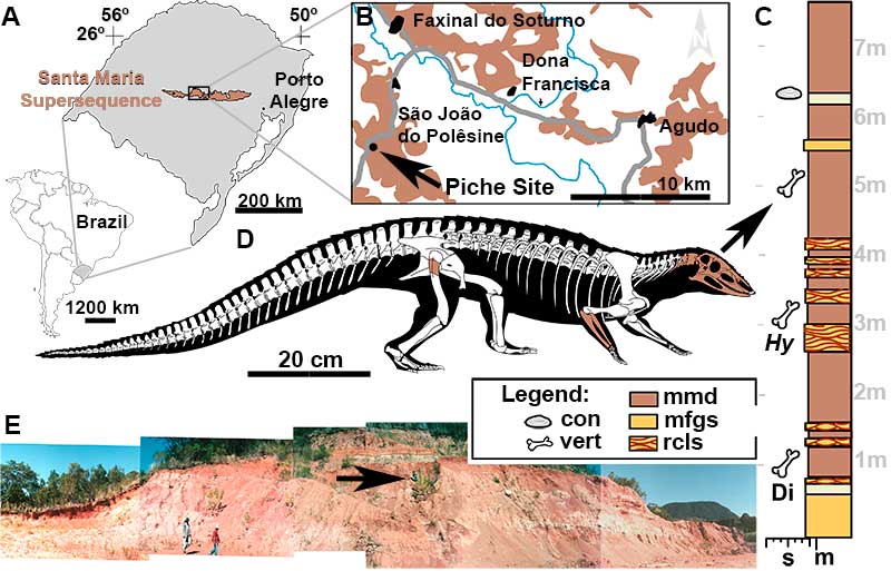
FIGURE 2. Skull of Aetosauroides scagliai (MCN-PV 2347) in dorsal view. A, photograph. B, interpretative drawing with matrix shown in brown. Abbreviations: aof, antorbital fenestra; cv, cervical centra; dep, nasal depression; f, frontal; for, frontal elevated orbital rim; j, jugal; la, lacrimal; lg, lateral groove; mx, maxilla; n, nasal; o, orbit; of, overhanging flange; op, paramedian osteoderm; pa, parietal; po, postorbital; pof, postfrontal; prf, prefrontal; r, ridge; stf, supratemporal fenestra; stfo, supratemporal fossa; tpmx, thorn-like lateral projection of the premaxilla; ts, transverse groove.
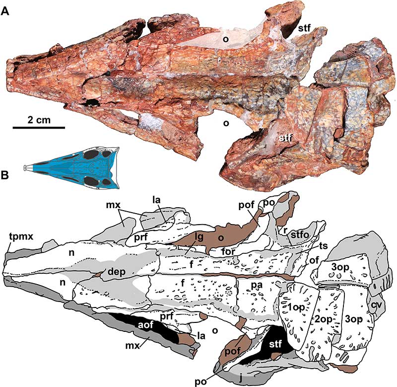
FIGURE 3. Skull of Aetosauroides scagliai (MCN-PV 2347) in left lateral view. A, photograph. B, interpretative drawing. Abbreviations: aof, antorbital fenestra; aofo, antorbital fossa; bb, braincase bones; de, dentary; en, external naris; f, frontal; fo, foramina; for, frontal elevated orbital rim; itf, infratemporal fenestra; j, jugal; la, lacrimal; lg, lateral groove; lo, dorsal lateral osteoderm; mx, maxilla; n, nasal; ns, nasal suture; ode, osteoderm dorsal eminence; o, orbit; op, dorsal paramedian osteoderm; pdmx, posterodorsal process of the maxilla; pmx, premaxilla; pmxn, premaxilla notch; prf, prefrontal; prf.fo, prefrontal foramen; po, postorbital; pof, postfrontal; pvpj, posteroventral process of the jugal; q, quadrate; qj, quadratojugal; r, ridge; rpar, retroarticular process of the articular; sq, squamosal; st, stapes; stfo, supratemporal fossa; su, surangular; sufo, surangular foramen.
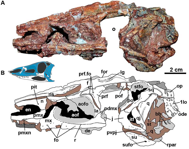
FIGURE 4. Skull of Aetosauroides scagliai (MCN-PV 2347) in right lateral view. A, photograph. B, interpretative drawing with matrix shown in brown. Abbreviations: ang, angular; aof, antorbital fenestra; aofo, antorbital fossa; de, dentary; en, external naris; fo, foramina; j, jugal; la, lacrimal; mf, mandibular fenestra; mx, maxilla; n, nasal; o, orbit; pa, parietal; par, prearticular; pdmx, posterodorsal process of the maxilla; pmx, premaxilla; pmxn, premaxilla notch; prf, prefrontal; po, postorbital; pof, postfrontal; pvpj, posteroventral process of the jugal; q, quadrate; qj, quadratojugal; r, ridge; stf, supratemporal fenestra; stfo, supratemporal fossa; su, surangular; sufo, surangular foramen; tpmx, thorn-like lateral projection of the premaxilla.
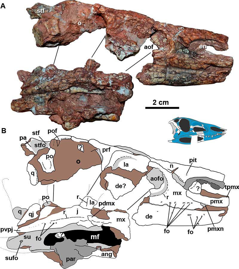
FIGURE 5. Three-dimensional reconstruction based on μCT-scan images of the rostrum of Aetosauroides scagliai (MCN-PV 2347). A, right lateral view. B, left lateral view. C, dorsal view. D, dorsal view without the nasals. E, ventral view without the dentary. Abbreviations: aof, antorbital fenestra; aofo, antorbital fossa; ch, choana; d, depression; de, dentary; en, naris; fi, foramen incisivum; fo, foramina; i.mxt, isolated maxillary teeth; idp, interdental plates; l., left; ls, lateral socket of the nasal; mg, Meckelian groove; mx, maxilla; mxt, maxillary alveoli/tooth; n, nasal; ns, nasal suture; pmx, premaxilla; pmxm, medial process of the premaxilla; pmxn, premaxillary notch; pmxt, premaxillary alveoli/tooth; pp, posterior process; r, ridge; r.,right; sy, symphysis; tpmx, thorn-like lateral projection of the premaxilla.
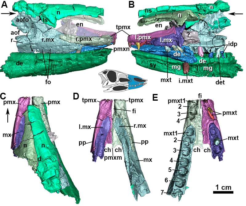
FIGURE 6. 3D Reconstruction based on µCT-scan images and photograph of Aetosauroides scagliai maxillae (MCN-PV 2347). A, right maxilla in medial view. B, left maxilla fragment in medial view. Abbreviations: aof, antorbital fenestra; ap, ascending process; idp, interdental plates; mxs, maxilla medial shelf; mxt, maxilla alveoli; rec.ch, choanal recess; spmx, slot for the premaxilla.
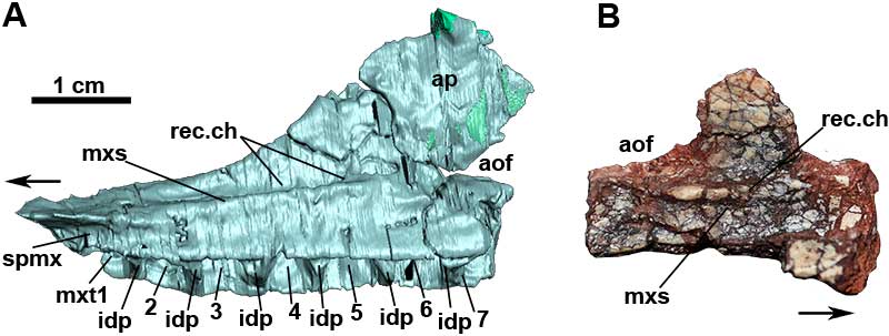
FIGURE 7. Three-dimensional reconstruction based on µCT-scan images of the right posterior portion of the skull and mandible of Aetosauroides scagliai (MCN-PV 2347). A, in lateral view. B, in posterior view. C, in dorsal view, without palatal and mandible elements. D, in dorsal view. E, in ventral view, without palatal and mandibular elements. F, in medial view, without palatal and mandibular elements. Abbreviations: ang, angular; aof, antorbital fenestra; ar, articular; fo, foramina; itf, infratemporal fenestra; j, jugal; jb, jugal ventral bend; la, lacrimal; mf, mandibular fenestra; mx, maxilla; o, orbit; par, prearticular; pdmx, middle finger-like projection of the posterior end of the maxilla; po, postorbital; pr, prootic; pt, pterygoid; pvpj, posteroventral process of the jugal; q, quadrate; qfo, quadrate foramen; qj, quadratojugal; qmw, quadrate medial wing; r, ridge; su, surangular; sufo, surangular foramen.
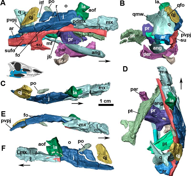
FIGURE 8. A, 3D Reconstruction based on µCT-scan images of the posterior portion of the left side of the skull of Aetosauroides scagliai (MCN-PV 2347). B, isolated broken left jugal and quadratojugal, in lateral view. C, µCT-scan images of the isolated broken jugal and quadratojugal, in dorsolateral view; Abbreviations: apqj, natural cast of the anterior projection of the quadratojugal; ar, articular; dqlw, depression for quadrate lateral wing; itf, infratemporal fenestra; j, jugal; nc, natural cast of the quadratojugal; pa, parietal; pdpj, posterodorsal process of the jugal; po, postorbital; pvpj, posteroventral process of the jugal; q, quadrate; qj, quadratojugal; rap, retroarticular process; sq, squamosal; su, surangular; stf, supratemporal fenestra; stfo, supratemporal fossa.
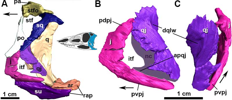
FIGURE 9. Fragments of the skull of MCP-3450-PV and right jugal of ULBRAPV003T. A, Photograph of the posterior process of the jugal of MCP-3450-PV in lateral view, also visible is the left surangular, in lateral view, and a fragmented left quadrate, in anterior view. B, interpretative drawing of MCP-3450-PV, matrix shown in brown. C, jugal of ULBRAPV003T in lateral view. D, jugal of ULBRAPV003T in medial view. Abbreviations: a.la, articulation with the lacrimal; b.mw, broken medial wing of the quadrate; e, elevation; fa, flat area; fo, foramen; qc, quadrate crest; qfo, quadrate foramen; itf, infratemporal fenestra; pvpj, posteroventral process of the jugal; lw, lateral wing; o, orbit; r, ridge; rap, retroarticular process; su, surangular; sufo, surangular foramen.
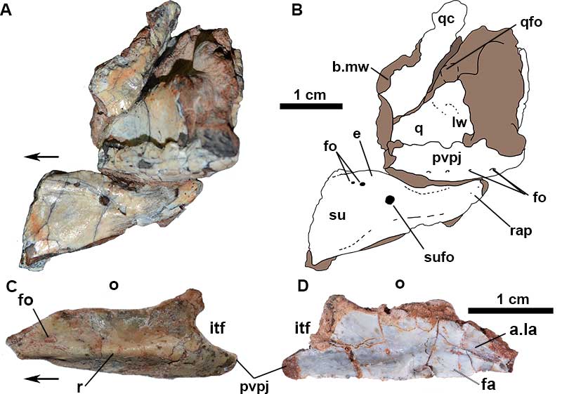
FIGURE 10. Inner surface of the partial skull of Aetosauroides scagliai (MCN-PV 2347). A, photograph in ventral view. B, anterior left orbital margin in posterior view. C, medial surface of the left side of the skull at the antorbital fenestra region. D and E, interpretative drawings with polyethylene glycol resin in dark gray and matrix in brown. Abbreviations: aof, antorbital fenestra; d, depression; de, dentary; f, frontal; j, jugal; la, lacrimal; mx, maxilla; n, nasal; n.s, nasal suture; o, orbit; pa, parietal; prf, prefrontal; prfvp, prefrontal ventral process; po, postorbital; pof, postfrontal.
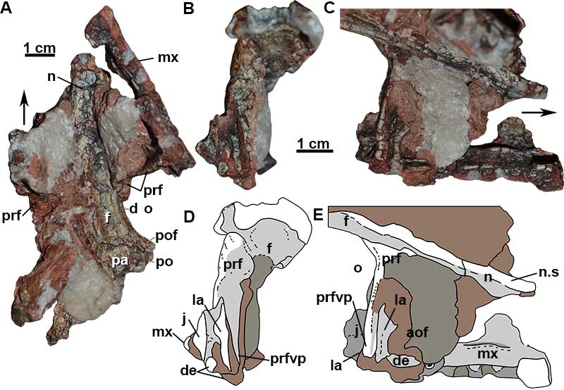
FIGURE 11. Three-deminsional reconstruction based on μCT-scan images of the left postorbital (A-D) and squamosal (E-H) of Aetosauroides scagliai (MCN-PV 2347) in lateral (A and E), posterior (B), medial (C and H), anterior (D), poserodorsal (F) and ventral (G) views. Abbreviations: aop, articulation surface for the otoccipital; apa, articulation surface of the parietal; b.tsp, triangular spur; d, depression; mk, medial keel; o, orbit; pp, posterior process; qs, quadrate socket; sqop, occipital process; stf, supratemporal fenestra; stfo, supratemporal fossa; vp, ventral process. Gray areas indicate missing portions.
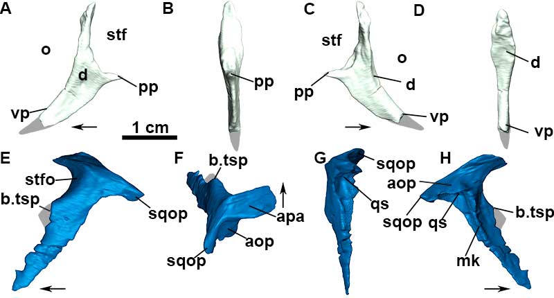
FIGURE 12. 3D Reconstruction based on µCT-scan images of the left quadrate of Aetosauroides scagliai (MCN-PV 2347). A, posterior view; B, medial view; C, anterior view; D, lateral view; E, dorsal view; F, ventral view. Abbreviations: lc, lateral condyle; lw, lateral wing; mc, medial condyle; mw, medial wing; pta, pterygoid articulation; qc, quadrate crest; qfo, quadrate foramen; qh, quadrate head; qp, quadrate pit; pta, pterygoid articulation.
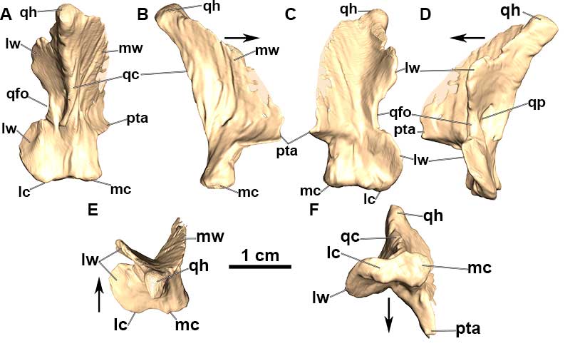
FIGURE 13. Parietal of Aetosauroides scagliai, MCN-PV 2347 (A, D and E) and MCP-3450-PV (B); and ‘Polesinesuchus aurelioi’ (ULBRAPV003T; C, F, G and H). A, photograph of posterior region of the skull of MCN-PV 2347 in dorsal view. B, photograph of the left parietal of MCP-3450-PV in dorsal view. C, photograph of the right parietal of ‘P. aurelioi’ in dorsal view. D, 3D reconstruction based on µCT-scan images of a fragment of the left parietal of MCN-PV 2347 in dorsal view. E, 3D reconstruction based on µCT-scan images of a fragment of the left parietal of MCN-PV 2347 in lateral view (mirrored). F, photograph of the right parietal of ‘P. aurelioi’ in lateral view. G, interpretative drawing of the right parietal of ‘P. aurelioi’ in lateral view. H, interpretative drawing of the right parietal of ‘P. aurelioi’ in dorsal view. The gray lines in E and F depict the inclination angle of the occipital portion of the parietal. Abbreviations: lg, lateral groove; o, orbit; of, overhanging flange; opt, occipital portion of the parietal. or, ornamentation; pof, postfrontal; pop, paraoccipital process of the parietal; r, ridge; stfo, supratemporal fossa; tr, transverse ridge; ts, transverse sulcus.
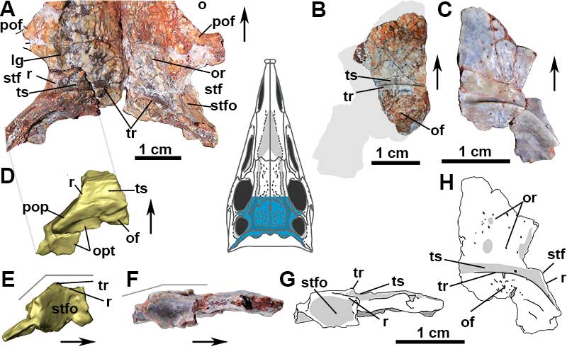
FIGURE 14. 3D Reconstruction based on µCT-scan images of the left pterygoid of Aetosauroides scagliai (MCN-PV 2347). A, dorsal view. B, lateral view. C, ventral view. D, medial view. E, posterior view. Abbreviations: b.qc, broken quadrate contact; bptc, basipterygoid process contact; ectc, ectopterygoid contact; lwpt, lateral wing of the pterygoid; pppt, palatal process of the pterygoid; r, ridge.
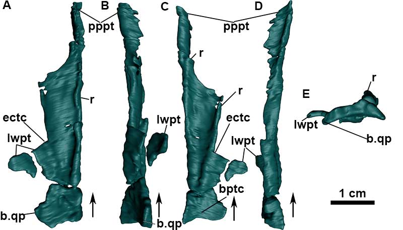
FIGURE 15. 3D Reconstruction based on µCT-scan images of the anterior portion of the dentary of Aetosauroides scagliai (MCN-PV 2347). A, right dentary in lateral view, with reconstructed tip. B, anterior tip of the left dentary in lateral view. C, anterior tip of the left dentary in medial view. D, right dentary in medial view. E, combined anterior portions of the dentary of MCN-PV 2347 in dorsal view. F, combined anterior portions of the dentary in anterior view. G, combined anterior portions of the dentary of MCN-PV 2347 in ventral view. H, mandible reconstruction with elements preserved in MCN-PV 2347. Asterisk (*) tooth number indicates that the teeth have resorption pits. Abbreviations: ae, anterior lateral expansion; afo, anterior foramen; dt, dentary teeth or alveolus; ed, edentulous portion; fo, foramen; idp, interdental plates; nf, nutrient foramen; sy, symphysis; mg, Meckelian groove.
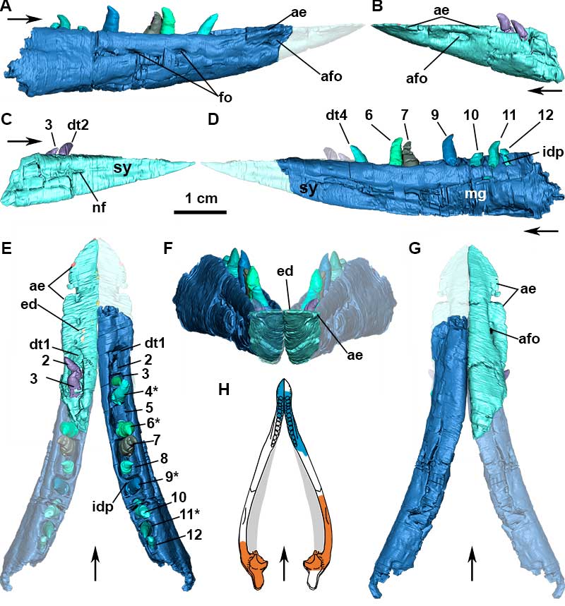
FIGURE 16. 3D Reconstruction based on µCT-scan images of the posterior portion of the mandible of Aetosauroides scagliai (MCN-PV 2347). A, articulation between the left quadrate and left mandible in postero-lateral view (including the position of the stapes). B, left articular in posterior view. C, left articular in dorsal view. D, left mandible in posterior view. E, left articular in lateroventral view. F, left articular in ventral view; G, left mandible in lateral view. H, left mandible in medial view. I, left mandible in dorsal view. J, left surangular in medial view. Abbreviations: ar, articular; as, glenoid; asar, articulation facet for the articular; b.su, broken surface of the surangular; e, elevation; fo, foramen; lw, lateral wing of the quadrate; par, prearticular; parc, prearticular contact of the articular; qc, quadrate crest; qfo, quadrate foramen; qh, quadrate head; rap, retroarticular process; st, stapes; su, surangular; suas, surangular articulation with quadrate; suc, surangular contact of the articular; sufe, surangular foramen medial exit; sufo, surangular foramen; sus, medial shelf of the surangular; vr, ventral ridge.
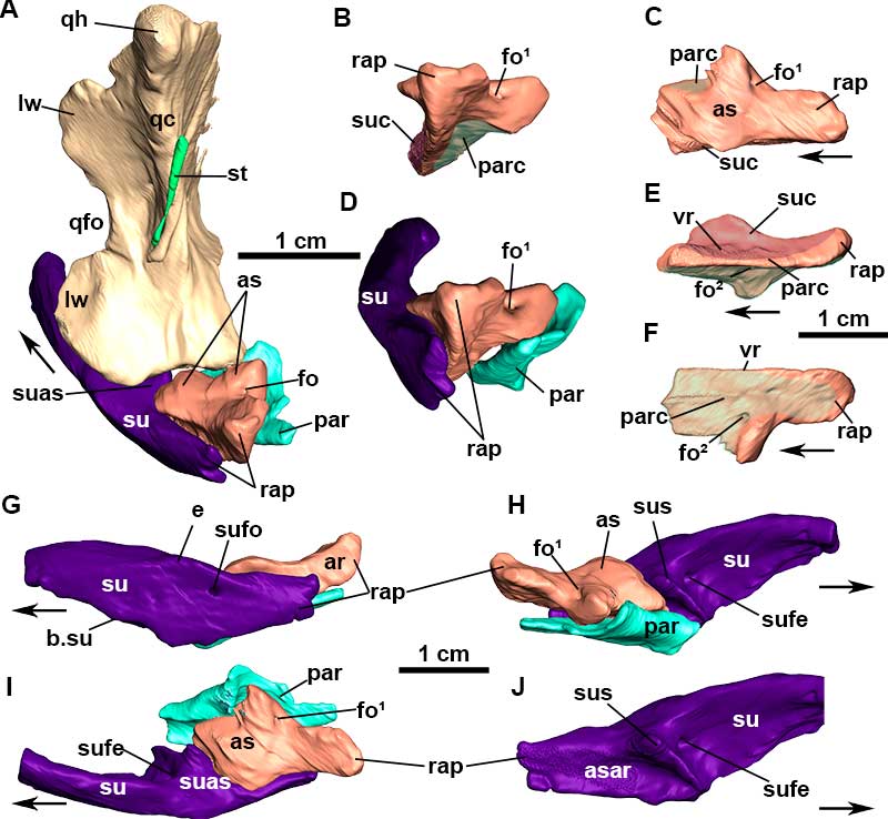
FIGURE 17. 3D Reconstruction based on µCT-scan images of the right angular (A-E), prearticular (F-H), and surangular (I-K) of Aetosauroides scagliai (MCN-PV 2347) in lateral (A, H, and I), antero-medial (B and J), medial (D, G, and K), dorsal (F), posterior (C), and ventral (E) views. Abbreviations: aang, articulation surface with the angular; asar, articulation surface of the articular; asp, articulation with the splenial; asu, articulation with the surangular; e, elevation; mf, mandibular fenestra; mk, internal meckelian groove; rap, retroarticular process; sufe, surangular foramen medial exit; sufo, surangular foramen; sup, surangular process; sus, medial shelf of the surangular; tu, surangular process; vs, ventral shelf of the angular.
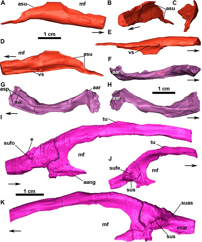
FIGURE 18. Dental morphology of Aetosauroides scagliai (MCN-PV 2347, MCP-3450-PV and UFSM 11505). A, Transversal section of the premaxilla of MCN-PV 2347 showing the premaxillary tooth in mesial view. B-C, 3D reconstruction based on µCT-scan images of the premaxillary tooth in labial (B), distal (C), and basal (D) views. E, in situ maxillary tooth of UFSM 11505 in labial view. F, 3D reconstruction based on µCT-scan images of the maxillary tooth in basal view. G, medial view of maxilla and dentary of MCN-PV 2347. H, isolated dentary teeth of MCP-3450-PV. I-M, 3D reconstruction based on µCT-scan images of the dentary tooth of MCN-PV 2347 in basal (I), distal (J), labial (K), distal (L), and lingual (M) views. Abbreviations: cr, crown; es, enamel striation; i.ct, incipient constriction; itp, interdental plate; kn, knee; nt, new tooth; ro, root; rp, resorption pit.
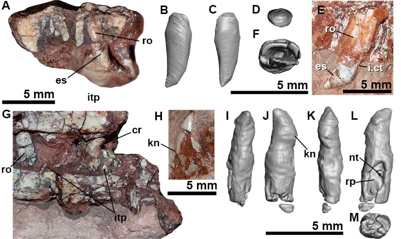
FIGURE 19. Skull reconstruction of Aetosauroides scagliai, based mainly on MCN-PV 2347 (scaled to this specimen), UFSM 11505 and PVL-2059. A, skull and mandible in lateral view. B, skull in dorsal view. C, skull in ventral view. Gray areas indicate depth. Abbreviations: ang, angular; aof, antorbital fenestra; ar, articular; ch, choana; de, dentary; itf, infratemporal fenestra; f, frontal; fi, foramen incisivum; itf, infratemporal fenestra; j, jugal; la, lacrimal; mf, mandibular fenestra; mx, maxilla; n, nasal; ne, external nares; o, orbit; pa, parietal; pmx, premaxilla; po, postorbital; pof, postfrontal; prf, prefrontal; pt, pterygoid; q, quadrate; qj, quadratojugal; rap, retroarticular process; sq, squamosal; sbf, suborbital fenestra; sbtf, subtemporal fenestra; stf, supratemporal fenestra; su, surangular.
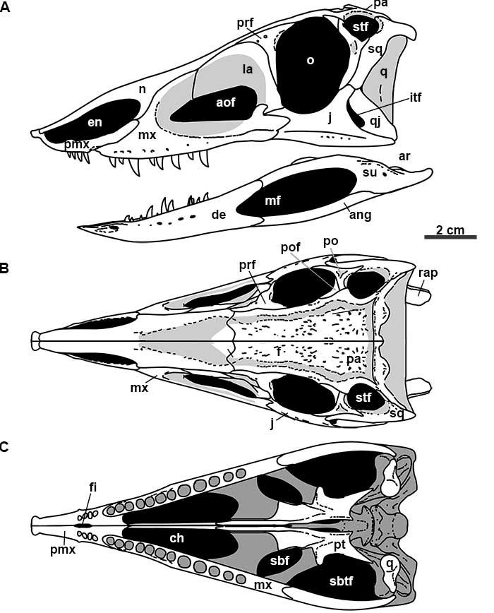
FIGURE 20. Jugal and quadratojugal of Aetosauria. Selected skulls of aetosaurs in lateral view and respectively close-ups: A and A1, Aetosauroides scagliai (MCN-PV 2347), with interpretative drawing. B and B1, Stenomyti huangae (DMNH 60708). C and C1, Aetosaurus ferratus (SMNS 5770 S-21). D and D1, Paratypothorax andressorum (SMNS 19003). E and E1, Desmatosuchus smalli (TTU P-9023). F and F1, Neoaetosauroides engaeus (PULR 4363). Abbreviations: aof, antorbital fenestra; itf, infratemporal fenestra; j, jugal; mf, mandibular fenestra; en, external nares; o, orbit; pvpj, posteroventral process of the jugal; q, quadrate; qj, quadratojugal; stf, supratemporal fenestra.
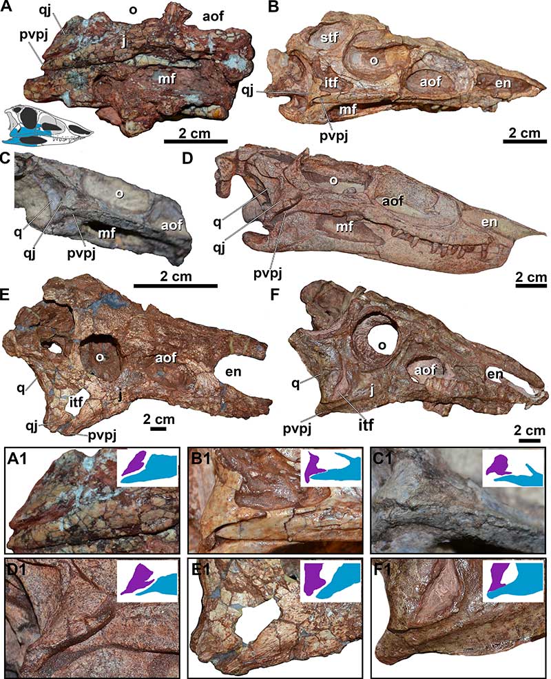
FIGURE 21. Comparisons of maxillae, jugals, and quadratojugals of Aetosauria and non-aetosaur pseudosuchians. A, the non-stagonolepidide aetosaur Aetosauroides. B, Revueltosaurus (redrawn from Nesbitt, 2011). C, the erpetosuchid Tarjadia (redrawn from Ezcurra et al., 2018). D, the aetosaur Desmatosuchus (redrawn from Small, 2002). E, the aetosaur Stagonolepis robertsoni (redrawn from Sulej, 2010). F, the aetosaur Neoaetosauroides (redrawn from Desojo and Báez, 2007). G, the aetosaur Stenomyti huangae (redrawn from Small and Martz, 2013). H, the aetosaur Paratypothorax andressorum (redrawn from Schoch and Desojo, 2016 with modifications, see text). I, the aetosaur Aetosaurus ferratus (redrawn from Schoch, 2007 with modifications, see text). J, the basal phytosaur Diandongosuchus (redrawn from Stocker et al., 2017). K, the loricatan Postosuchus (redrawn from Nesbitt, 2011). L, the loricatan Prestosuchus (based on UFRGS-PV-0152-T and Mastrantonio et al., 2019). M, the ornithosuchid Riojasuchus (redrawn from Bakzco et al., 2018). N, the gracilisuchid Gracilisuchus stipanicicorum (redrawn from Nesbitt, 2011 and observations of Butler et al., 2014). O, the crocodylomorph Dromicosuchus (redrawn from Nesbitt, 2011). Colors indicate bone elements: orange, maxilla; blue, jugal; purple, quadratojugal. Not to scale.
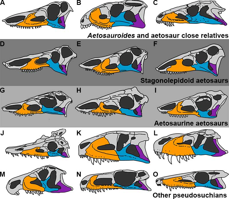
FIGURE 22. Maxillary teeth diversity of Revueltosaurus callenderi (A, based in PEFO 34561), Acaenasuchus geoffreyi (B, based in Marsh et al., 2020), and Aetosauria: C, Paratypothorax andressorum (SMNS 19003, mirrored). D, Typothorax coccinarum (based in Reyes et al., 2021). E, Aetosauroides scagliai (UFSM 11505). F, Aetosaurus ferratus (SMNS 5772 S-18). G, c.f. Stenomyti huangae (DMNH 60708). H, Neoaetosauroides engaeus (PULR 108). I, Desmatosuchus smalli (TTUP 9420); J, Stagonolepis robertsoni (ELGNM 38, mirrored); K, Stagonolepis olenkae (ZPAL AbIII 2752). Abbreviations: ct, constriction of the base; dc, large denticles; es, enamel striations (faint gray line); g, grooves or flutes (black line); r, ridge (bold black line); rp, resorption pit; t, longitudinal ridge; wf, wear facet.
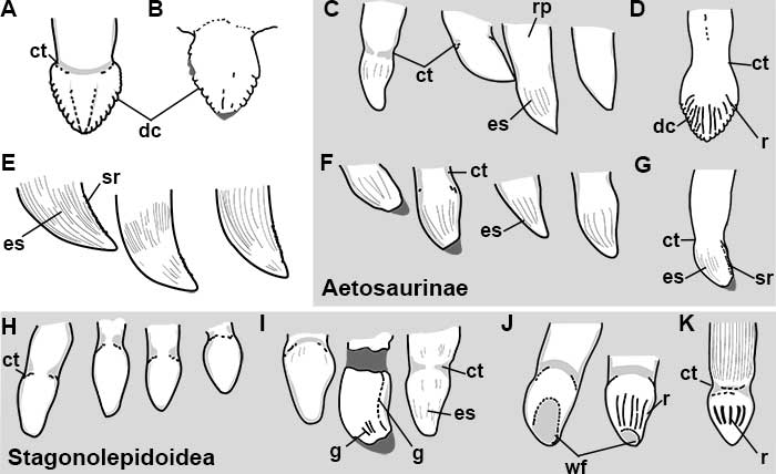
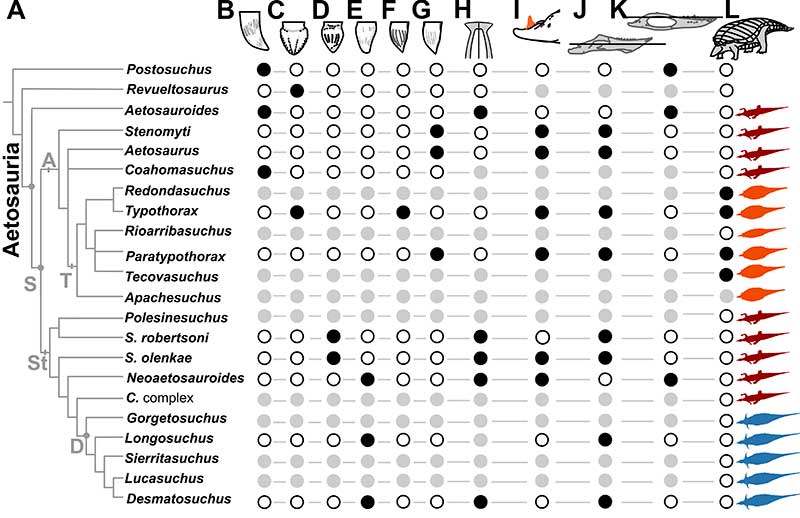
FIGURE 23. Morphological features related to feeding strategies in Aetosauria in a phylogenetic perspective. A, simplified genus level phylogeny of Reyes et al. (2020). We coalesced the clade formed by Calyptosuchus, Adamanasuchus and Scutarx (C. complex) as no dental information is available for these taxa and they all share the same overall body-shape. B, ziphodont teeth. C, folidont teeth with marked serrations. D, folidont tooth with tooth wear. E, elongated folidont teeth with tooth wear. F, folidont to bulbous teeth with grooves/flutes. G, bulbous teeth with enamel striations. H, shovel-shaped premaxilla. I, medial dorsal process of the articular; J, glenoid placed ventral to the tooth line in the mandible. K, glenoid placed near or dorsal to the teeth line in the mandible. L, wide-body morphology indicating large gastric cavities. Dark circles indicate presence, white circles indicate absence and gray circles indicate missing data. Silhouettes at the right side show the overall body shape of aetosaurs as suggested by Desojo et al. (2013), in red the ‘narrow-body’, in orange the ‘wide-body’ and in blue the ‘spinose’. Abbreviations: A, Aetosaurinae; D, Desmatosuchini; S, Stagonolepididae; St, Stagonolepidoidea; T, Typothoracinae.