FIGURE 1. Geography and geology at the Finis Shale outcrop, TXV-200, spillway section northeast of Jacksboro, Texas, USA. A - location of Jacksboro (Jack County), approximately 100 km northwest of Fort Worth, Texas. B - TXV-200, the horseshoe-shaped outcrop at the spillway section at Lost Creek Lake. Greyish area in the middle of the horseshoe is a bedding plane, conulariids are usually found here; numbers 1-3 refer to the numbers in the landscape photo in Figure 1C. C - landscape photo of the spillway section toward the South, view when standing on the bedding plane; 1-3 inserted to compare positions as indicated in Figure 1B, white arrows with B and C indicate the location of the sections sampled (also compare Figure 2B). D - yellowish, weathered Finis Shale. E - greyish Finis Shale. D, E - sampling transects (compare Figure 1C and Figure 2B) were prepared to be able to collect fresh, in situ shale. F - top of the outcrop (above sampled section B) represented by the Jacksboro Limestone.
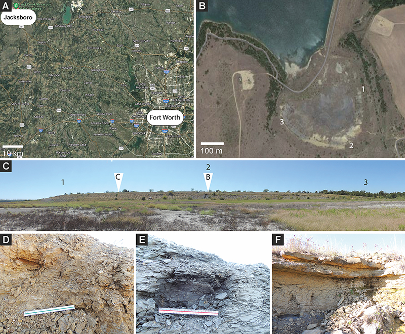
FIGURE 2. Stratigraphy and sampled sections of the Finis Shale at TXV-200. A - stratigraphic position of the Finis Shale Member modified after Yang and Kominz, 2003 (fig. 2). B - measured sections, not from the base of the Finis Shale; base represented by accessible part of the Finis at individual section (compare with Figure 1); profile C is marked by exclusively yellowish shale, profile B by grey shale that only mixes with yellowish shale in the uppermost part; the latter section is capped by the Jacksboro Limestone. Ss - sandstone.
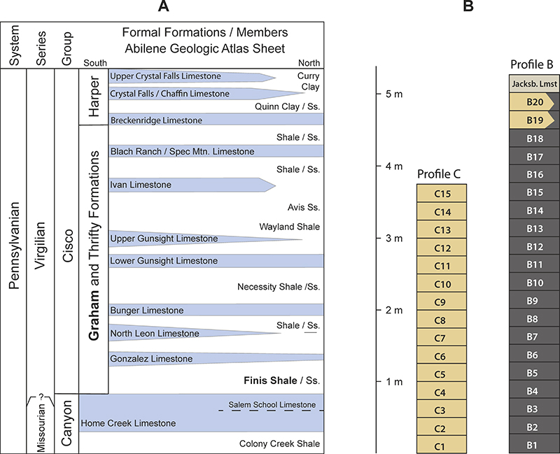
FIGURE 3. Picked bryozoan fragments from the disaggregated, sieved samples from the Finis Shale. A - profile B15; B - profile B16; C - profile C5; D - profile C13.
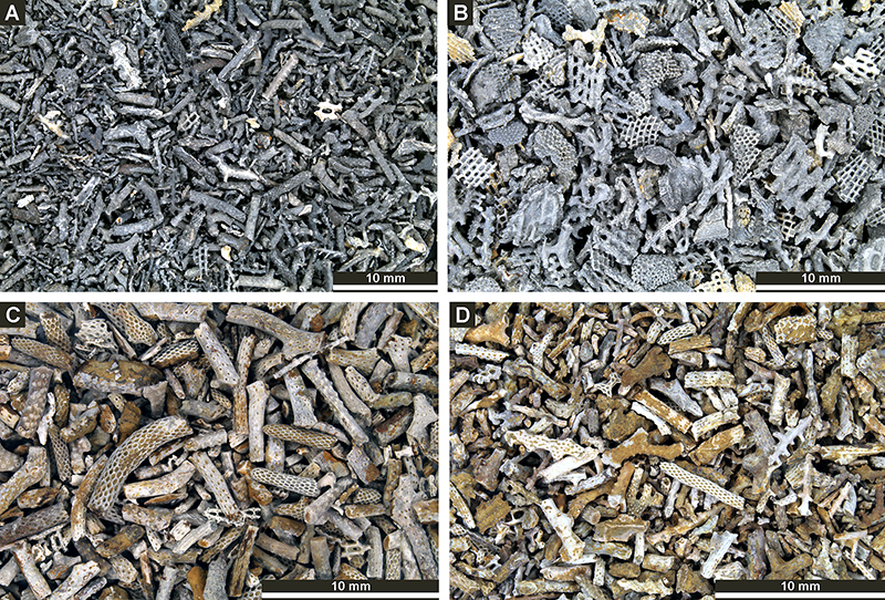
FIGURE 4. Fistulipora nodulifera Meek, 1872 (A-C): A - colony surface with autozooecial apertures and lunaria (XCI 97); B - tangential thin section showing autozooecial apertures with lunaria and vesicles (XCI 29); C - longitudinal thin section showing autozooecial chambers and vesicles (XCI 56b). Eridopora beilensis Perkins and Perry in Perkins et al., 1962 (D-F): discoidal colony showing autozooecial apertures with triangular lunaria (XCI 98).
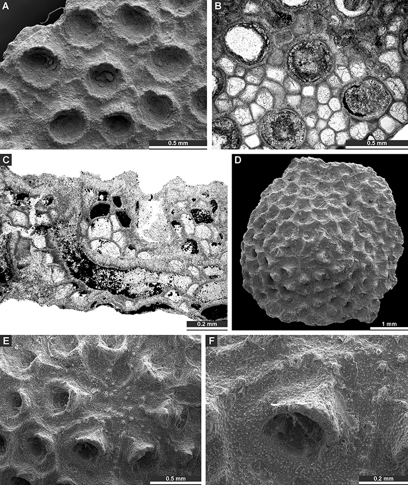
FIGURE 5. Eridopora beilensis Perkins and Perry in Perkins et al., 1962 (A-B): A - tangential thin section showing autozooecial apertures and vesicles (XCI 36); B - longitudinal thin section of a colony on echinoderm fragment showing autozooecial chambers and vesicles (XCI 59a). Cystodictya formosa Moore, 1929 (C-G): C, D - branch fragment with autozooecial apertures and lunaria (XCI 99); E-F: tangential thin section showing autozooecial apertures with lunaria (XCI 23a); G - deep tangential section showing autozooecial chambers with hemisepta and vesicular skeleton (XCI 23a).
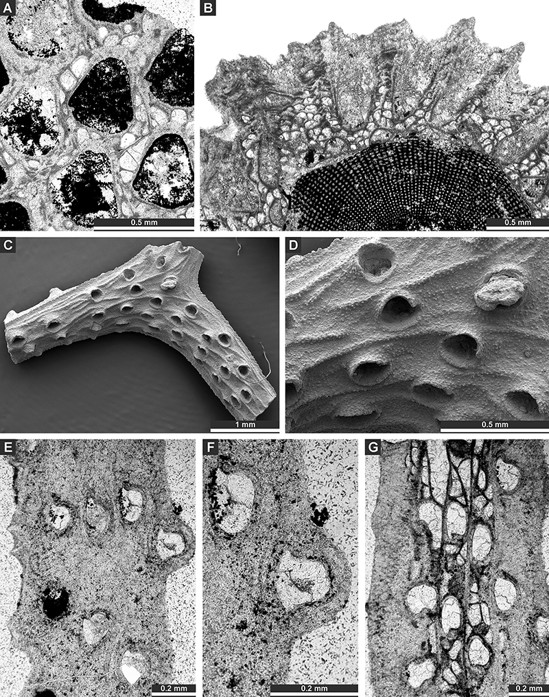
FIGURE 6. Cystodictya formosa Moore, 1929 (A-B): A - deep tangential section showing autozooecial chambers with hemisepta and vesicular skeleton (XCI 27); B - branch transverse section showing mesotheca, autozooecial chambers and vesicles (XCI 23c). Goniocladia grahamensis Moore, 1929 (C-E): branch fragment showing the shape of fenestrule, ridges on branches and autozooecial apertures with lunaria (XCI 100). Dyscritella felixi n. sp. (F-H): F - colony encrusting a brachiopod spine (holotype XCI 101); G - colony surface showing autozooecial apertures, exilazooecia, and acanthostyles; H - tangential thin section showing autozooecial apertures, exilazooecia, and acanthostyles (paratype XCI 86).
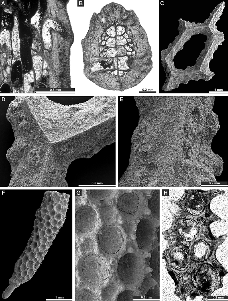
FIGURE 7. Dyscritella felixi n. sp. (A-B): longitudinal thin section of a colony encrusting a brachiopod spine (paratype XCI 87). Rhombopora lepidodendroides Meek, 1872 (C-H): C - branch fragment (XCI 102); D, F - colony surface with autozooecial apertures, acanthostyles and aktinotostyles (XCI 102); E - colony surface with autozooecial apertures, acanthostyles and aktinotostyles (XCI 103); G, H - tangential thin section showing autozooecial apertures, acanthostyles and aktinotostyles (XCI 80b).
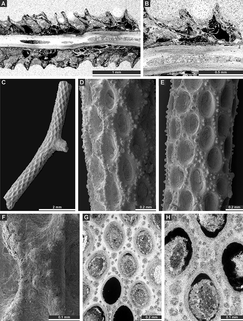
FIGURE 8. Rhombopora lepidodendroides Meek, 1872 (A-C): A - branch transverse section (XCI 58a); B, C - branch longitudinal section showing autozooecial chambers and aktinotostyles in autozooecial wall (XCI 45b). Streblotrypa (Streblotrypa) multipora Warthin, 1930 (D-G): D, E - branch fragment showing autozooecial apertures and metazooecia (XCI 104); F - branch transverse section showing autozooecial chambers and axial bundle (XCI 39b); G - branch longitudinal section showing branch transverse section showing autozooecial chambers with hemisepta (arrows) and axial bundle (XCI 40). Rhombocladia delicata Rogers, 1900 (XCI 105) (H-I) - branch fragment (H) and colony surface showing autozooecial apertures, acanthostyles and paurostyles (I).
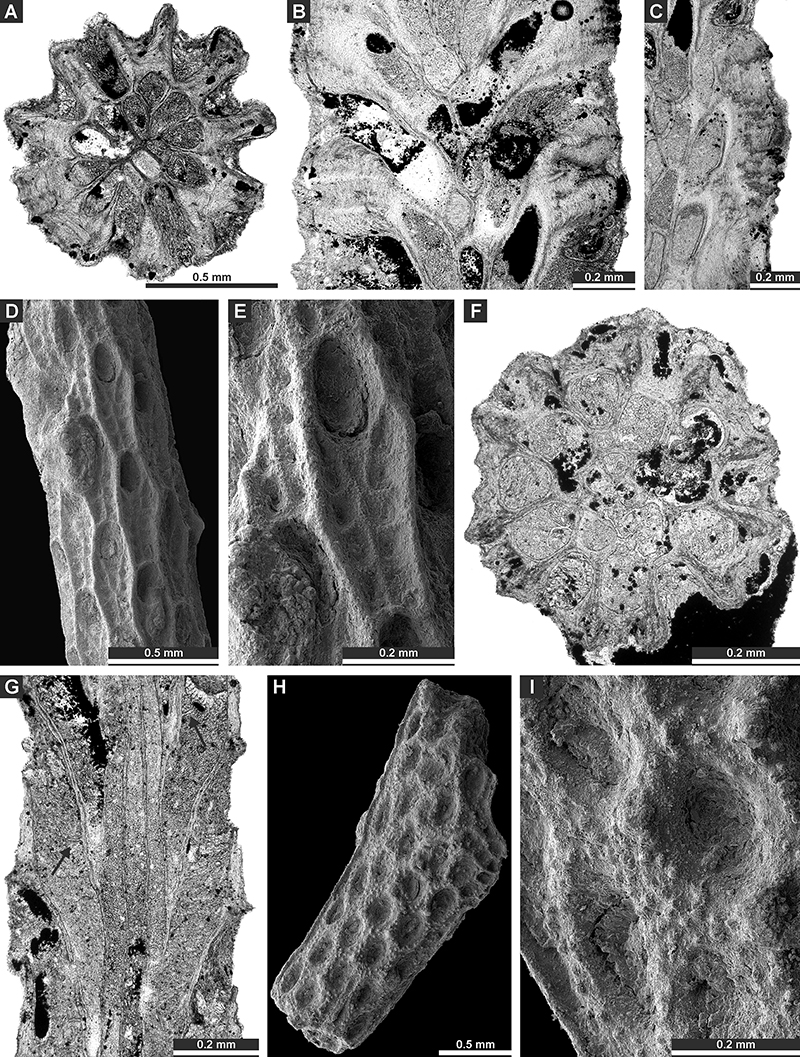
FIGURE 9. Rhombocladia delicata Rogers, 1900: (A) - transversal thin section (XCI 22c), (B) - branch longitudinal thin section (XCI 22b). Chainodictyon minor Ulrich, 1890, (C-G): C - branch fragment and colony surface showing autozooecial apertures (XCI 106); D - branch reverse side showing transversal striation (XCI 125); E-G - tangential thin section showing fenestrules, autozooecial apertures and chambers, and a leptozooecium (arrow) (XCI 37).
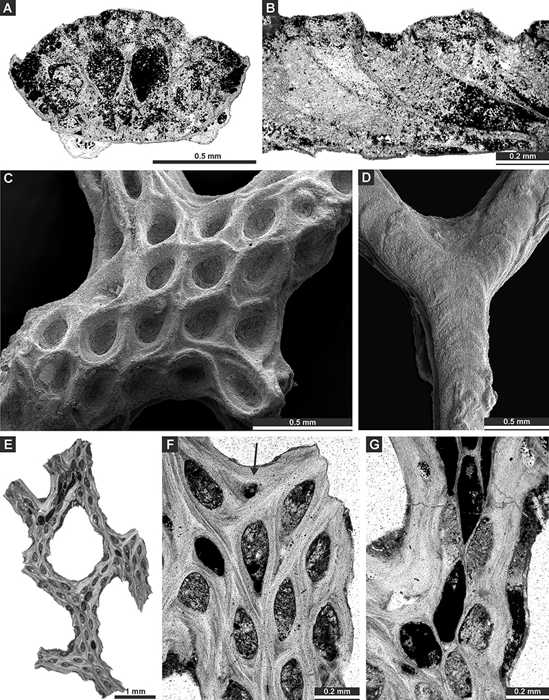
FIGURE 10. Chainodictyon minor Ulrich, 1890, (A): longitudinal thin section showing autozooecial chambers (XCI 42). Fabifenestella compactilis (Condra, 1902) (B-G): B - tangential section showing autozooecial apertures and chambers with hemisepta (XCI 49); C-E - fragment of colony showing fenestrules, autozooecial apertures and keels with nodes (arrow - apertural pore) (XCI 107); F, G - tangential section showing autozooecial apertures and chambers with hemisepta, and keels with nodes (XCI 62). Laxifenestella placida Moore, 1929 (H) - deep tangential section showing autozooecial chambers with hemisepta (XCI 65).
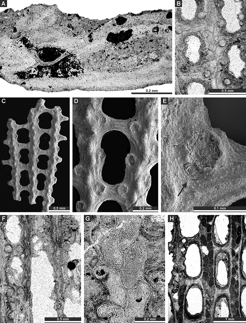
FIGURE 11. Laxifenestella placida Moore, 1929 (A-D): A - tangential section showing autozooecial apertures and keel with nodes (XCI 35); B - deep tangential section showing autozooecial chambers with hemisepta (XCI 65); C, D - colony fragment showing fenestrules, autozooecial apertures and keels with nodes (XCI 108). Laxifenestella texana n. sp. (E, F) - tangential section showing fenestrules, autozooecial apertures and chambers, keels with nodes, and reproductive heterozooecia (arrows), holotype (XCI 81).
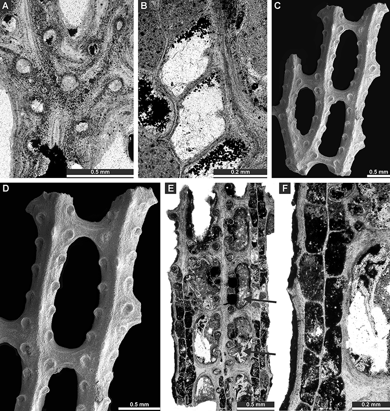
FIGURE 12. Laxifenestella texana n. sp. (A-G): A-C - tangential section showing autozooecial apertures and chambers, and reproductive heterozooecia (arrows) (holotype XCI 81). D - colony fragment showing fenestrules, autozooecial apertures divided by keels with nodes (paratype XCI 109); E - colony fragment showing reproductive heterozooecia (arrows) (paratype XCI 109); F - almost intact chamber of a reproductive heterozooecium (paratype XCI 110); G - colony fragment showing secondary nanozooecia (arrows) (paratype XCI 111). Cavernella praecavifera (Schulga-Nesterenko, 1951) (H) - colony fragment showing fenestrules, autozooecial apertures divided by keels with nodes (XCI 112).
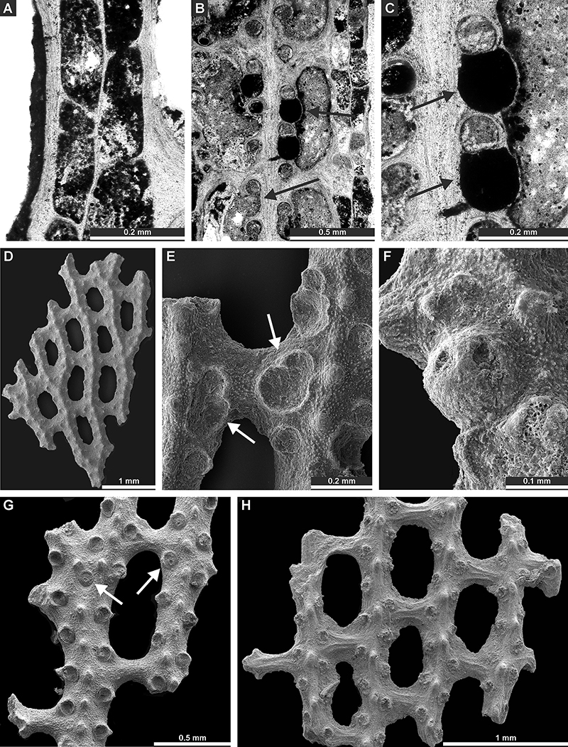
FIGURE 13. Cavernella praecavifera (Schulga-Nesterenko, 1951) (A-F): A-B - colony fragment with broken autozooecial chambers (XCI 126); C - autozooecial aperture with preserved stellate structure (XCI 127); D - colony fragment showing a weathered cavernozooecium (arrow) (XCI 128); E, F - tangential section showing autozooecial apertures and chambers (XCI 69).
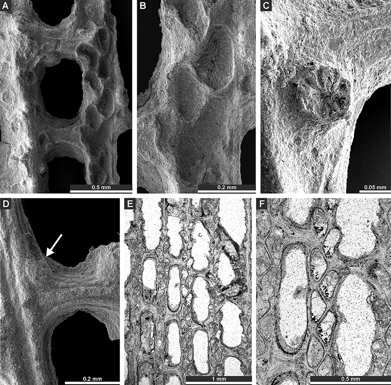
FIGURE 14. Cavernella praecavifera (Schulga-Nesterenko, 1951) (A-D): A - tangential section showing autozooecial apertures and chambers (XCI 69); B-D - tangential section showing cavernozooecia (arrows) (XCI 73). Acupipora elliptica (Rogers, 1900) (E) - colony fragment showing fenestrules, autozooecial apertures and nodes (XCI 113).
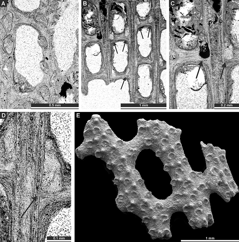
FIGURE 15. Acupipora elliptica (Rogers, 1900) (A-D): A - branch fragment with autozooecial apertures, nodes and nanozooecium (arrow) (XCI 113). B, C - deep tangential section showing autozooecial chambers with hemisepta (XCI 70); D - tangential section showing autozooecial apertures (XCI 70). Polypora triangularis Rogers, 1900 (E-H): E, F - tangential section showing autozooecial apertures and chambers (XCI 50); G, H - tangential section showing autozooecial apertures and reproductive heterozooecia (arrows) (XCI 26).
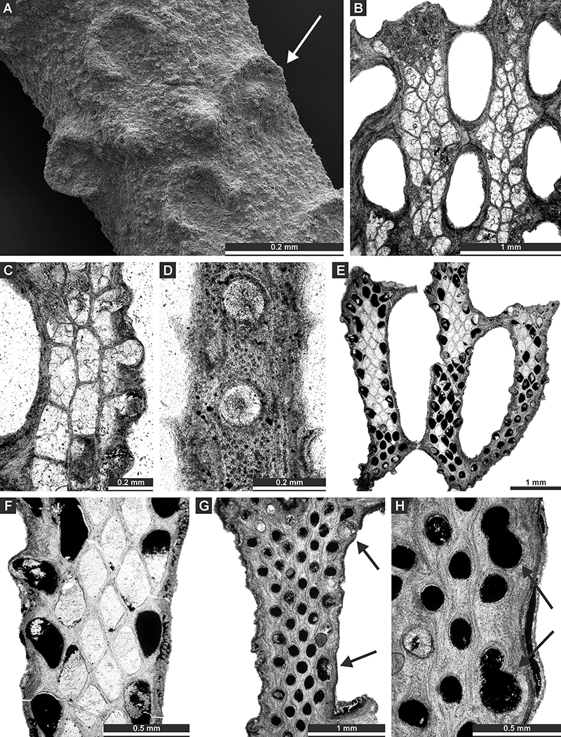
FIGURE 16. Polypora triangularis Rogers, 1900 (A-G): A-C - colony fragments with intact chambers of reproductive heterozooecia (XCI 114); D - branch fragment with nanozooecia (XCI 115); E, F - branch fragment with autozooecial apertures with proximal pores (arrows) (XCI 116); G - branch fragment with nodes on the reverse side (XCI 117). Polypora aff. hexagona Moore, 1929 (H, I) - colony fragment with fenestrules, autozooecial apertures, and nodes (XCI 118).
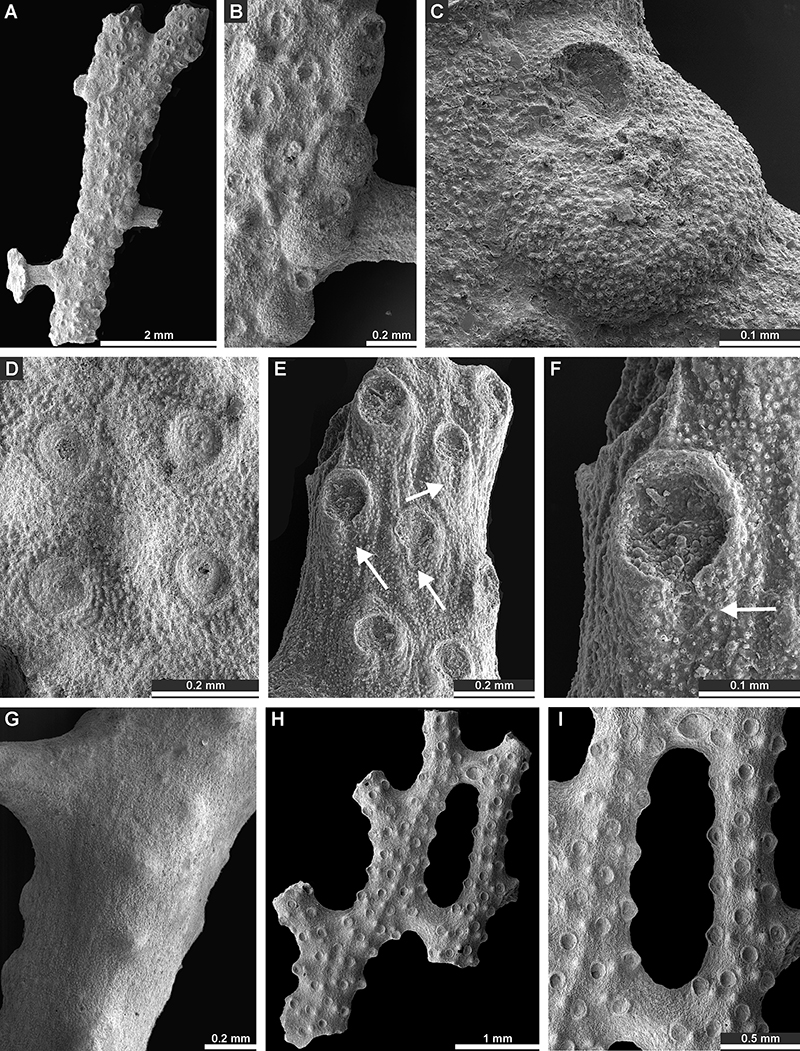
FIGURE 17. Polypora aff. hexagona Moore, 1929 (A-E): tangential section showing autozooecial apertures and chambers, nodes, microstyles, and reproductive heterozooecia (arrows) (XCI 55). Septopora blanda Moore, 1929 (F-I): F - tangential section showing autozooecial chambers and cyclozooecia (XCI 54); G - colony fragment with cyclozooecia on the reverse side (arrows) (XCI 119). H, I - colony fragment with autozooecial apertures, keel nodes, and cyclozooecia (arrows) (XCI 120).
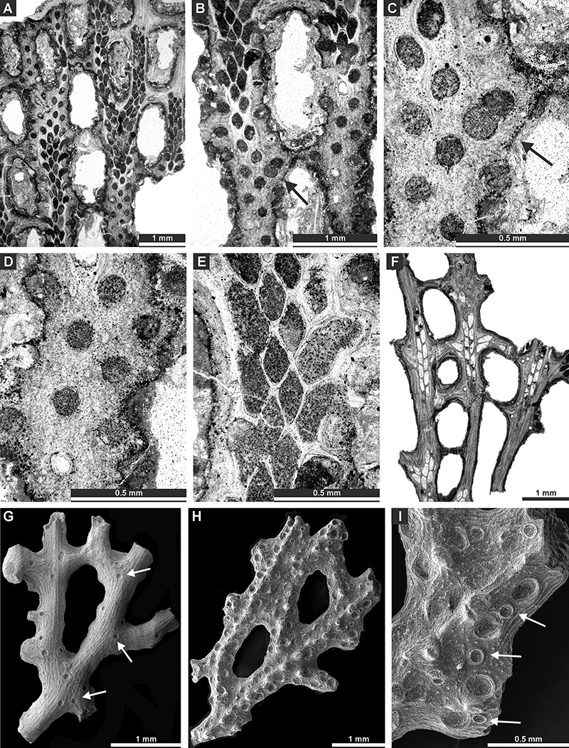
FIGURE 18. Septopora blanda Moore, 1929 (A-C): A, B - tangential section showing autozooecial apertures and chambers, and cyclozooecia (arrows) (XCI 47); C - tangential section showing autozooecial chambers and cyclozooecia (XCI 54). Penniretepora flexistriata Richards, 1959 (D-J): D-F - colony fragments with autozooecial apertures with apertural pores (arrows) and stellate structures, divided by low undulating keel (D: (XCI 121), E-F: (XCI 122); G - autozooecial aperture with apertural pore (arrow) and stellate structure (XCI 123); H, I - thin section showing autozooecial chambers (XCI 83); J - thin section showing autozooecial apertures with apertural pore (arrow) (XCI 82).
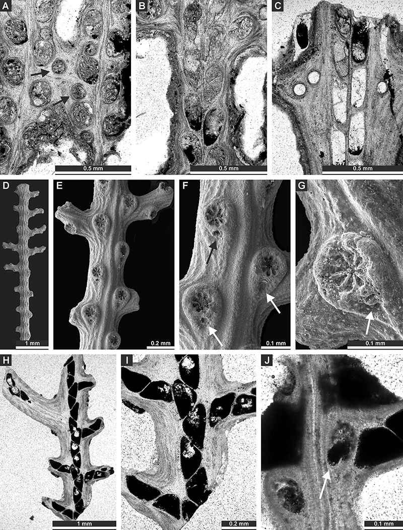
FIGURE 19. Penniretepora oculata Moore, 1929 (A-E): A, B - branch fragment with autozooecial apertures divided by keel with nodes (XCI 124); C, D - tangential section showing autozooecial chambers (XCI 96); E - tangential section showing autozooecial apertures (arrow: nanozooecium) (XCI 96).
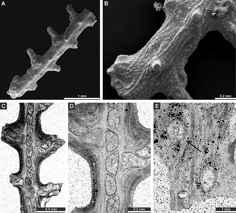
FIGURE 20. Distribution of bryozoan growth forms (number of fragments) within the profiles B and C (samples B1-B4 contained almost no bryozoans).

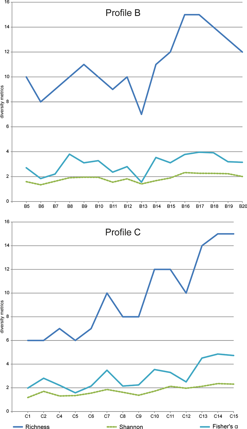
FIGURE 21. Bryozoan diversity indices in profiles B and C (species richness, Shannon index, and Fisher’s α). Diversity indices were counted using PAST version 1.81 (Hammer et al., 2001).