FIGURE. 1. The site along Calvert Cliffs where the two pathological cetacean vertebrae (CMM-V-10108) and associated Otodus megalodon tooth (CMM-V-8522) were found in situ in Shattuck-Zone 12. Looking north along the cliffs at Warrior’s Rest. Photo by M. Ellwood.
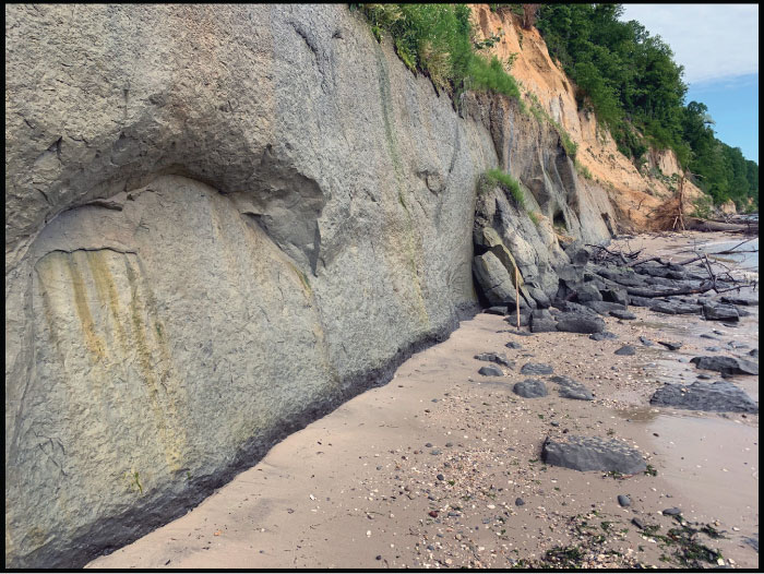
FIGURE 2. CMM-V-10108, a Miocene pathological cetacean vertebra associated with the one shown in Figure 5 and Figure 6. A. Anterior view showing a major shear-compression fracture with comminution. B. right lateral view, and C. posterior view showing the intact fused epiphysis.
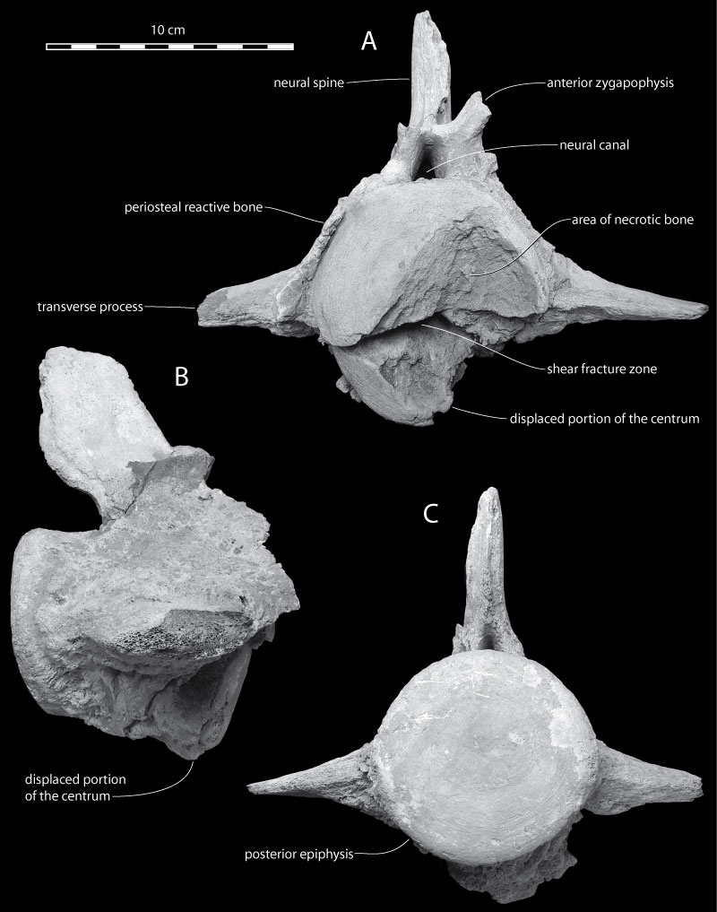
FIGURE 3. CMM-V-10108, shear-fractured Miocene cetacean lumbar vertebra in three transverse CT-scan images. These CT-scan images cut through the vertebra in an anterodorsal-posteroventral direction. A. CT-scan image through the anterior portion of the vertebra showing the wide-open lumen of the shear-compression fracture. B. CT-scan image from approximately 1 cm behind A, showing the thickness of the periosteal reactive bone layer. C. CT-image at about the midpoint in the length of the vertebra showing the posterior-most part of the sheared base of the centrum compressed (telescoped) into the body of the centrum.
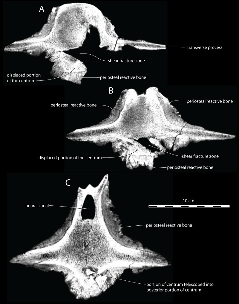
FIGURE 4. CMM-V-10108, a single CT-scan image in the sagittal plane of a Miocene pathological cetacean vertebra in left lateral view showing the broken lower portion of the centrum, the displaced piece of bone, and the new bone growth (periosteal reactive bone ventrally).
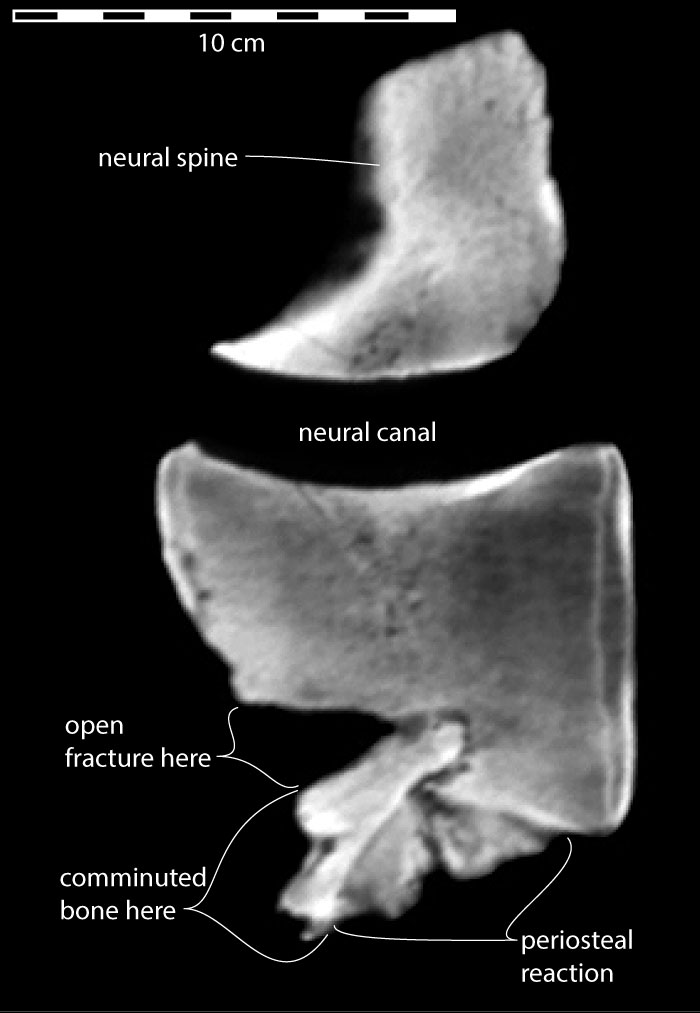
FIGURE 5. CMM-V-10108, a second Miocene pathological cetacean vertebra (CT-scans shown in Figure 6) associated with the one shown in Figure 2, Figure 3, Figure 4. A. Posterior view showing that the neural spine is incomplete and that the sides of the centrum are covered with periosteal reactive bone. B. ventral view to highlight the periosteal reactive bone. In B, the anterior end of the centrum is up.
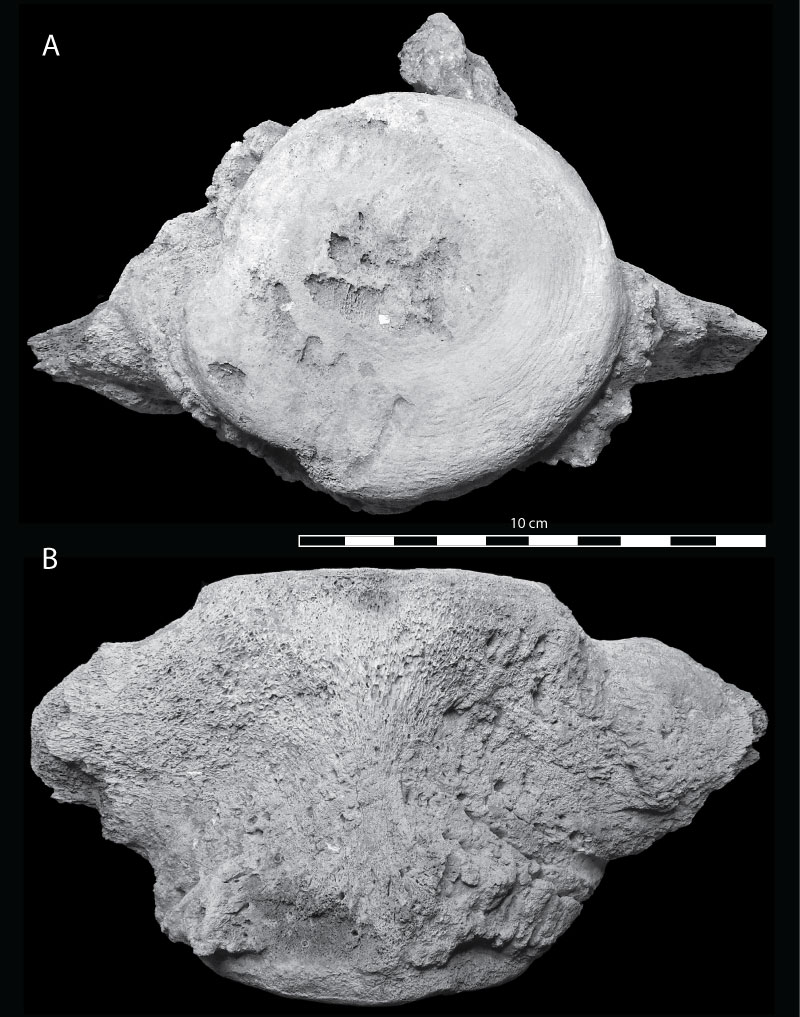
FIGURE 6. CMM-V-10108, a second Miocene pathological cetacean vertebra (also shown in Figure 5) associated with the one shown in Figure 2, Figure 3, Figure 4. A. CT-scan image towards the anterior end of the vertebra. B. CT-scan image at about the midpoint in the length of the vertebra showing the thickness of the periosteal reactive bone.
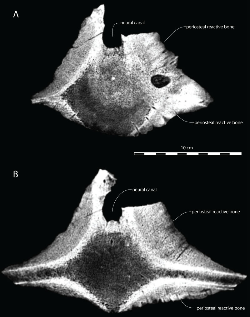
FIGURE 7. CMM-V-8522, Otodus megalodon lower anterior tooth in labial view. This tooth was found touching one of the two pathological vertebrae (CMM-V-10108). Notice the spall-fracture marking the tip of the tooth. White scale bar equals 10 mm.
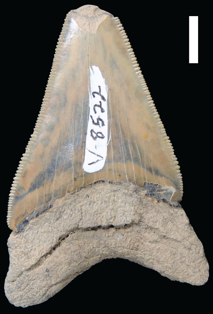
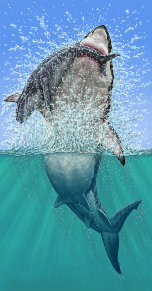
FIGURE 8. One possible way in which the shear-compression fracture occurred in CMM-V-10108. The posterior vertebral column was severely hyperflexed to such a degree that at least one of its vertebrae experienced a shear-compression fracture, and the periosteum was pulled away from most of the sides of both vertebrae. Artwork by Clarence (Shoe) Schumaker (CMM).