FIGURE 1. Location and geologic position of the Woodbine Group. A, General stratigraphic sequence and timescale for the Cretaceous of central and north central Texas showing the position of the Woodbine Group. Terrestrial deposits represented by stippled intervals. Time scale based on Gradstein et al. (2004) and Denne et al., 2016, and modified from Noto et al. (2022). B, Map of Woodbine surface exposures in the study area showing position of localities where fossils were discovered. Exposures are stippled, water bodies are solid gray. 1 = Arlington Archosaur Site, 2 = Grapevine Lake southwest shore.
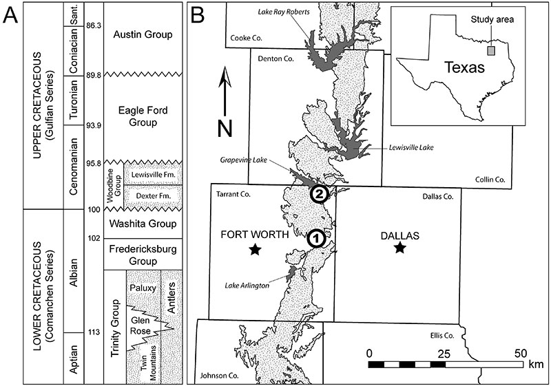
FIGURE 2. DMNH 2013-07-1942, Gehennachelys maini comb. nov. skull: A, dorsal photograph; B, dorsal line drawing; C, ventral photograph; D, ventral line drawing; E, right lateral photograph; F, right lateral line drawing; G, left lateral photograph; H, left lateral line drawing. Arrow indicates anterior orientation for A-D. For E and F, anterior is to the right. Abbreviations: ac= acromion; cm= condylus mandibularis; ct= cavum tympani; fr= frontal; fst= foramen stapediotemporale; ju= jugal; mx= maxilla; or= orbit; pa= parietal; po= postorbital; pm=premaxilla; pr= prootic; qj= quadratojugal; qu= quadrate; so= supraoccipital; sq= squamosal.
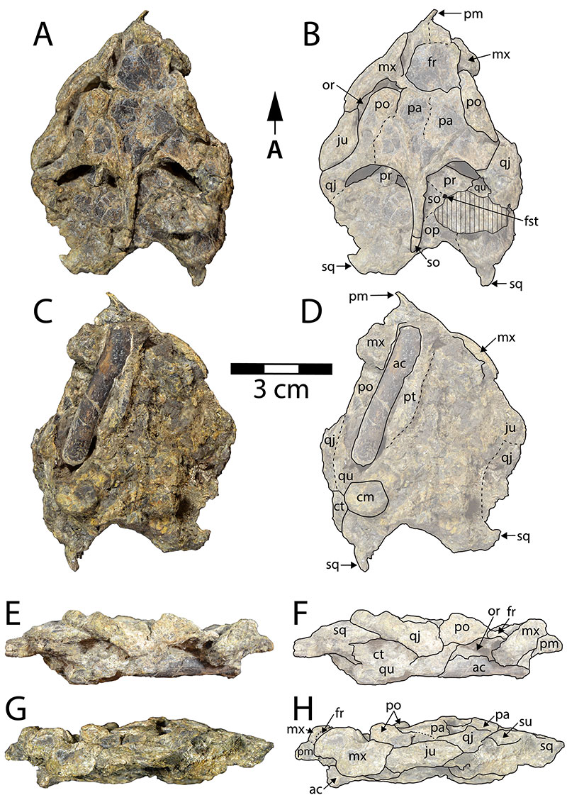
FIGURE 3. DMNH 2013-07-1942 carapace in A, dorsal, and B, ventral views. Plastron in C, dorsal, and D, ventral views. E corresponds with the red box in C, showing the left humerus, rotated 180°. Arrow indicates anterior orientation for A-D. Scale increments in mm for E.
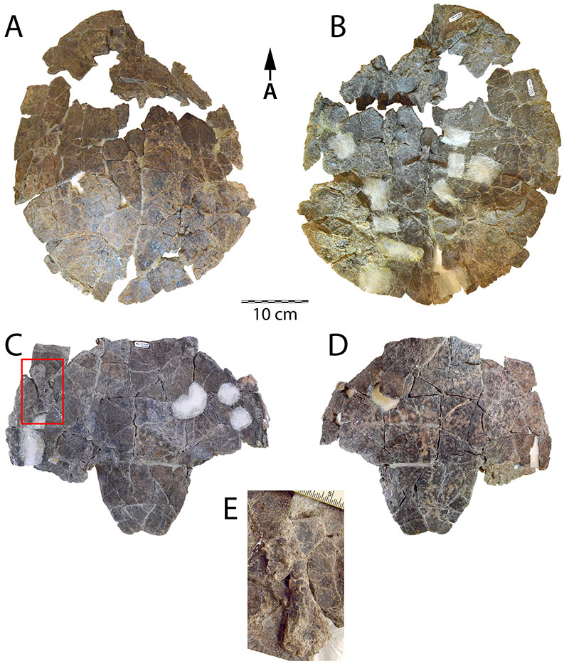
FIGURE 4. HMNS-10-TM, shell of Gehennachelys maini comb. nov., in A, dorsal, B, ventral, C, right lateral, D, anterior, and E, posterior views. Note separate scales for A-B, C, and D-E.
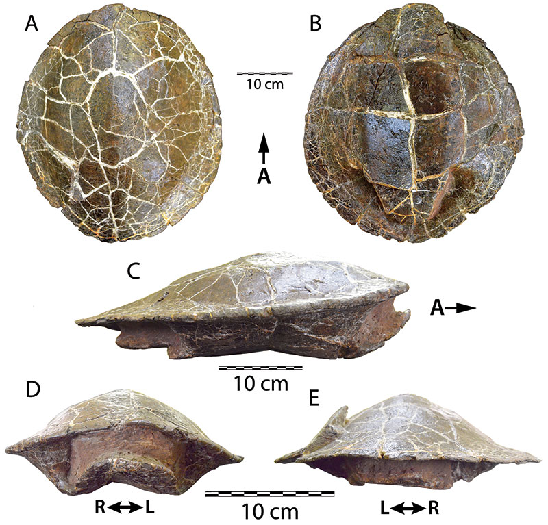
FIGURE 5. DMNH 2013-07-1942 vertebrae. Cervical vertebrae in A, left lateral, B, anterior, and C, posterior views. Ventral view of indeterminate dorsal vertebrae in D, with anterior to the right.
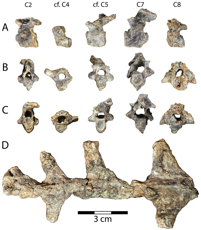
FIGURE 6. DMNH 2013-07-1942, Gehennachelys maini comb. nov. shoulder girdle and forelimb bones. Partial left scapula of DMNH 2013-07-1942 in A, posteromedial, and B, anterolateral views. Partial right scapula of DMNH 2013-07-1942 in C, posteromedial, and D, anterolateral views. Partial right scapula of DMNH 2013-07-0601 in E, posteromedial, and F, anterolateral views. Right coracoid of DMNH 2013-07-1942 in G, dorsal, and H, ventral views. Partial left coracoid of DMNH 2013-07-1942 in I, dorsal, and J, ventral views. Right humerus in K, posterior, L, dorsal, M, proximal, N, distal, O, ventral, and P, posterior views. M and N are oriented with B.
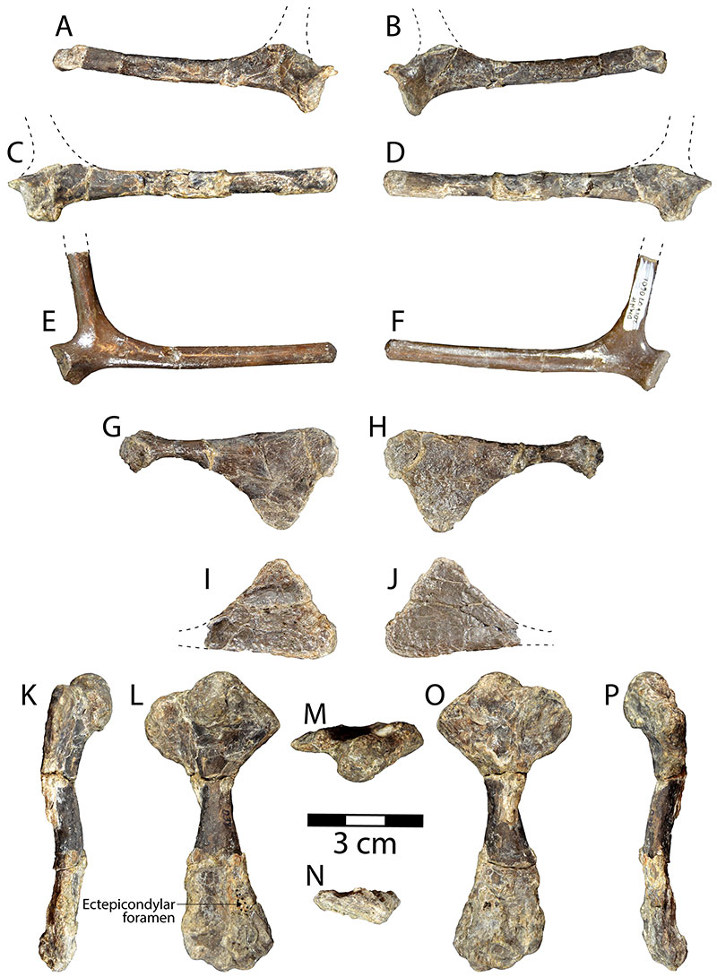
FIGURE 7. DMNH 2013-07-1942 pelvic girdle and hind limb bones. Partial pelvis in A, right lateral view, oriented with anterior to the right. Partial pubic plate in B, ventral, and C, dorsal views. Left femur in D, anterior, E, ventral, F, proximal, G, distal, H, dorsal, and I, posterior views. Partial right tibia in J, dorsal, K, medial, L, ventral, and M, lateral views. Dotted line in G indicates margin of acetabulum.
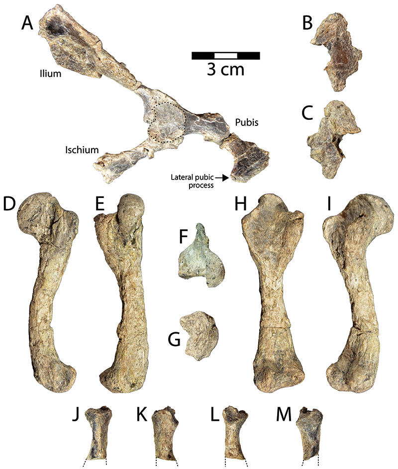
FIGURE 8. Gehennachelys maini comb. nov. shell reconstruction, based on DMNH 2013-07-0696, DMNH 2013-07-0704, DMNH 2013-07-0712, DMNH 2013-07-0784, DMNH 2013-07-1703, DMNH 2013-07-1708, DMNH 2013-07-1942, and HMNS-10-TM. Red lines indicate estimated sulci. Abbreviations: ab= abdominal scale, an= anal scale, ce= cervical scale, eg= extragular scale, fe= femoral scale, gu= gular scale, hu= humeral scale, im= inframarginal scale; ma= marginal scale, pec= pectoral scale, pl= pleural scale, ve= vertebral scale.
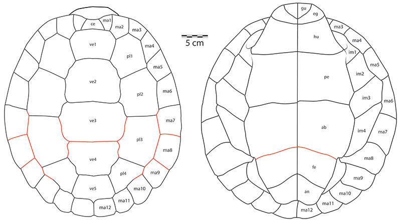
FIGURE 9. Plot of Plastron Length/Carapace Length by Anterior Plastral Lobe Length/Posterior Plastral Lobe Length for baenid taxa and Glyptops, which have associated carapaces and plastra. Proportions of Gehennachelys maini comb. nov. from HMNS-10-TM. Data on other taxa from Archibald (1977, table 57), Lyson and Joyce (2009a, b), Larson et al. (2013), Sullivan et al. (2013), Lively (2015), and Lyson et al. (2019). See Table 3 for raw data. Graph generated in Microsoft Excel. Grouping abbreviations: EUB= Eubaeninae; NEU= Neurankylus spp.; PLT= Palatobaeninae/ Plesiobaena grade. Squares indicate phylogenetically ungrouped taxa (non-baenodd baenids and Glyptops), circles indicate phylogenetically grouped taxa (Neurankylus and baenodds), and red square indicates Gehennachelys maini comb. nov.
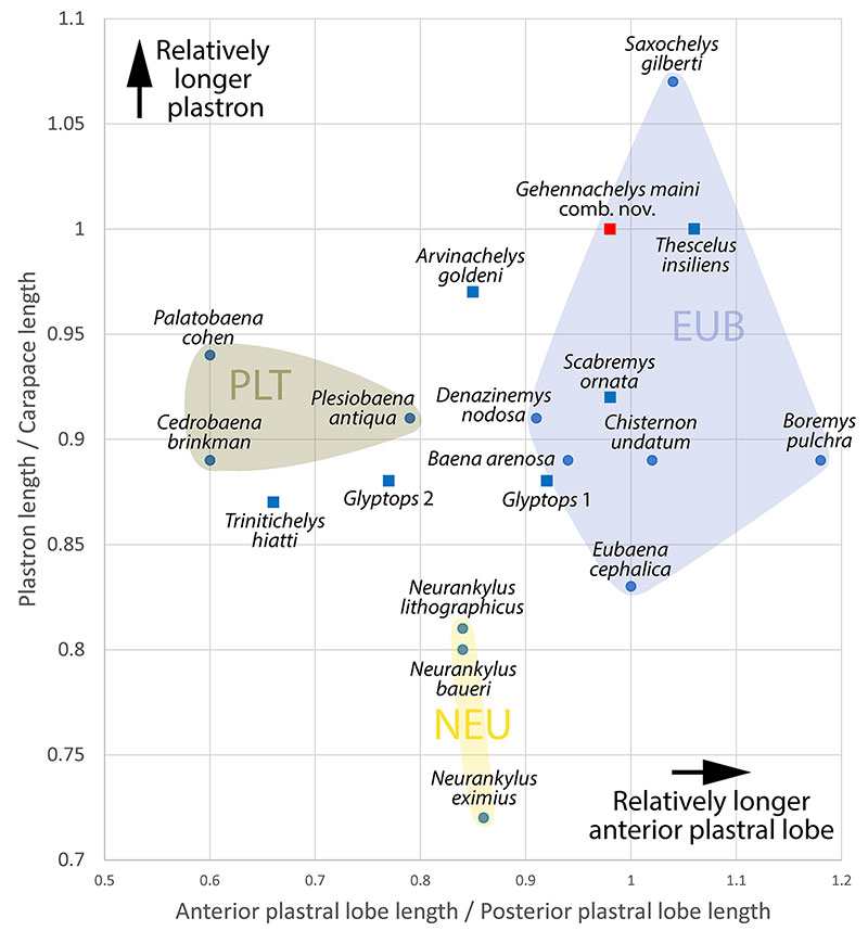
FIGURE 10. Gehennachelys maini comb. nov. histological thin sections: A, DMNH 2013-07-1703, an indeterminate juvenile costal, and B, DMNH 2013-07-0588, a probably fourth costal. Scale bars equal 1 mm. C, closeup of vascularized external cortex and transition to cancellous bone, with growth marks indicated by blue arrowheads, D, internal cortex with intercalated vascular rows from DMNH 2013-07-0588, and E, sutural sockets of DMNH 2013-07-1703. Abbreviations: CB= cancellous bone, ECO= external cortex, ICO= internal cortex.
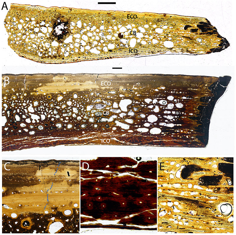
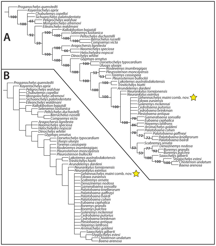
FIGURE 11. Results of phylogenetic analyses for Gehennachelys maini comb. nov., produced from the modified matrix Rollot et al. (2022b), with 170 minimum trees and 367 steps (CI= 0.35; RI= 0.71): A) Consensus 50% majority-rule phylogenetic tree of Baenidae; B) Strict consensus tree. Yellow stars indicate Gehennachelys maini comb. nov.