FIGURE 1. The Sactórum-JAO2 site (= IGM-loc 3875). A) General view. B-D) Close-up views of the millimetric and laminated fossiliferous siltstone strata details. E) Permineralized wood in situ.
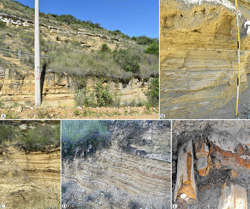
FIGURE 2. Map of the Santa María de Amajac-Sanctórum region at Atotonilco El Grande Municipality, Hidalgo, Mexico, and the lithostratigraphic section of the locality IGM-loc 3875 (Sanctórum-JAO); the yellow areas show the lacustrine deposits of the Paleolake Amajac (modified from Arellano-Gil et al., 2005, figure 2).
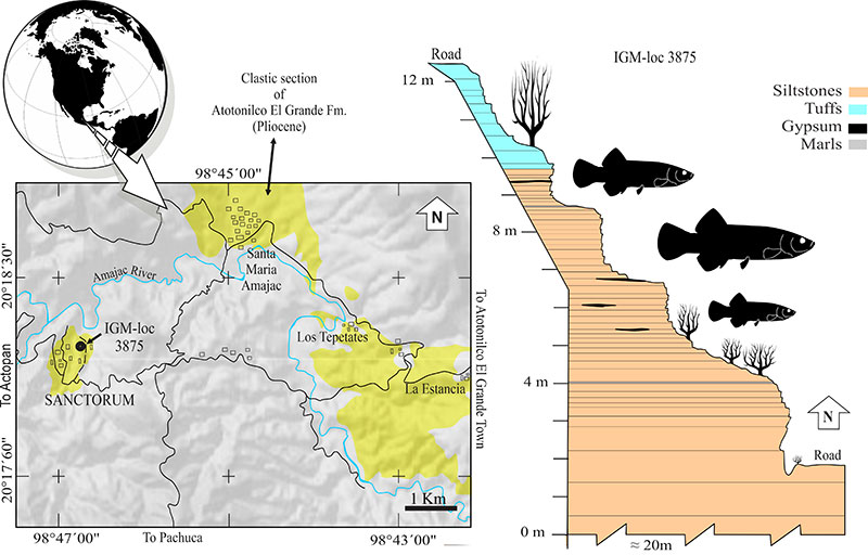
FIGURE 3. IGM 13117, holotype of Paleocharacodon guzmanae gen. and sp. nov., a pregnant female specimen from the Pliocene lacustrine sediments of the Paleolake Amajac, Sanctórum, Atotonilco El Grande Municipality, Hidalgo, central Mexico. A) Part of the specimen observed in white light. B) Part of the specimen observed under UV light. C) Counterpart of the specimen observed under UV light.
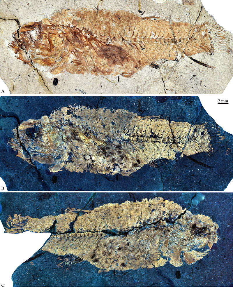
FIGURE 4. Paratypes of Paleocharacodon guzmanae gen. and sp. nov., from the lacustrine sediments of the Paleolake Amajac, in Sanctórum, Atotonilco El Grande Municipality, Hidalgo, central Mexico, observed under UV light. A) Part of IGM 13120, a male specimen. B) Part of IGM 13121, a male specimen. C) Part of IGM 13125, a female specimen.
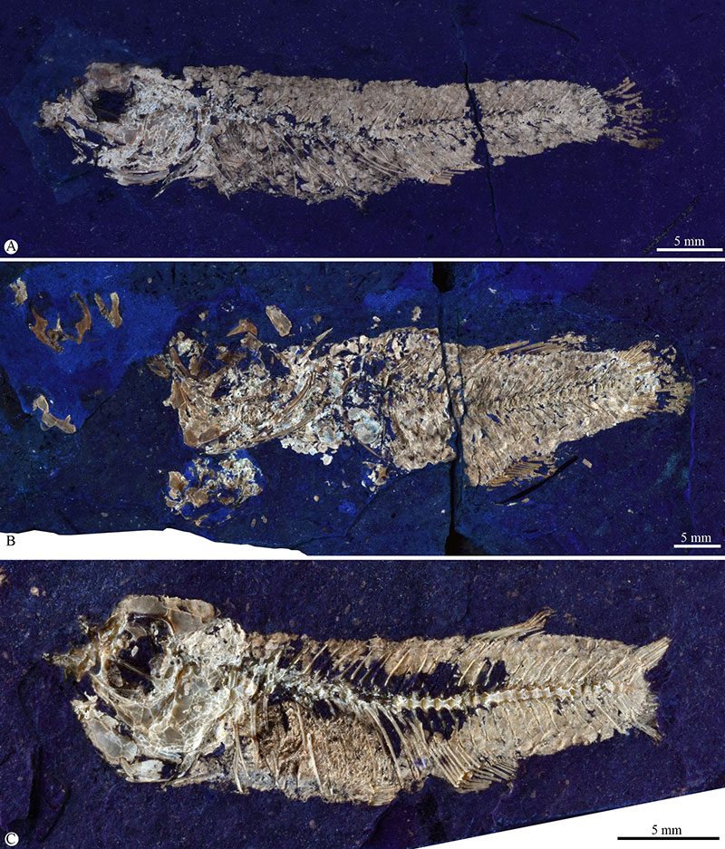
FIGURE 5. Details of IGM 13125 (part), paratype of Paleocharacodon guzmanae gen. and sp. nov., from the lacustrine deposits of the Paleolake Amajac, in Sanctórum, Hidalgo, central Mexico. A) Head observed under white light. B) Unborn fishes preserved in the abdominal cavity, observed under UV light. Abbreviatures: fr, frontal; la, lacrimal; let, lateral ethmoid; psp, parasphenoid (note its anterior end, bifid and with no lateral expansion); black arrows show articulated vertebrae of unborn fishes; red arrows show the pectoral fin of an unborn fish.
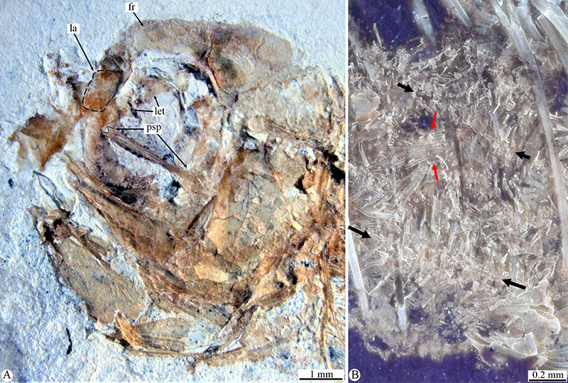
FIGURE 6. μCT imagens of IGM 13127, paratype of Paleocharacodon guzmanae gen. and sp. nov., from the lacustrine sediments of the Paleolake Amajac, in Sanctórum, Atotonilco El Grande Municipality, Hidalgo, central Mexico. A) Left side of the head and anterior part of the trunk. B) Quadrate and bones of both jaws exposed on the right side. C) Close-up of bones involved in the articulation quadrate-lower jaw showing the bilobed head of the quadrate and the articular facet of the anguloarticular in which there is a central participation of the retroarticular (here disarticulate and displaced from its normal position) and two enveloping projections of the anguloarticular. D) Right view of the hyoid arch. The scale only applies to A, B and D. Abbreviations: 1, 2a, 2b, 3, pores of the supraorbital sensory canal; aa, anguloarticular; bh, basihyal; br, brachiostegal ray; bst, bicuspid sharp tooth; cha, ceratohyal anterior; chp, ceratohyal posterior; cl, cleitrum; co, coronomeckelian; id; cor, coracoid; d, dentary; dsp, dermosphenotic; entp, entopterygoid; epi, epioccipital; fr, frontal; hy, hyomandibula; ih, interhyal; iop, infraopercle; let, lateral ethmoid; mx, maxilla; na, nasal; op, opercle; pa, parietal; pcli, postcleitrum inferior; pmx, premaxilla; pop, preopercle; psp, parasphenoid; ptt, posttemporal; q, quadrate; r, rib; rar, retroarticular; scl, supracleitrum; soc, supraoccipital; sp, sphenotic; sop, subopercle; sy, symplectic; uh, urohyal; ust, unicuspid sharp tooth; utt, unicuspid truncated tooth; vhh, ventral hypohyal; dubious elements; within parenthesis: i, internal view (if it is not indicated, the structures show the right view); l, left; r, right; va, ventroanterior view; vp, ventroposterior view.
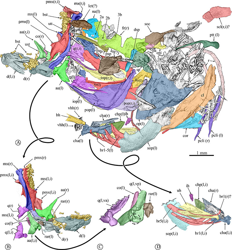
FIGURE 7. Close-up of IGM 13130, a specimen of Paleocharacodon guzmanae gen. and sp. nov. of unknown sex. A) Disarticulated bones of the head under white light. B) Line drawing based on A. Abbreviations: fr, frontal (dorsal surface); let, lateral ethmoid (lateral surface); mx, maxilla (dorsal surface of the proximal end); psp, parasphenoid (dorsal surface of the ocular part); 2b, 3a, 3b, 4a,4b, 5a, 5b, 6, and 7 are the openings of the supraorbital canal preserved in the frontal.
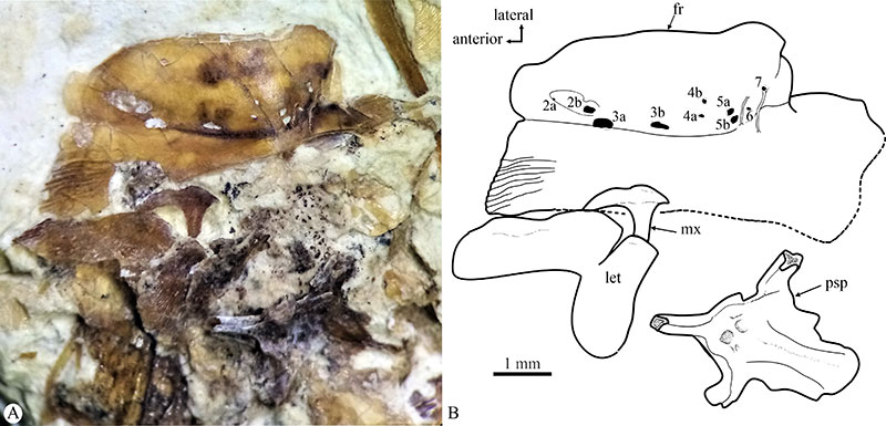
FIGURE 8. Osteological details of Paleocharacodon guzmanae gen. and sp. nov., from the Paleolake Amajac deposits, Sanctórum, Mexico. A) Close-up of the right side of the head of IGM 13118 showing the medial surface of the left otoliths. B) Left anguloarticular bone of IGM 13121 showing the articular facet for the quadrate formed by a surrounding C-shaped process (note this in the shadow below), as well as the central participation of the retroarticular bone that here is empty (see the red line) because this bone is not preserved. C) Right dentary bone and associated teeth of the part of IGM 13121. D) Suspensorium of IGM 13123. E) Dorsal surface of the lateral process of right sphenotic, preserved in IGM 13130. F) Abdominal region of IGM 13130. G) Close-up of pharyngeal teeth preserved in the counterpart of IGM 13121. Abbreviations: aa, anguloarticular; aadp, anguloarticular dorsal process; aapp, anguloarticular anterior process;ast, asteriscus otolith; bst, bicuspid sharp tooth; cor, coracoid; d, dentary; d, dentary; entp, entopterygoid; epn, epineural; faq, articular facet for the quadrate; hy, hyomandibula; lap, lapillus otolith; mx, maxilla; pal, palatine (note the anterolateral projection in the head); pmx, premaxilla; pmx, premaxilla; pop, preopercle; q, quadrate; r, rib; sag, sagittal otolith; sc, scale; sop, subopercle; sp, sphenotic; sy, symplectic; ust, unicuspid sharp tooth. The red arrow in B shows the space in the articular facet for the square (which should be occupied by the retroarticular bone) surrounded by the pair of curved processes of the anguloarticular that also forms part of such facet (see also the shadow of the process)
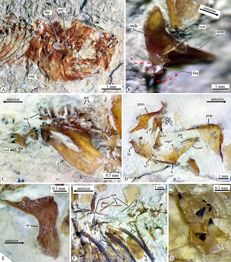
FIGURE 9. Left sagittal otolith of Paleocharacodon guzmanae gen. and sp. nov., IGM 13119, a male specimen from the Pliocene Paleolake Amajac deposits in Sanctórum, Hidalgo, Mexico, observed through SEM. Abbreviations: AH, antirostrum height; AL, antirostrum length; DL, dorsal length; ea, exisura angle; maximum height; ML+RL, total length; ML, medial length; pa, posterior angle; pva, posteroventral angle; RH, rostrum heigh; RL, rostrum length; VL, ventral Length (See Table 3).
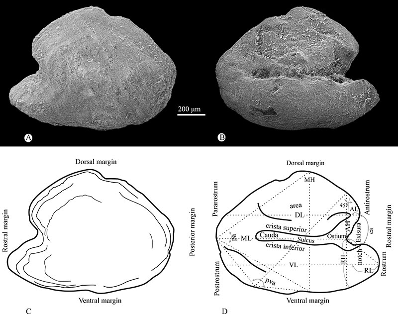
FIGURE 10. Bones of the jaws preserved in IGM 13121, a female specimen of Paleocharacodon guzmanae gen. and sp. nov., from the Paleolake Amajac deposits, Sanctórum, Mexico. A) closeup of the anterior part of the head under white light. B) Line drawing based on A. Abbreviations: aa, anguloarticular; bst, sharp bicuspid tooth; co, coronomeckelian; d, dentary; mx, maxilla; pl, palatine; pmx, premaxilla; sop, subopercle; ust, unicuspid sharp tooth. In parenthesis i, internal view; l, left; r, right.
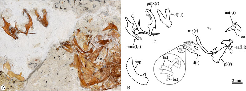
FIGURE 11. Jaw teeth of Paleocharacodon guzmanae gen. and sp. nov., IGM 13130, a male specimen from the Pliocene Paleolake Amajac deposits in Sanctórum, Hidalgo, Mexico, observed under UV light. A) Short acute bifid tooth from a lateral position of the principal row of premaxilla; B) long acute bifid tooth from the anterior position of the principal row of the dentary; C) villiform tooth from posterior rows on the dentary anterior region.
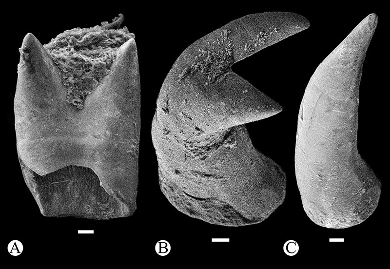
FIGURE 12. Pelvic fin of Paleocharacodon guzmanae gen. and sp. nov., from the Pliocene Paleolake Amajac deposits in Sanctórum, Hidalgo, Mexico. A) Preserved in IGM 13129, observed under UV light. B) Preserved in part of IGM 13121, observed under white light. Abbreviations: afpvr, articular facet for pelvic fin rays; isp, ischial process; lp, lateral process of pelvic fin bone; mp, medial pelvic; pvb, pelvic bone; pvfr, right pelvic fn ray; r, rib; (l), left; (r), right.
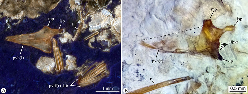
FIGURE 13. Impaired fins of Paleocharacodon guzmanae gen. and sp. nov., from the Pliocene Amajac Paleolake deposits in Sanctórum, Hidalgo, Mexico, observed under UV light. A) Dorsal fin of IGM 13129 (sex undefined; the distal ends branched and segmented of the dorsal rays are covered). B) Anal fin of IGM 13121 (male with anal rays 2 and 13 only preserved as impressions; since the bone of tips of these rays is not-preserved, the arrows show the anterior short rays forming the andropodium and the posterior rays forming a rounded fin). Abbreviations: afr, anal fin ray; amp, anal medial pterygiophore; app, anal pterygiophore; dfr, dorsal fin ray; dmp, dorsal medial pterygiophore; dpp, dorsal proximal pterygiophore.
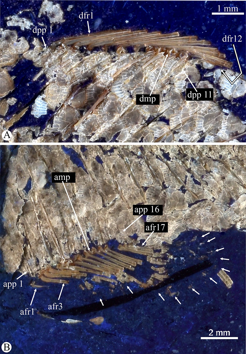
FIGURE 14. Caudal skeleton of Paleocharacodon guzmanae gen. and sp. nov., IGM 13119, from the Pliocene Paleolake Amajac deposits in Sanctórum, Hidalgo, Mexico. A) Specimen observed under UV light. B) Idealized line drawing of A. Abbreviations: cc, caudal complex (= preural centrum 1+ ural 1 and 2); ep, epural; hspu, hemal spine of preural centrum; ihf, interhypural foramen; ksp, keel-shaped process; nspu, neural spine of preural centrum; ph, parhypural; pu, preural centrum; sc, scale; ssp, spine-shaped process; ur, urostyle.
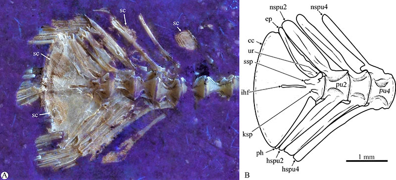
FIGURE 15. Phylogenetic hypotheses of the family Goodeidae. The numbers indicate the distribution of osteological features. Underlined numbers are synapomorphies; not underlined numbers are homoplasies; bold characters are those shared by Paleocharacodon guzmanae gen. and sp. nov.; italic characters are those which P. guzmanae shows an alternative condition or state; characters in normal font are those unknown in this new species.
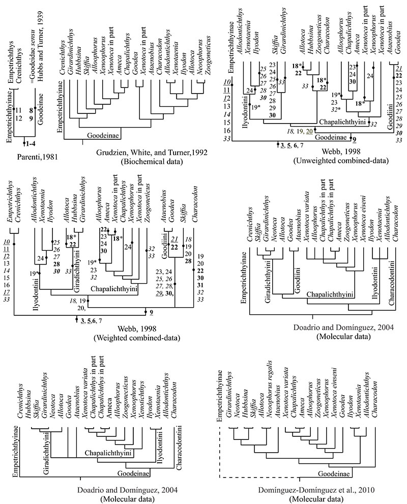
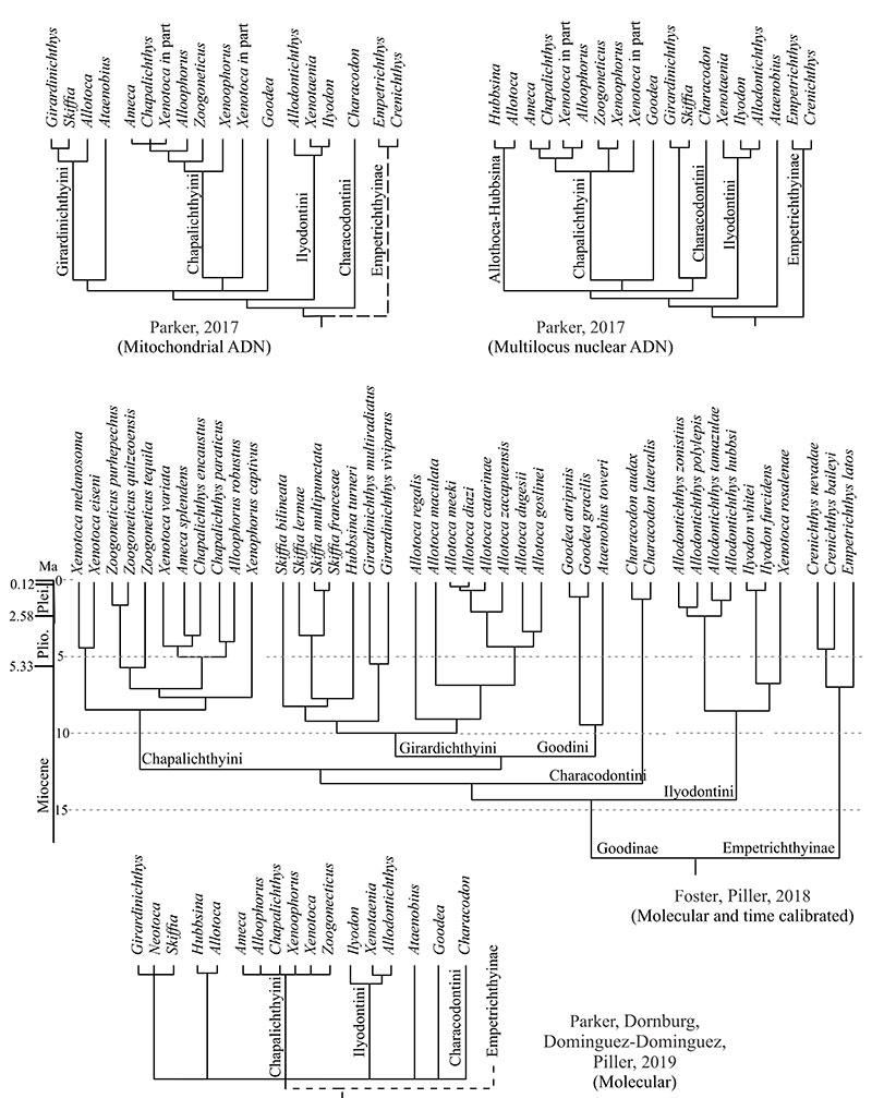
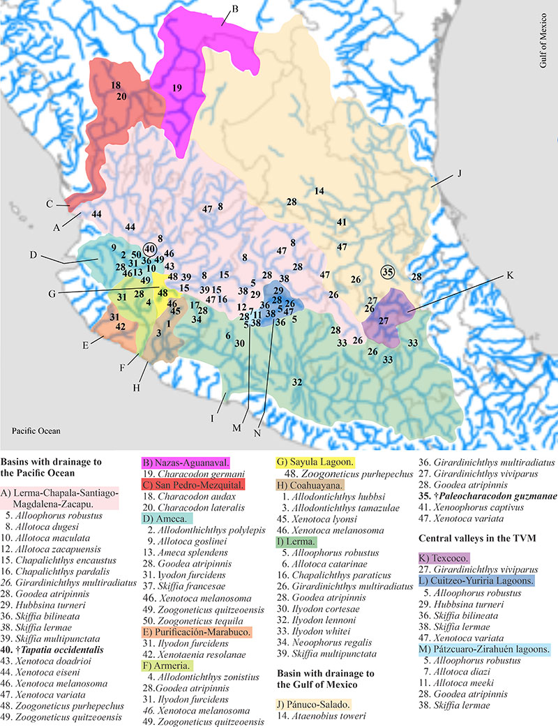
FIGURE 16. General distribution of recent and fossil Goodeinae species in the hydrological basins of Mexico (based on Domínguez-Domínguez et al., 2006, 2010)