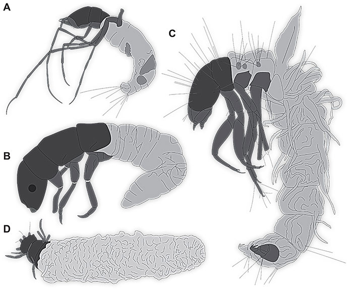
FIGURE 1. Amber piece PED 1383 with assemblage of different fossils. A, Overview. B-D. Caddisfly larva morphotype 1, Lepidostomatidae. B, Anterior region of specimen 1. C, Ventral view of specimen 2. D, Another specimen (Caddisfly larva morphotype 1, Lepidostomatidae; specimen 2 from amber piece PED 1383) not seen in overview from this direction. E, F, Non-biting midge larva (Diptera: Chironomidae) sitting on the case of a caddisfly specimen 3. E, Overview. F. Colour-marked version of F. Images obtained with digital microscopy, white transmitted light.

FIGURE 2. Caddisfly larva morphotype 1, Lepidostomatidae; specimen 2 from amber piece PED 1383, volume renders of µCT-scan A, Habitus, antero-ventral view. B, Colour-marked version of A. C, Head in frontal view. D, Thorax and head in dorsal view. E, Head in ventral view with mouthparts. F, Head in lateral view. Abbreviations: ad = abdomen; an = antenna; hc = head capsule; la= labium; lb = labrum; md = mandible; mp = maxillary palps; pt = prothorax; t3 = trunk appendage 3.
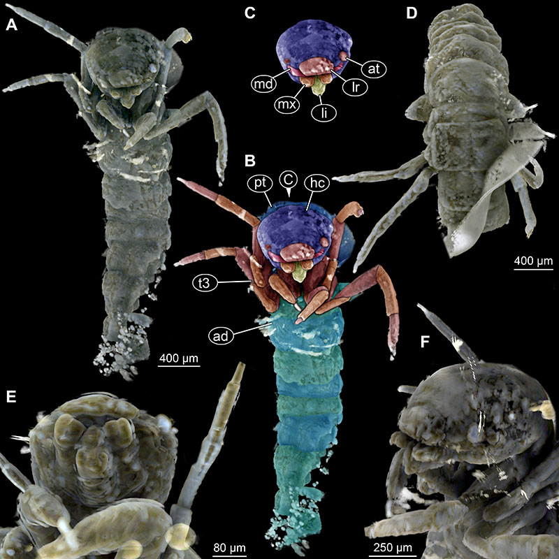
FIGURE 3. Caddisfly larva morphotype 1, Lepidostomatidae; specimen 1 from amber piece PED 1383. Colourmarked version of Figure 1B. Abbreviations: at = antenna; frc = frontoclypeus; lr = labrum; md = mandible.. Small brown dots on the labrum are marking labral setae bases. Images obtained with digital microscopy, white transmitted light.
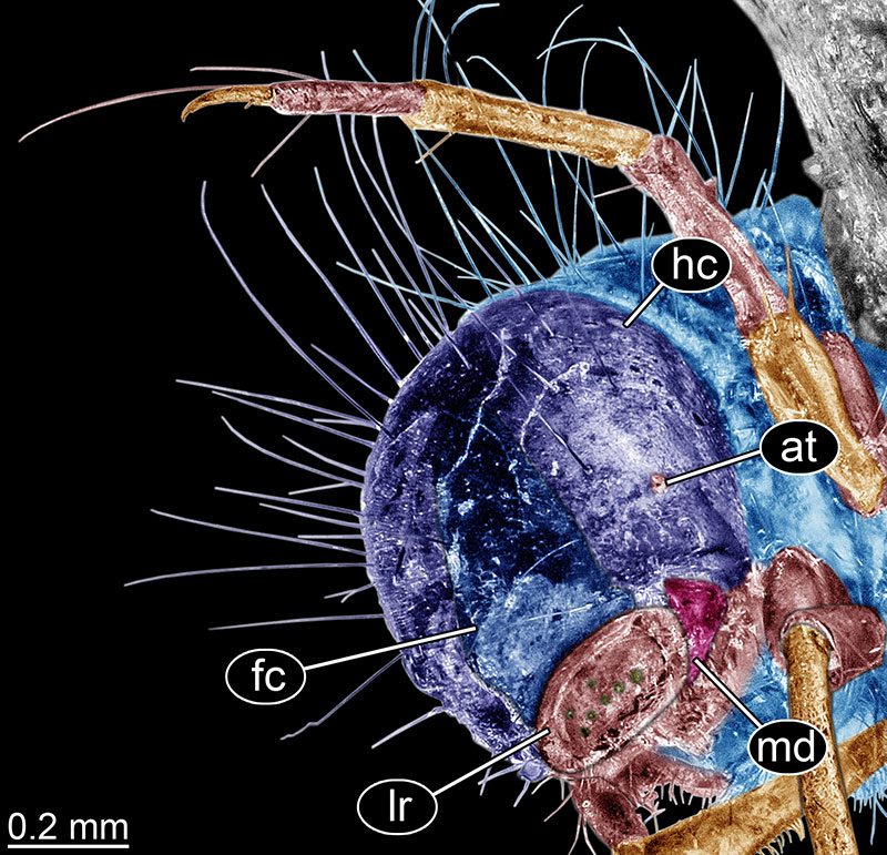
FIGURE 4. Caddisfly larva morphotype 1, Lepidostomatidae; specimen 3 from amber piece PED 1383. A-D. Volume renders of µCT-scan. A, Head in lateral view. B, Frontal view of the larva in the case. C, Dorsal view, “roof” of the case digitally removed. D, Frontal view slightly different angle than in B. E–G, Surface reconstruction of µCT-scan, case removed. E, Dorsal view. F, Lateral view. G, Antero-lateral view. Abbreviations: lh = lateral hump of abdomen unit 1.
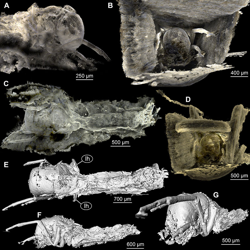
FIGURE 5. Additional caddisfly larvae in the Baltic amber. A, B. Collection Gröhn L7698, Leptoceridae. A, Overview. B, Close-up on head in oblique lateral view. C, D, Representatives of Integripalpia (probably related to Leptoceridae). E, Representative of Annulipalpia. F. PED 1635, Leptoceridae. C-F, image courtesy of Jonas Damzen, used with permission
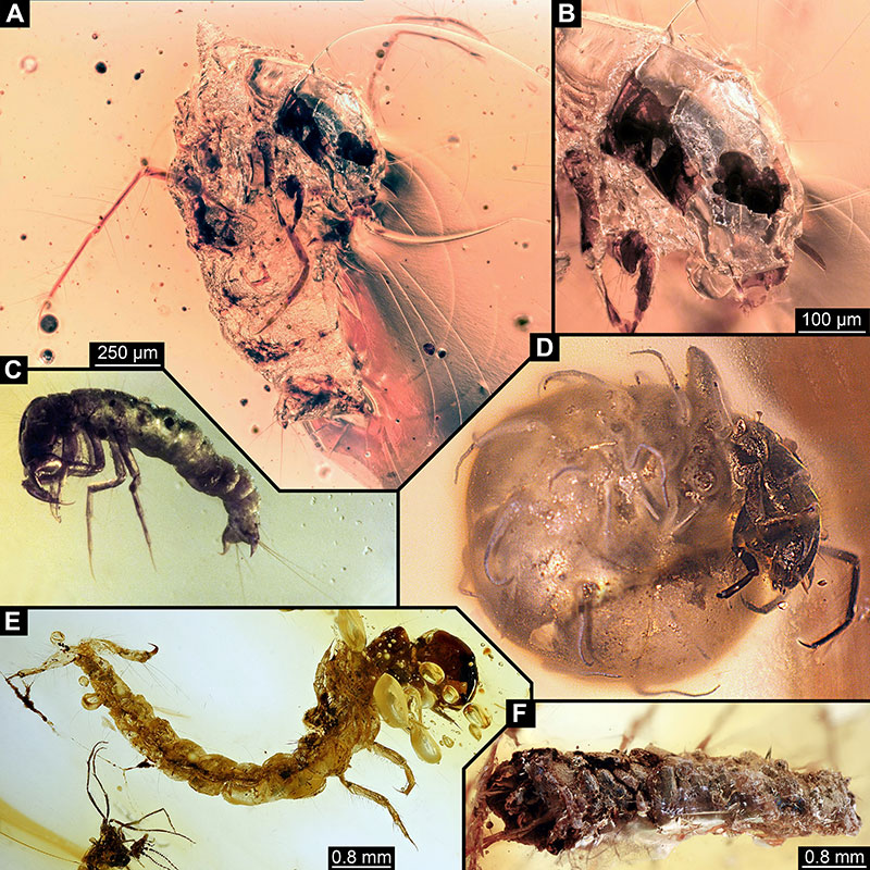
FIGURE 6. Caddisfly larva morphotype 3, Leptoceridae, PED 1635, volume renders of µCT-scans. A, Lateral view. B, Lateral view, other side. C, Fronto-lateral view, case partially removed. D, Frontal view. E, Colour-marked version of D. F, labeled head. Abbreviations: at = antenna; md = possible mandibles.
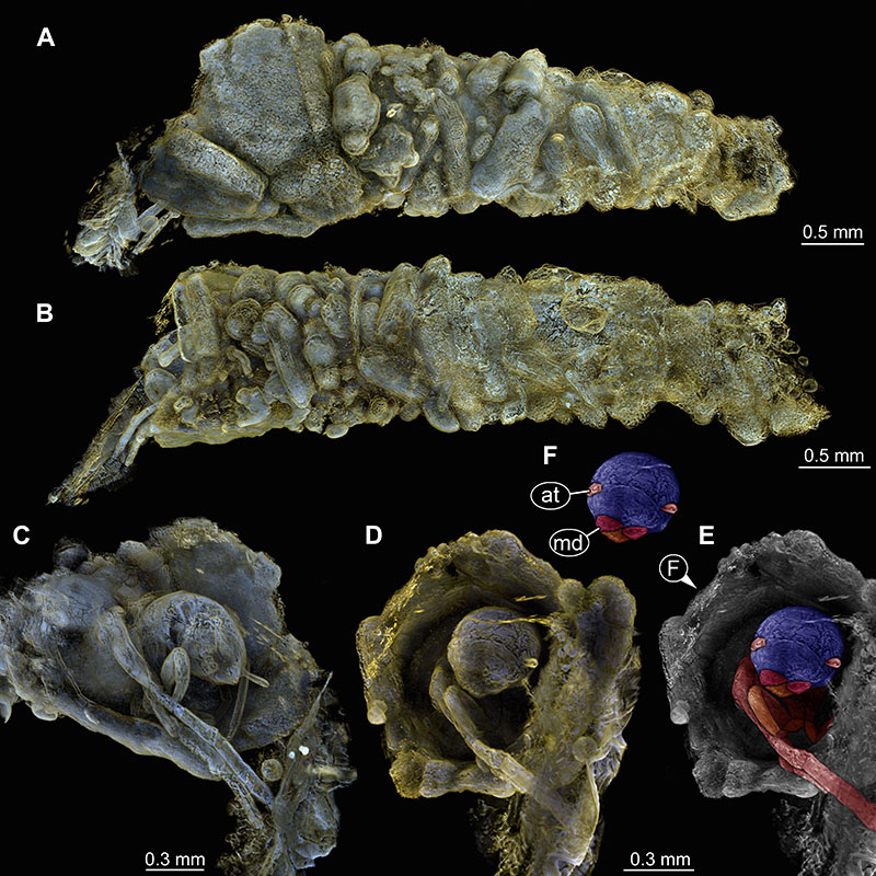
FIGURE 7. Details of non-biting midge larva (Diptera: Chironomidae: Chironominae) in the amber piece PED 1383. A, Head in dorsal view B, Colour-marked version of A. C, Close-up on labrum region. D, Colour-marked version of C. A, B recorded with digital microscope with overhead light source, C, D recorded with digital microscopy, white transmitted light.. Abbreviations: at = antennae; hc = head capsule; lo Lauterborn organ; lr = labrum; sI-II = labral setae I-II.
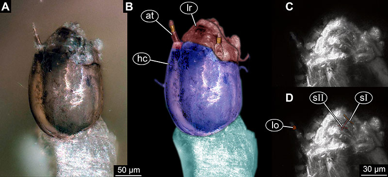
FIGURE 8. Details of non-biting midge larva (Diptera: Chironomidae: Chironominae) in the amber piece PED 1383; volume renders of µCT-scan. A, Habitus, dorsal view. B, Habitus, dorso-posterior view. C-G, Anterior body. C, In ventral view. D, Colour-marked version of C. E, Lateral view. F. Colour-marked version of D. G, Head capsule in ventral view. H, Colour-marked version of G. Abbreviations: at = antennae; lr = labrum; me = mentum; mep = mental plates, hc = head capsule.
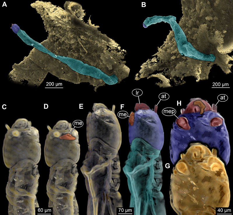
FIGURE 9. Caddisfly larvae preserved in Baltic amber from the literature, all simplified. A, Leptoceridae representative larva (Wichard et al., 2009 their fig 09.04a, b). B, Collection Hoffeins 144.1, probably Ecnomidae (Wichard et al., 2009, their fig 09.05a, b). C, Collection Gröhn 3230, Phryganidae (likely Hagenella) (Wichard et al., 2009, their fig 09.06). D, First caddisfly larva preserved with its case (Gröhn, 2015, his fig 7665).