FIGURE 1. Simplified drawings of larvae of Haliplus and Brychius, stage 3 larvae or larvae of unknown stage, based on various sources. A-N. Haliplus. A. H. confinis, Böving and Craighead (1931). B. Peterson (1957). C-I. Stage 3 larvae. C-F. Vondel (2012). C. H. halsei. D. H. pilbaraensis. E. H. fortescueensis. F. Haliplus pinderi. G-H. Vondel (2004). G. H. testudo. H. H. timmsi. I. H. variomaculatus, Vondel (2011b). J. Spangler (1991). K-N. Stage 3 larvae. K. H. laminatus, Vondel (1986). L. H. varius, Vondel (1996). M. H. subseriatus, Vondel (2001). N. H. kamiyai, Watanabe and Yamasaki (2020). O. Haliplus variegatus, stage 3 larva, Vondel (1997). P. Brychius elevatus, Bertrand (1933).
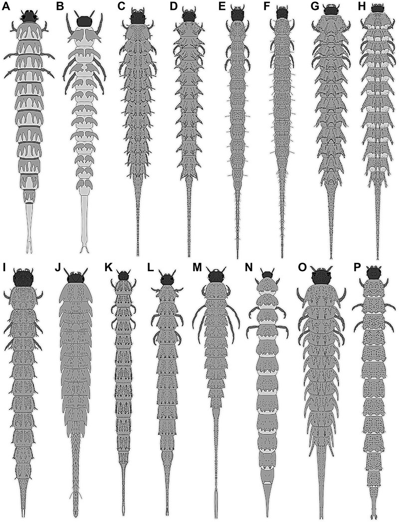
FIGURE 2. Simplified drawings of larvae of Haliplus, partially representing ontogenetic sequences, based on various sources. A-D. Vondel (2011a). A, B. H. abbreviatus or H. maculatus. A. Stage 1 larva. B. Stage 2 larva. C, D. Stage 3 larvae. C. H. maculatus. D. H. abbreviatus. E-G. H. lineatocollis, Bertrand (1933). E. Stage 1 larva. F. Stage 2 larva. G. Stage 3 larva. H-J. H. apicalis. H, I. Vondel (1997). H. Stage 1 larva. I. Stage 2 larva. J. Stage 3 larva, Vondel (1995). K, L. Stage 1 larvae. K. H. indistinctus, Michat et al. (2020). L. H. halsei, Vondel (2012). M. H. fulvus, Klausnitzer (1978).
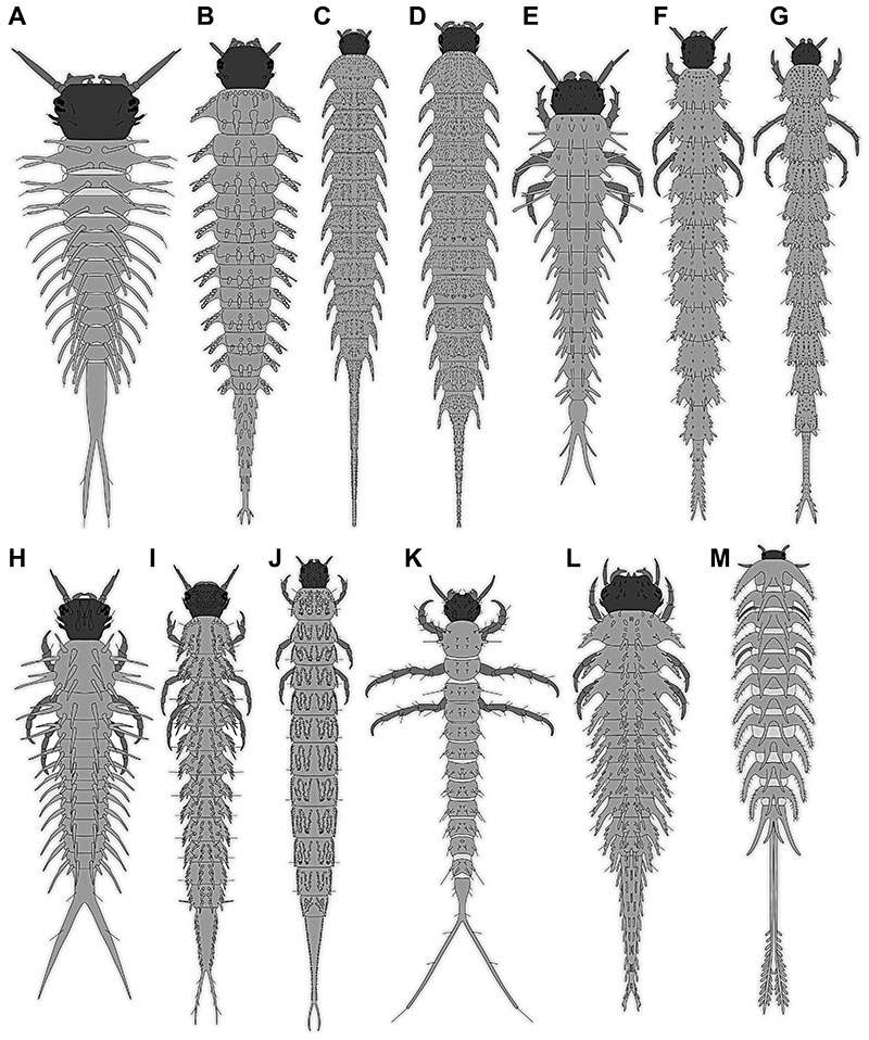
FIGURE 3. Simplified drawings of larvae of Peltodytes, based on various sources. A. P. caesus, stage 3 larva, Vondel (2011b). B. Spangler (1991).
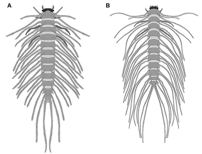
FIGURE 4. Fossil larvae BUB 4436, specimens 1 and 6. A-D. Specimen 1. A. View on one side. B. Colour-marked version of A. C. View on other side. D. Very terminal end, with different contrasting to highlight the setae. E-G. Specimen 6. E. Overview. F. Colour-marked version of E. G. Close-up on very terminal end. Abbreviations: ad = abdomen; at = antenna; hc = head capsule; ms = mesothorax; mt = metathorax; pt = prothorax; te = trunk end.
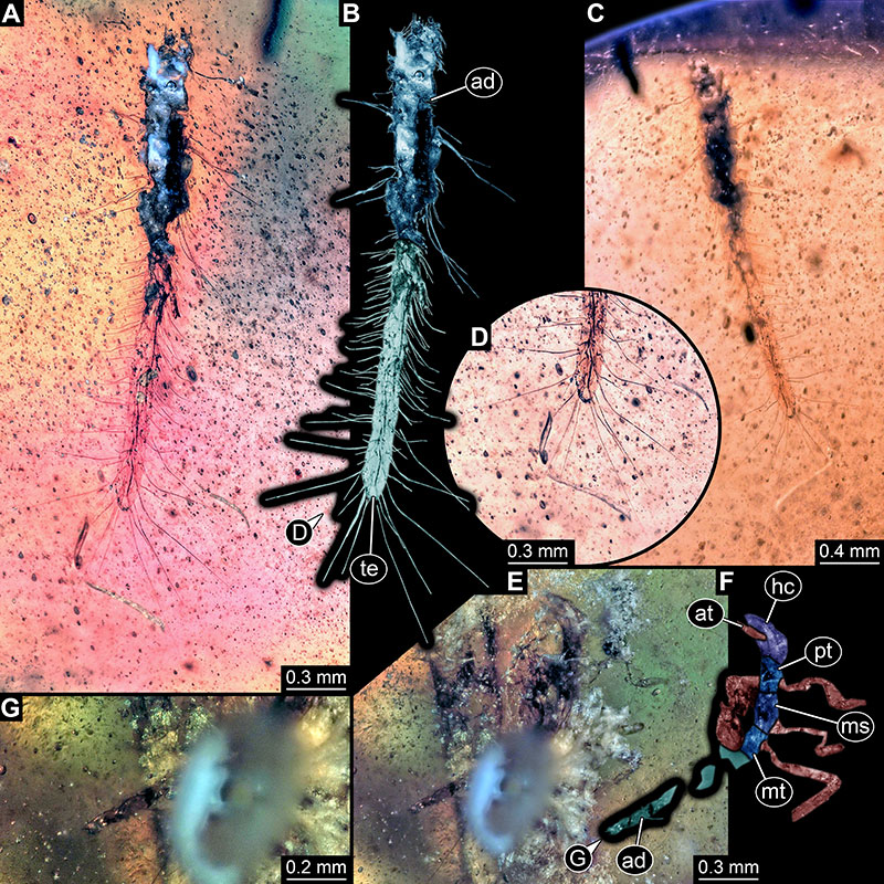
FIGURE 5. Fossil larva BUB 4436, specimen 2. A. Overview on lateral side. B. Colour-marked version of A. C. Other side. D. Close-up on trunk end. E. Close-up on thorax. Abbreviations: ad = abdomen; at = antenna; hc = head capsule; ms = mesothorax; mt = metathorax; pt = prothorax.
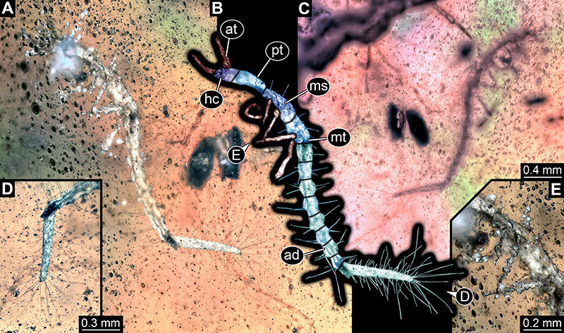
FIGURE 6. Fossil larva BUB 4436, specimen 3. A. Overview dorsal side. B. Colour-marked version of B. C. Ventral side. D. Close-up on anterior region in ventral view. E. Close-up on head. F. Close-up on very terminal end. Abbreviations: ad = abdomen; at = antenna; hc = head capsule; ms = mesothorax; mt = metathorax; pt = prothorax; te = trunk end.
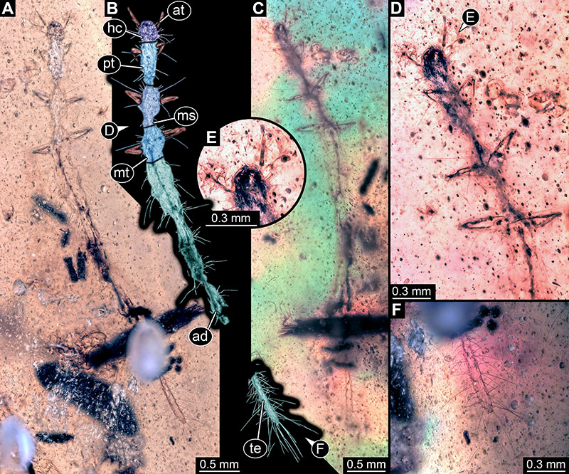
FIGURE 7. Fossil larva BUB 4436, specimen 4. A. Overview dorsal side. B. Colour-marked version of B. C. Ventral side. D. Close-up on trunk end. E. Close-up on head. Abbreviations: ad = abdomen; at = antenna; hc = head capsule; pt = prothorax; te = trunk end.
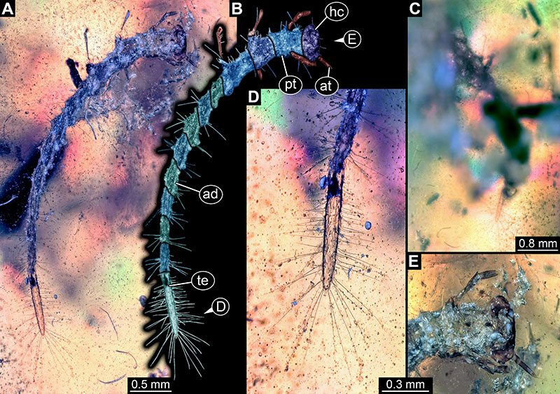
FIGURE 8. Fossil larva BUB 4436, specimen 5. A. Overview ventral side. B. Colour-marked version of B. C. Dorsal side. D. Close-up on head. E. Close-up on leg. F. Terminal end in different contrasting to highlight setae. Abbreviations: ad = abdomen; at = antenna; hc = head capsule; th = thorax.
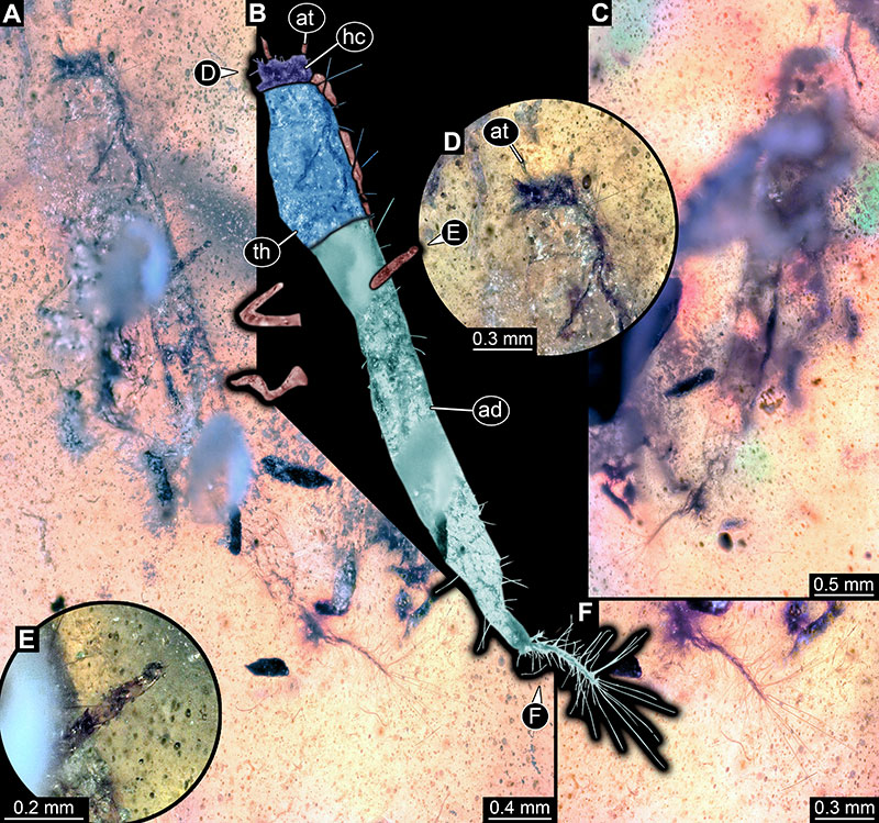
FIGURE 9. Fossil larva BUB 4436, specimen 7. A. Overview dorsal side. B. Colour-marked version of B. C. Ventral side. D. Close-up on thorax; arrows point to claws. E. Close-up trunk end. F. Close-up on head. Abbreviations: ad = abdomen; at = antenna; fe = femur; hc = head capsule; ms = mesothorax; mt = metathorax; pt = prothorax.
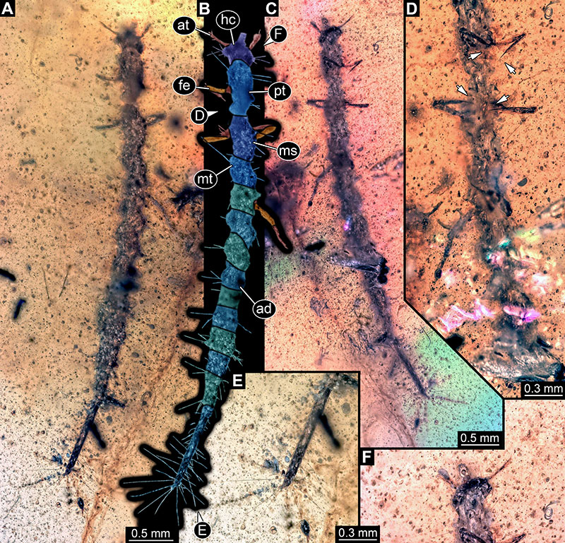
FIGURE 10. Fossil larva BUB 4436, specimen 8. A. Overview ventral side. B. Colour-marked version of C. C. Dorsal side. D. Close-up on head. E. Close-up trunk end. F. Close-up on leg. G. Close-up on tergites. Abbreviations: ad = abdomen; at = antenna; hc = head capsule; ms = mesothorax; mt = metathorax; pt = prothorax.
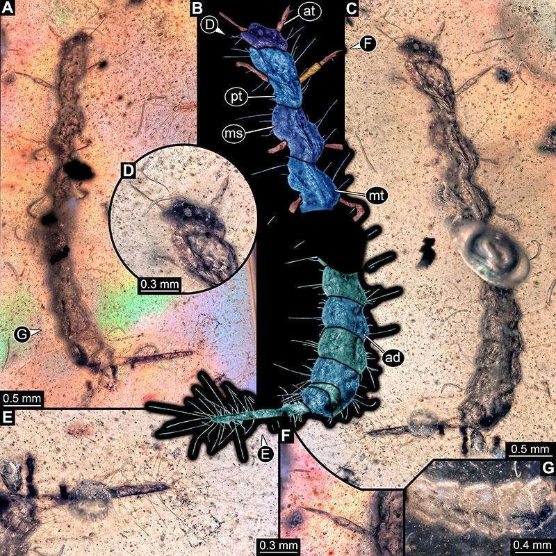
FIGURE 11. Fossil larva BUB 4436, specimen 9. A. Overview dorsal side. B. Colour-marked version of A. C. Ventral side. D. Close-up trunk end. E. Close-up on head. F. Close-up on thorax. Abbreviations: ad = abdomen; at = antenna; fe = femur; hc = head capsule; ms = mesothorax; mt = metathorax; pt = prothorax.
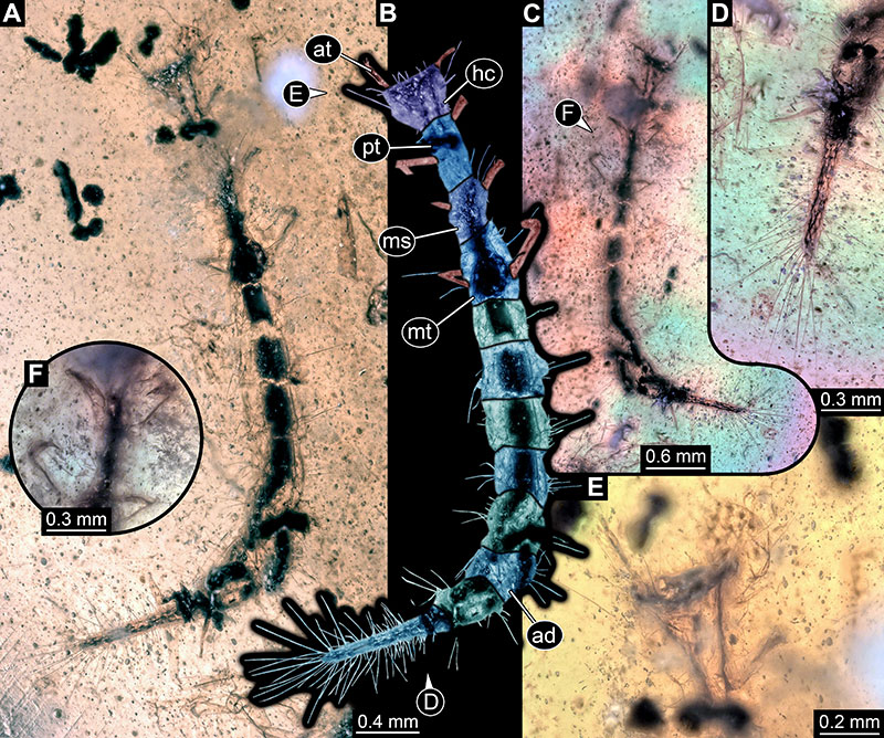
FIGURE 12. Fossil larvae BUB 4436, specimens 10 and 11. A-F. Specimen 10. A. Overview dorsal side. B. Colour-marked version of A. C. Ventral side. D. Close-up on head. E. Close-up trunk end. F. Close-up on leg. G-K. Specimen 11. G. Overview ventral side. H. Colour-marked version of G. I. Dorsal side. J. Close-up on head. K. Close-up on trunk end. Abbreviations: ad = abdomen; at = antenna; hc = head capsule; ms = mesothorax; mt = metathorax; pt = prothorax; th = thorax; ti = tibia.
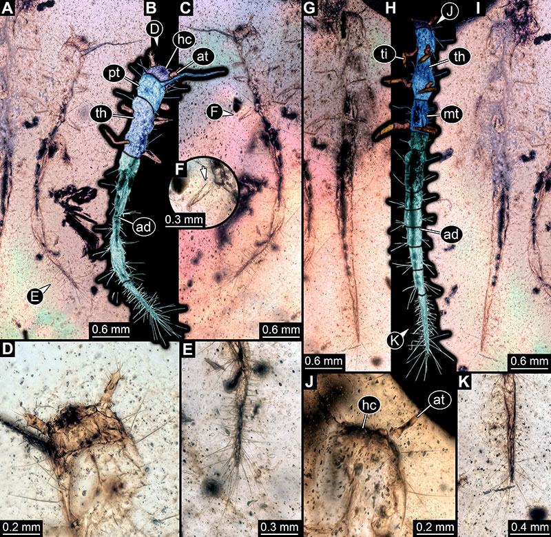
FIGURE 13. Fossil larva BUB 1222. A. Overview ventral side. B. Colour-marked version of A. C. Dorsal side. D. Close-up on head. E. Close-up trunk end. F. Close-up on leg; arrow points to claw. Abbreviations: ad = abdomen; at = antenna; fe = femur; hc = head capsule; ms = mesothorax; mt = metathorax; pl = palp; pt = prothorax.
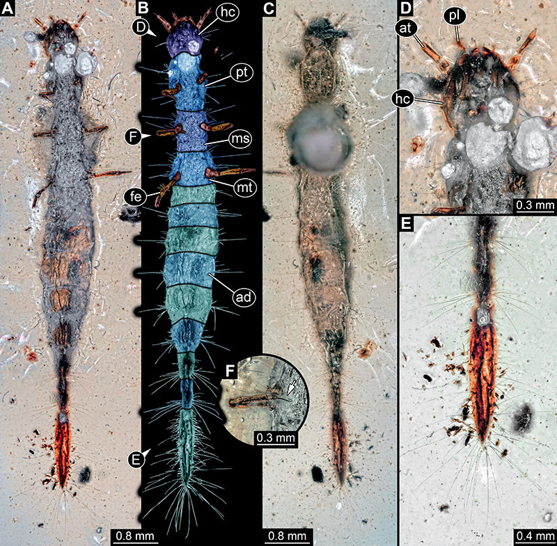
FIGURE 14. Fossil larva PED 1859. A. Overview ventral side. B. Colour-marked version of A. C. Dorsal side. D. Close-up on head. E. Close-up on leg; arrows point to claws. F. Close-up trunk end. Abbreviations: ad = abdomen; at = antenna; hc = head capsule; ms = mesothorax; mt = metathorax; pt = prothorax.
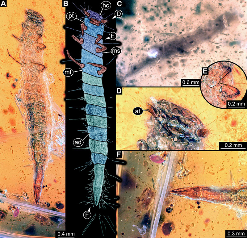
FIGURE 15. Scatter plot of head length over total length of larvae of Haliplidae and the new fossils. The differentiated stages are all representatives of Haliplus. Note the possible three stages of the fossils: a single one on the lower left might represent a stage 1 larva; those clustering in the middle might represent stage 2 larvae, the single fossil far up right might represent a stage 3 larva.
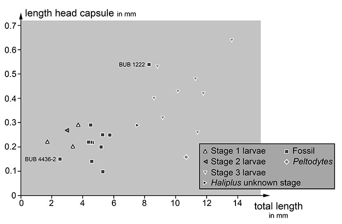
FIGURE 16. Scatter plot of PC2 vs. PC1 of body outlines of larvae of Haliplidae and the new fossils. The differentiated stages are all representatives of Haliplus. Note how tightly together the fossils cluster, indicating a very similar overall shape. This is different for the larvae of the extant forms that show quite some variation, especially over ontogeny.
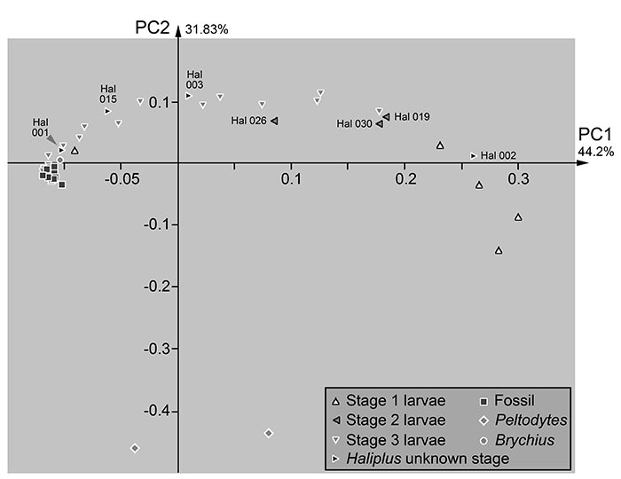
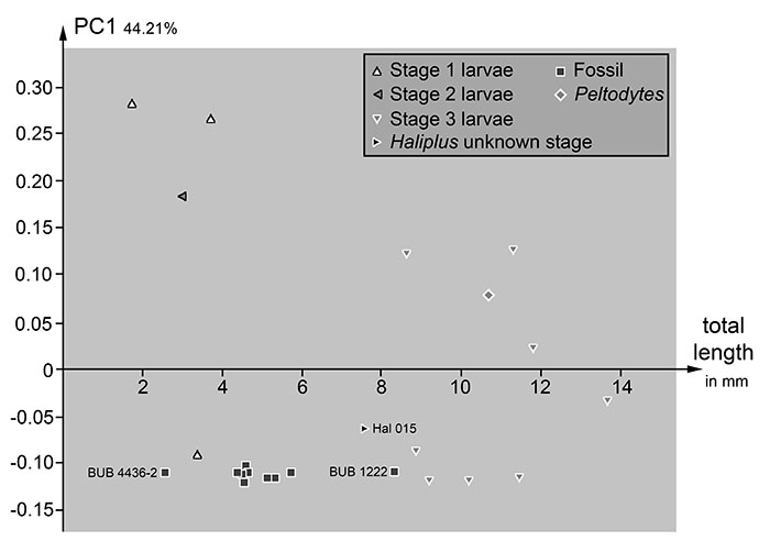
FIGURE 17. Scatter plot of PC1 versus total length of larvae of Haliplidae and the new fossils. The differentiated stages are all representatives of Haliplus. Note that modern day stage 1 and 2 larvae plot away from the smaller fossils, with a single exception. For the fossils, the outline seems to be very similar, independent of the size.