FIGURE 1. A. Reconstruction of “Sphenophyllum colombianum” (MONQ-602) from Huertas (2003). Note the apparent wedge-shaped structures that resemble leaves borne in whorls and having veins that radiate from their attachment point. B, C. Sphenophyllum emarginatum from the Paleozoic Mazon Creek Flora (IL, USA) for comparison (PP-16865, PP-58015). Scale bars equal 1 cm.
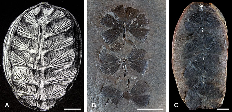
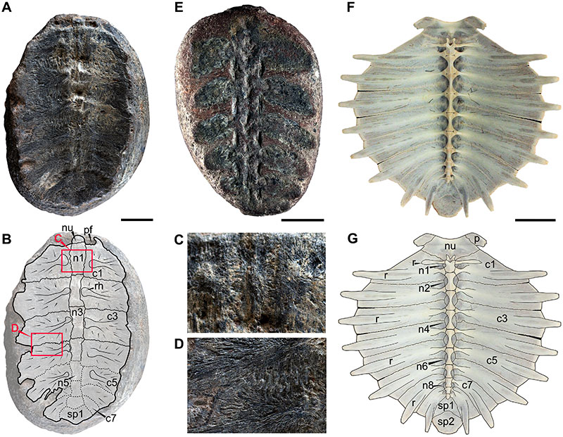
FIGURE 2. Pan-chelonioidea indet. turtles (A, B, C, D, E) and extant turtle carapace in ventral view (F, G). A. Partially preserved carapace in ventral view (MONQ-602). B. Outline of MONQ-602 specimen indicating the preserved bones and texture. C. Close-up of the serrated sutural contact between neural 1 and costals 1, see red rectangle in B for reference. D. Close-up of the bone growth showing radial pattern and sutures between left costal 3 and 4, see red rectangle in B for location. E. Partially preserved carapace in ventral view showing probable scars from the insertion of thoracic vertebrae into the neurals (LLC-65). F. Extant Lepidochelys olivacea showing the complete bone morphology of the carapace in ventral view (QM-J85545). G. Same from F showing outline of diagnostic bones and sutures. Abbreviations: c, costal bone; n, neural bone; nu, nuchal bone; pf, post-nuchal fontanel; p, peripheral bone; r, rib; rh, rib head; sp, suprapygal bone. Scale bars equal 1 cm (A, B, C, D, E), 9 cm (F, G).