FIGURE 1. Three-dimensional model visualizing the sequence of the wear stages (1.1-1.15) for the m1 of Pliospalax macoveii (ACA 858), from the locality of Çalta in Turkey (Şen, 1977). Labial and occlusal view. The scale bars apply to all panels of the figure.
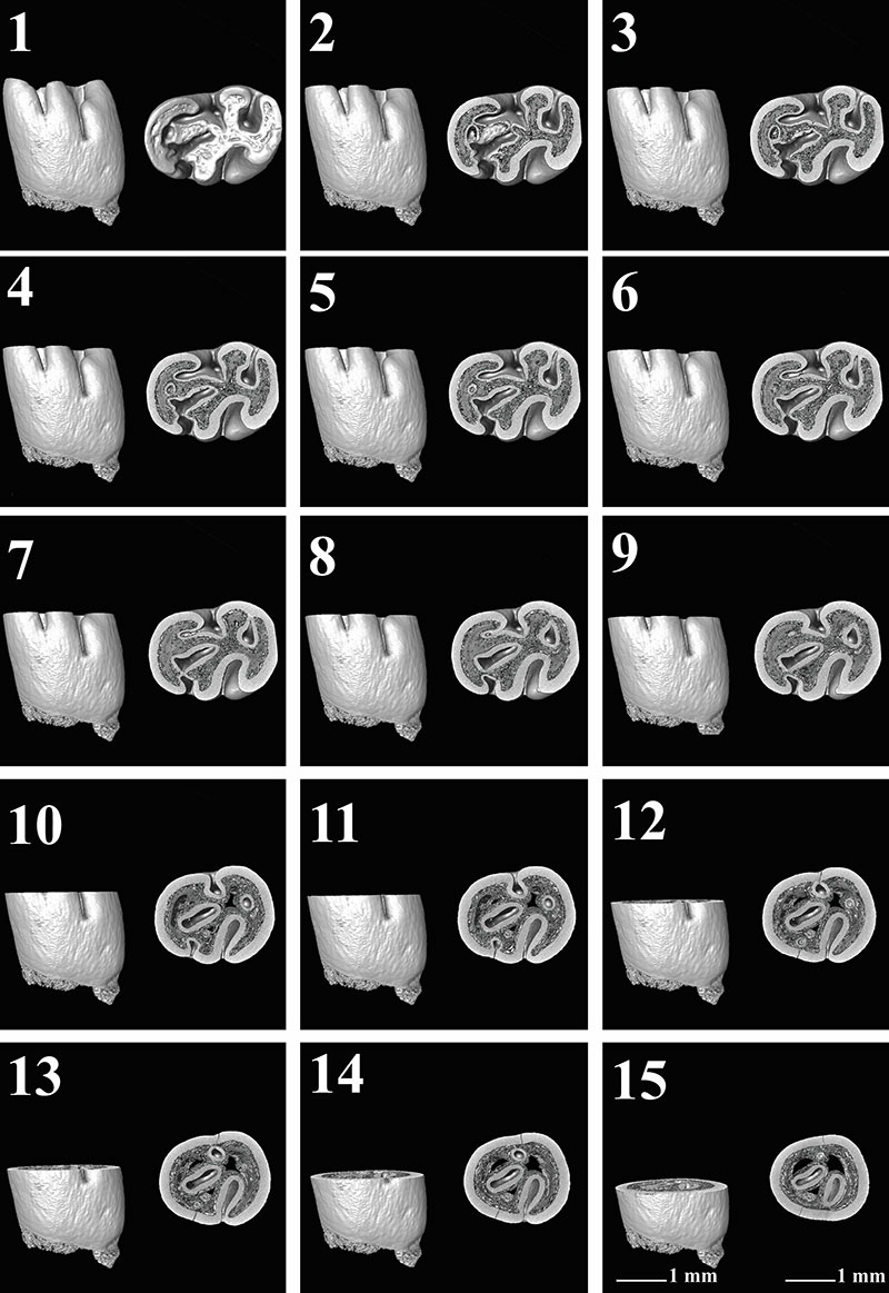
FIGURE 2. Three-dimensional model visualizing the sequence of the wear stages (2.1-2.12) for the m1 of Pliospalax macoveii (ACA 859) from the locality of Çalta in Turkey (Şen, 1977). Labial and occlusal view. Inverted figure of the specimen. The scale bars apply to all panels of the figure.
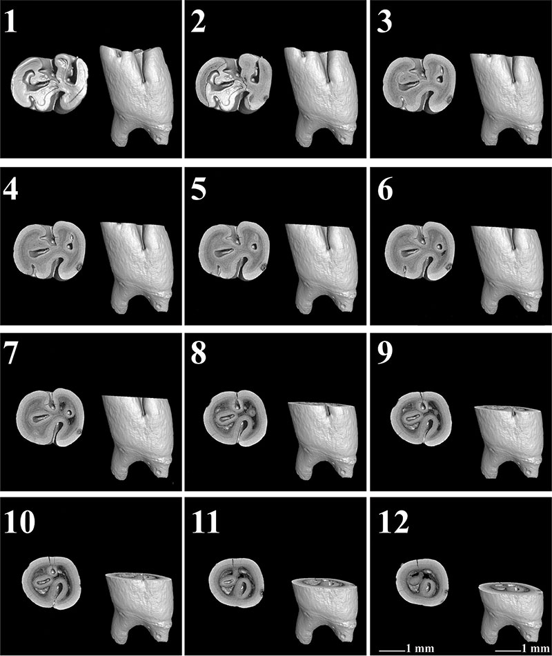
FIGURE 3. Three-dimensional model visualizing the sequence of the wear stages (3.1-3.12) for the right m1 of Pliospalax macoveii (ACA 881) from the locality of Çalta in Turkey (Şen, 1977). Labial and occlusal view. Inverted figure of the specimen. The scale bars apply to all panels of the figure.

FIGURE 4. Three-dimensional model visualizing the sequence of the wear stages (4.1-4.12) for the left m1 of Pliospalax macoveii (ACA 881) from the locality of Çalta in Turkey (Şen, 1977). Labial and occlusal view. The scale bars apply to all panels of the figure.
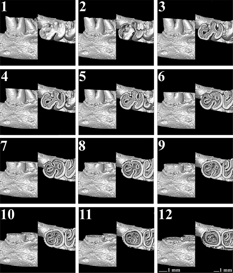
FIGURE 5. Three-dimensional model visualizing the sequence of the wear stages (5.1-5.9) for the holotype (m1) of Pliospalax tourkobouniensis (TB1 481), from the locality of Tourkobounia-1 in Greece (De Bruijn and Van der Meulen, 1975). Labial and occlusal view. The scale bars apply to all panels of the figure.
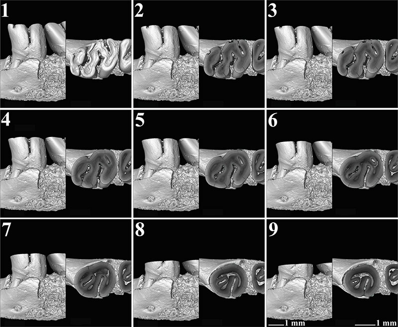
FIGURE 6. Three-dimensional model visualizing the sequence of the wear stages (6.1-6.12) for the m1 of Pliospalax tourkobouniensis (TB1-901), from the locality of Tourkobounia-1 in Greece (De Bruijn and Van der Meulen, 1975). Labial and occlusal view. The scale bars apply to all panels of the figure.
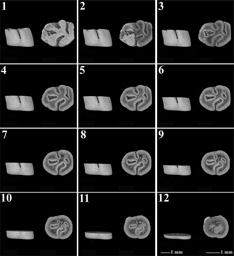
FIGURE 7. Three-dimensional model visualizing the sequence of the wear stages (7.1-7.12) for holotype (m1) of Pliospalax sotirisi (MR 1286) from Maritsa, in Greece (De Bruijn et al., 1970). Inverted figure of the specimen. The scale bars apply to all panels of the figure.
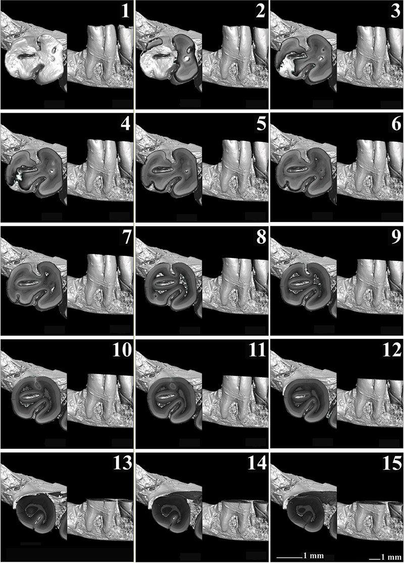
FIGURE 8. Three-dimensional model visualizing the sequence of the wear stages (8.1-8.9) for the right m1 of Spalax microphthalmus (REG 22726) from Romania. Labial and occlusal view. Inverted figure of the specimen. The scale bars apply to all panels of the figure.
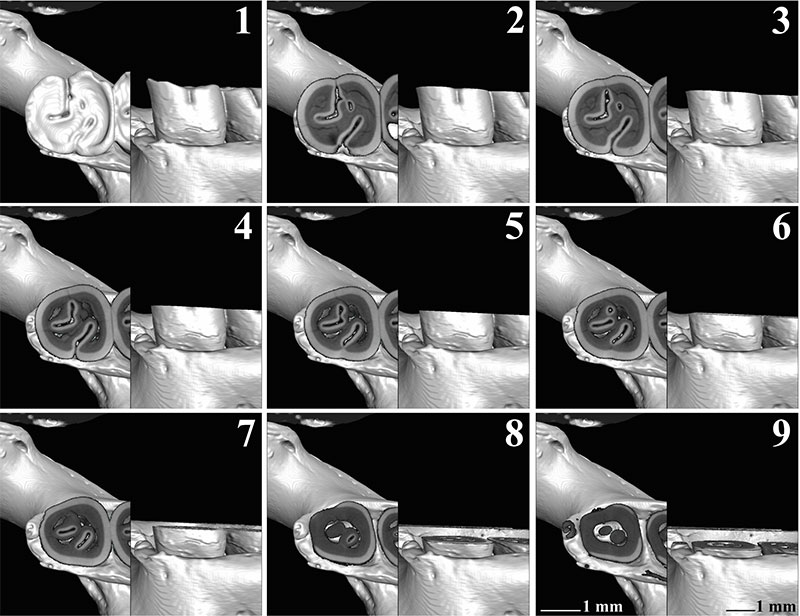
FIGURE 9. Three-dimensional model visualizing the sequence of the wear stages (9.1-9.9) for the left m1 of Spalax microphthalmus (REG 22726) from Romania. Labial and occlusal view. The scale bars apply to all panels of the figure.
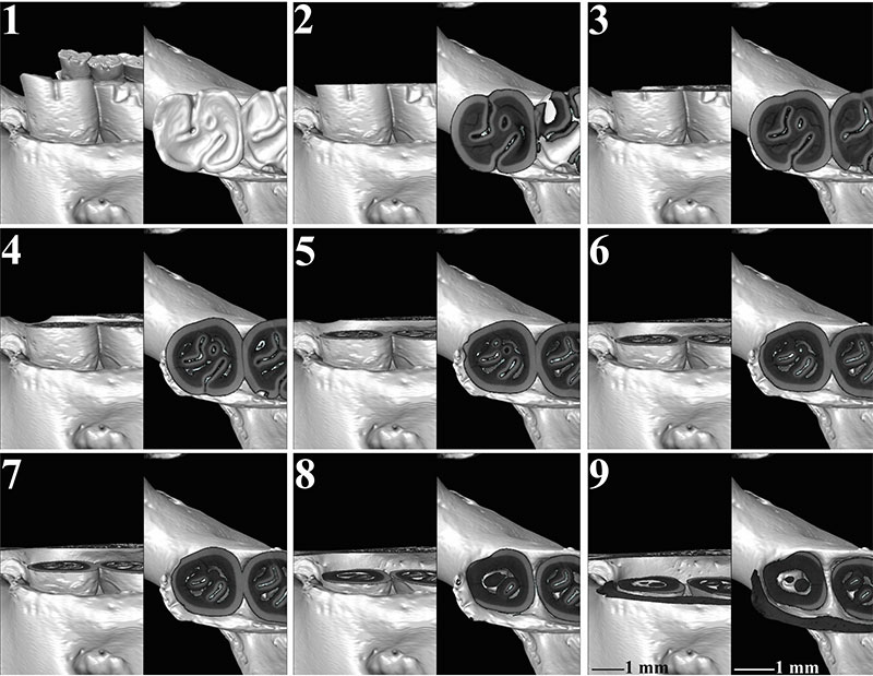
FIGURE 10. Three-dimensional model visualizing the sequence of the wear stages (10.1-10.12) for the right m1 of Spalax microphthalmus (REG 22729) from Hungary. Labial and occlusal view. Inverted figure of the specimen. The scale bars apply to all panels of the figure.
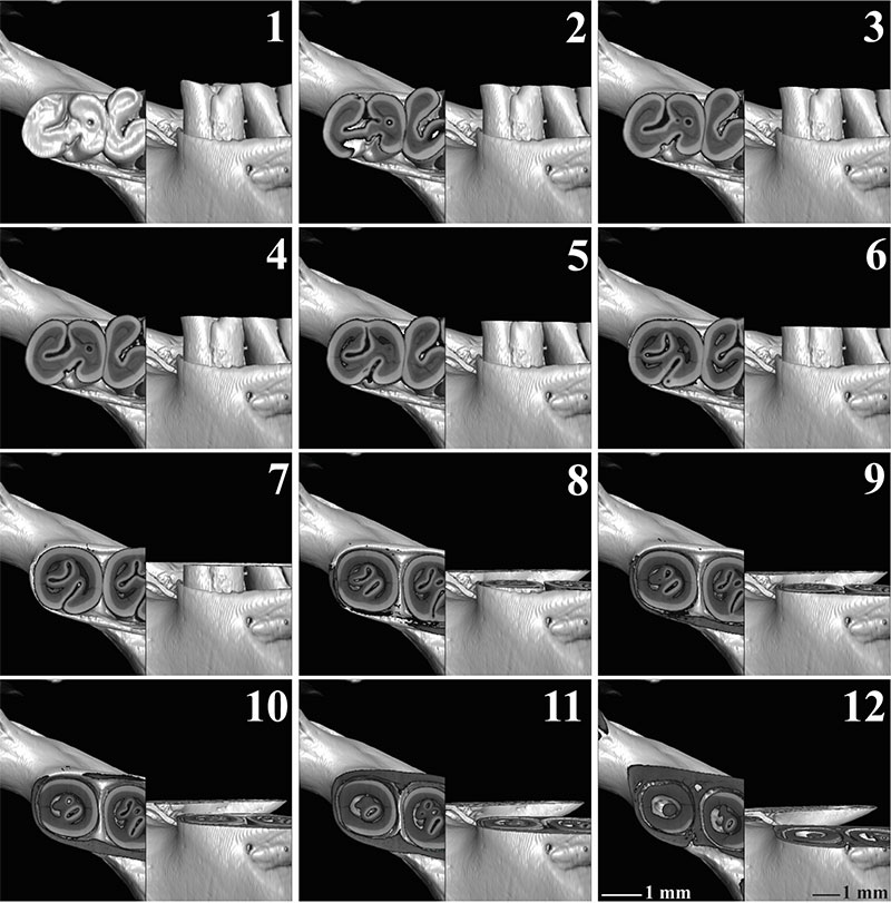
FIGURE 11. Three-dimensional model visualizing the sequence of the wear stages (11.1-11.9) for the left m1 of Spalax microphthalmus (REG 22729) from Hungary. Labial and occlusal view. The scale bars apply to all panels of the figure.
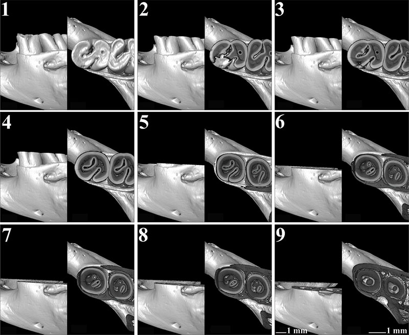
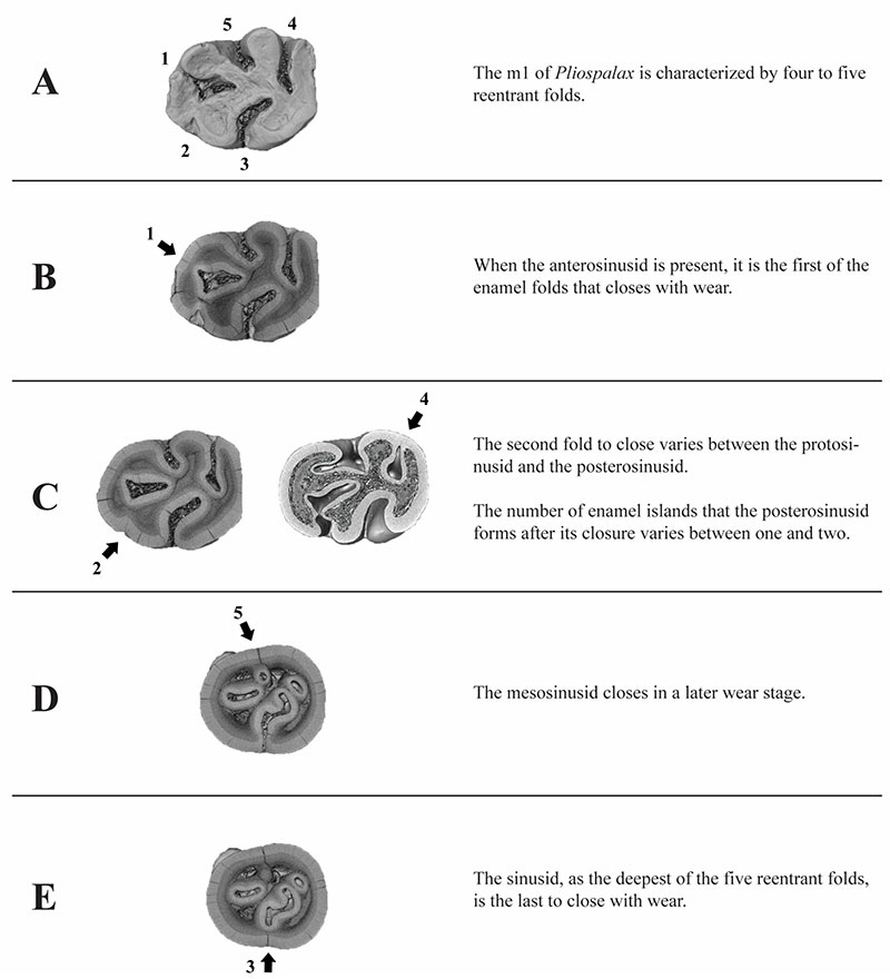
FIGURE 12. Three-dimensional models visualizing the sequence of the wear stages (A-E) for the m1 of Pliospalax. Occlusal view. (1) anterosinusid, (2) protosinusid, (3) sinusid, (4) posterosinusid, (5) mesosinusid.