

Lycopsids are one of the oldest groups of vascular plants. These ancient plants only have a few living relatives still around today, the clubmosses and quillworts. Lycopsids were some of the first plants to diversify on land and their long history stretches all the back to the late Silurian period, about 425 million years ago.
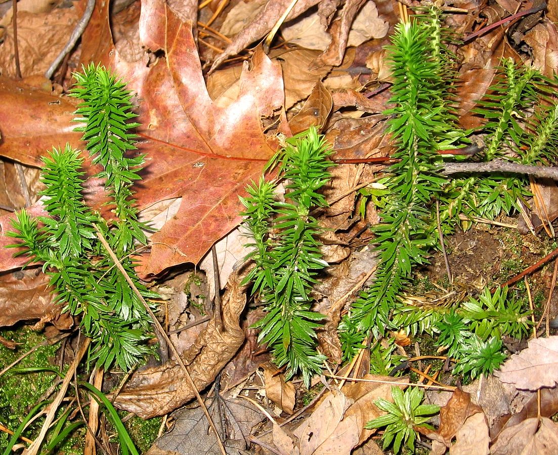 Shining clubmoss, Huperzia lucidula; Wikimedia Commons
Recently, authors Evreïnoff, Meyer-Berthaud, Decombeix, Lebrun, Steemans, and Tafforeau published an article in Palaeo Electronica about a new Late Devonian lycopsid from New South Wales, Australia. When asked about their most recent project, the authors wrote, “when studying how early land plants acquired the features that made them comparable to modern plants, Australia comes to a special place with the discovery of large leafy stems of the lycopsid Baragwanathia as early as the Late Silurian.”
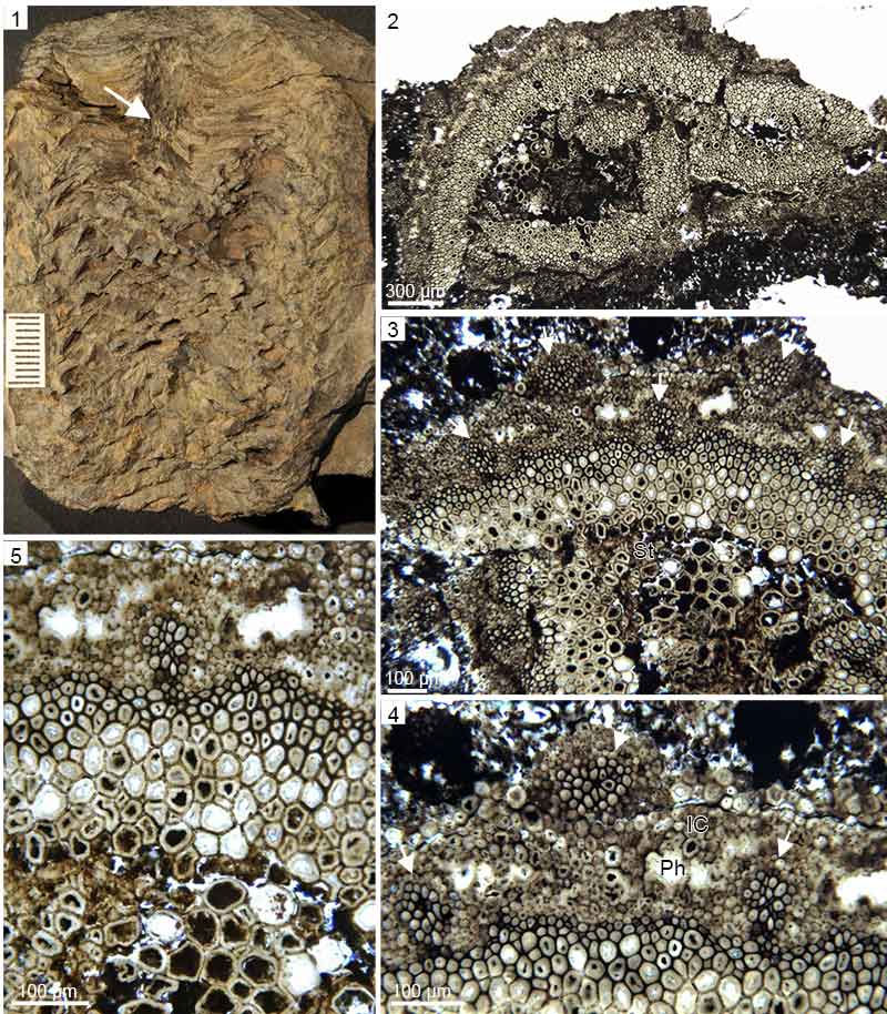 Cymastrobus irvingii gen. et sp. nov. NMVP 161998. 1, General view of the cone; cone axis at arrow. 2, Distal part of cone axis in transverse section. 3, Detail showing the stele (St) and five sporophyll traces at arrows. 4, Detail showing the wavy outline of the xylem ring, presumed location of the phloem (Ph), inner cortical cells (IC) and three sporophyll traces at arrows. 5, Detail showing the emission of a sporophyll trace from a groove of the primary xylem ring.l traces at arrows. 5, Detail showing the emission of a sporophyll trace from a groove of the primary xylem ring.
Not only is Australia home to the oldest known lycopsid, its also produces fossils of these early plants in abundance.
“The lycopsids are significant components of the earliest vegetation of Australia during the Paleozoic, and the study of these unique plants contributes to a better understanding about the patterns of colonization of vegetation on the continents.”
While lycopsid fossils remain abundant in the latest part of the Devonian of Australia, these specimens consist mainly of stems and branches of Leptophloeum, a genus that is found around the world. This could indicate that lycopsids are poorly diversified in Australia during the Devonian; however, a new lycopsid fossil from New South Wales is challenging that notion.
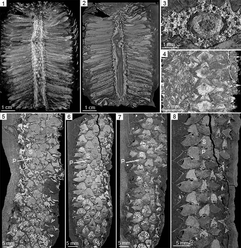 Cymastrobus irvingii gen. et sp. nov. NMVP 161998. Virtual sections, X-Ray synchrotron microtomography. 1, Cone in tangential section. 2, Cone in radial section; note the proximal position of the megasporangia. 3, Cone axis in transverse section. 4, Outer portion of the cone in tangential section showing four sporophyll-sporangium units in longitudinal row. 5-8, Inwards to outwards series of tangential sections through the cone showing the progressive changes in size of the sporophyll pedicels (P) and the sporangia (S).
In this recent PE paper, Evreïnoff and colleagues report a new genus of lycopsid represented by a cone that is structurally intact from a locality in New South Wales that is about 360 million years old. Not only is this a new lycopsid genus for Australia, the specimen also happens to be a cone, a complex reproductive organ, that’s been preserved for over 350 million years.
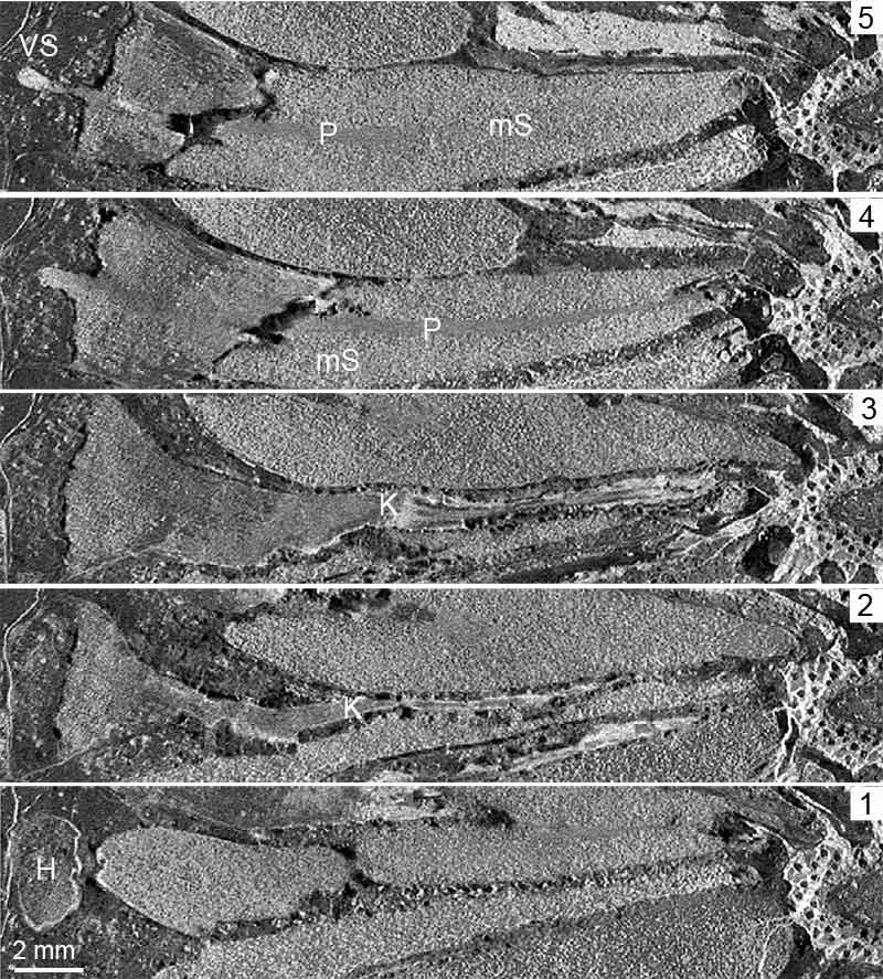 Cymastrobus irvingii gen. et sp. nov. NMVP 161998. Virtual sections, X-Ray synchrotron microtomography. 1-5, Proximal-distal series of longitudinal sections through a sporophyll-sporangium unit; note the heel (H) in 1, keel (K) in 2 and 3, microsporangium (mS) and longitudinal pad of tissue (P) in 4 and 5, vascular strand (VS) in 5.
How do you understand and visualize the structure of a 3-D fossil? The authors shared with PE that, “the 3-D structure of this complex reproductive organ and of its components has been reconstructed from virtual sections obtained from a non-destructive method, the X-ray synchrotron microtomography.”
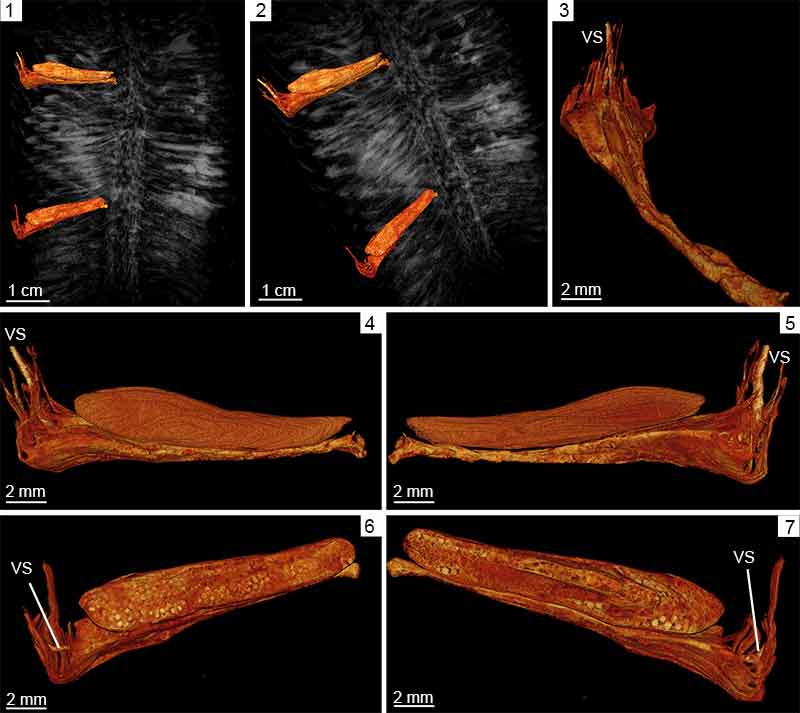 Cymastrobus irvingii gen. et sp. nov. NMVP 161998. Virtual reconstructions and volume rendering visualization of chosen anatomical units within the X-Ray synchrotron microtomography scan. 1-2, General view of the cone showing two reconstructed sporophyll-sporangium units, the proximal one producing megaspores, the distal one microspores. 3, Reconstructed sporophyll showing the enlarging pedicel and dissected lamina. 4-5, Two reconstructed sporophyll-microsporangium units in profile view. 6-7, Two reconstructed sporophyll-megasporangium units in profile view. VS: vascular strand.
This fossil is significant for many reasons, including an increase in known lycopsid diversity in Australia during the Devonian. The authors also point out, “this cone differs from most of the Late Devonian cones described to date. It represents one of the oldest representatives of a group that thrived during the Carboniferous and includes the extant genus Isoetes.”
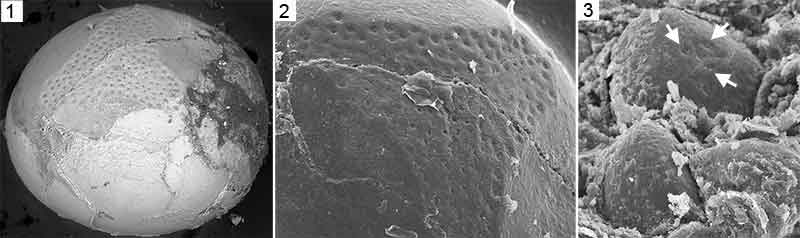 Cymastrobus irvingii gen. et sp. nov. NMVP 161998. 1, Cast of a megaspore central body showing numerous small circular pores arranged in several rows around the trilete mark. 2, Detail of previous view. 3, Casts of microspore central bodies; the largest one shows three pores between the rays of the trilete mark (arrows).
It is incredible to think how one single fossil can change what we know about paleontology and enhance our knowledge about how plants first adapted to land. While individual fossils play a major role in scientific investigations, the bigger picture is always in the back of a paleontologists’ mind. For the authors of the recent Australian lycopsids article, “this work is part of a larger project studying the diversity dynamics of the early vascular plants and the factors that may have contributed to the waxing and waning of the continental vegetation during a time interval of the Earth history that includes massive extinction events in the marine realm.”
Thank you to the authors for contributing to PE and the PE blog. To learn more about the new lycopsid fossil from Australia, check out the original article here.
