

Article Search
Volume 27.1
January–April 2024
Full table of contents
ISSN: 1094-8074, web version;
1935-3952, print version
Recent Research Articles
See all articles in 27.1 January-April 2024
See all articles in 26.3 September-December 2023
See all articles in 26.2 May-August 2023
See all articles in 26.1 January-April 2023
Glossary of CT terms
Beam Hardening Phenomenon referring to the effect of selective x-ray attenuation and scatter from polychromatic X-ray beams. An x-ray beam is composed of individual photons with a range of energies. As the beam passes through an object, it becomes "harder" (mean energy increases) because the lower-energy photons are selectively absorbed (or scattered) more rapidly, leaving behind only the high-energy photons. Both cupping and streak artefacts can occur as a result.
Cupping artifact
A ring of excessively bright voxels around the edge of the fossil reducing to excessively dark voxels at the centre – caused by beam hardening
CT numbers
Term coined by Godfrey Hounsfield to describe the voxel grey values
Electron volt (eV)
is a unit of energy equal to the amount of kinetic energy gained by a single unbound electron when it accelerates through an electric potential difference of one volt.
LUT plot
Image look up table, a bivariate plot of grey scale voxel values (CT numbers) versus the number of voxels
Noise
Unusually bright and dark voxels are superimposed over the CT data, which gives a CT scan a grainy or speckled appearance, leading to blurring of material boundaries.
Partial volume
Artefact resulting when numerous linear attenuation coefficients (i.e., material density) different within a single voxel being represented by an averaged grey value.
Segmentation
The process of defining regions of interest with a scan, which usually represent a fossil or part thereof. The regions of interest can be modelled and rendered independently. Segmentation invariably requires the user to threshold (i.e., define) material boundaries within an object e.g., fossil and matrix (note: the term segmentation refers to a different process in the SPIERS software suite).
Polychromatic
An X-ray beam composed of radiation of more than one photon energy (i.e., wavelength)
Steak artefacts
Dark bands or streaks between dense objects in ta CT slice image.
Threshold
The process of defining material boundaries within a scan. In its simplest form this entails selecting a grey value (threshold) above which any voxels are grouped in a region of interest and those, which fall below, are discarded or made transparent.
Reconstruction
is the process of transforming the set of 2D X-ray projections to a 3D volume.
Region of interest (ROI)
is a selected sub-region of a CT scan that can be identified e.g., a fossil tooth within a matrix or vascular voids with the tooth.
Rendering
the process of generating an image from a model
Surface Determination
defining the surface of an object to produce a hollow 3D model without internal features
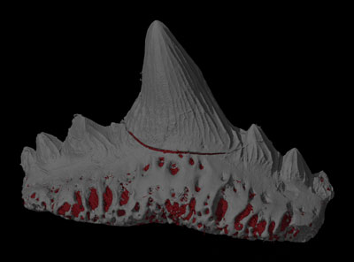
SUPPLEMENTARY FILE 1. Virtual fossil animation, rotating once about the long axis and revealing the vascular structure. The specimen is rendered with lights, colour and perspective.
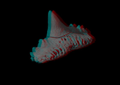
SUPPLEMENTARY FILE 2. Stereo-anaglyph virtual fossil animation, rotating once about the long axis and revealing the vascular structure. The specimen is rendered with lights, colour and perspective. The movie can be viewed in three-dimensions using red-cyan (or red green) spectacles.
SUPPLEMENTARY FILE 3. Stereolithographic (STL) model of the fossil shark tooth. The 3D model can be opened by readers using freeware (see text). and SUPPLEMENTARY FILE 4. Stereolithographic (STL) model of the dental vascular system. The 3D model can be opened by readers using freeware (see text) (zipped file).
TABLE 1. Ten step workflow for virtual fossil preparation.
|
Task |
Step |
Process |
|
Reconstruct |
1 |
Reduce blurring by applying noise reduction (see method) |
|
2 |
Reduce cupping by applying beam hardening correction (see method) |
|
|
3 |
Increase contrast by stretching floating point range of fossil grey values |
|
|
Render |
4 |
Segment fossil from matrix by applying global threshold (Fig.3A) |
|
5 |
Segment fossil form non-adjacent matrix using region grower tool (Fig. 3B) |
|
|
6 |
Remove adjacent matrix with a masking tool (Fig.3C) |
|
|
7 |
Repeat steps 4-7 to segment anatomical features (Fig. 3D) |
|
|
8 |
Apply lighting and false colour to create virtual specimen see (Fig. 4) |
|
|
9 |
Animate rendered 3D model using a keyframer (Fig. 6) |
|
|
Store |
10 |
Archive slice stack as 8 Bit BMP, 16 Bit DICOM or 32 Bit vol files. |
TABLE 2. Data processing software for voxel and surface based 2D and 3D data. The useful tools and applications included in the packages are highlighted.
|
Package |
Source |
Surface Determination |
Magic Wand |
Masking |
Rendering |
Stereo-Anaglyphs |
Movie Animation |
STL Files |
Traditional Morphometrics |
Geometric Morphometrics |
URL |
|
VG Studio Max |
Commercial |
3D |
3D |
2D |
3D |
3D |
3D |
3D |
3D |
3D |
www.volumegraphics.com |
|
SPIERS |
Freeware |
3D |
|
2D |
3D |
3D |
|
|
|
|
www.spiers-software.org/ |
|
DRISHTI |
Freeware |
|
|
|
3D |
3D |
3D |
|
|
|
www.sf.anu.edu.au/Vizlab/drishti/ |
|
ImageJ |
Open Source |
|
2D |
2D |
3D |
|
|
|
3D |
2D |
www.rsb.info.nih.gov/ij/ |
|
Mimics |
Commercial |
3D |
2D |
3D |
3D |
|
|
3D |
|
|
www.materialise.com/mimics |
|
Simpleware |
Commercial |
2D |
2D |
2D |
3D |
|
3D |
3D |
3D |
|
www.simpleware.com |
|
Amira |
Commercial |
3D |
2D |
2D |
3D |
|
3D |
3D |
|
|
www.amira.com/ |
|
Blender |
Open Source |
|
|
|
3D |
|
3D |
|
|
|
www.blender.org |
|
Stratovan Checkpoint |
Commercial |
|
|
|
|
|
|
|
3D |
3D |
www.stratovan.com |
|
Meshlab |
Commercial |
|
|
|
|
|
|
3D |
|
3D |
www.meshlab.sourceforge.net/ |
|
Simpleware |
Commercial |
2D |
2D |
2D |
3D |
|
3D |
3D |
3D |
|
www.simpleware.com |
FIGURE 1. Micro-CT can be used to" virtually" extract fossils. (1.1) Photo of the shark tooth in matrix and (1.2) sectioned micro- CT rendering after virtual preparation from rock matrix.
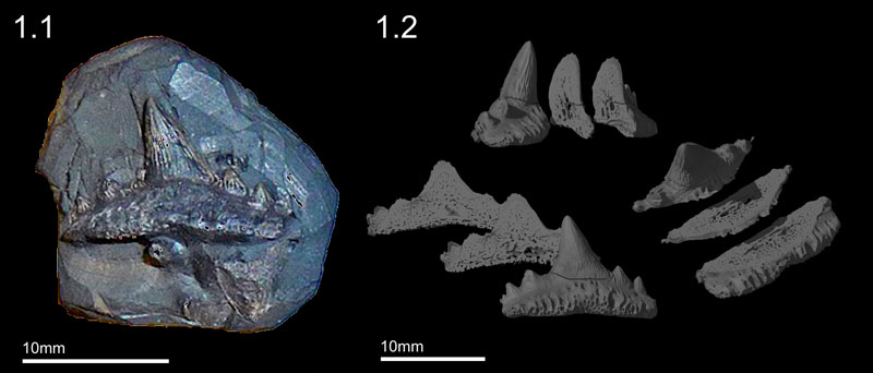
FIGURE 2. A simple example of back projection based on two digital projections at 90o to each other. Corresponding rows of pixels from each projection are used to create an X-ray transmission profile. Profiles are used to create digital images of the row that are then back projected (i.e., smeared) onto one another to create a 3D CT slice. In this example the cylindrical objects appear in the image as cubes, but the shape would be resolved if more projections were analysed. This process is repeated for every slice (pixel row) in the scan.
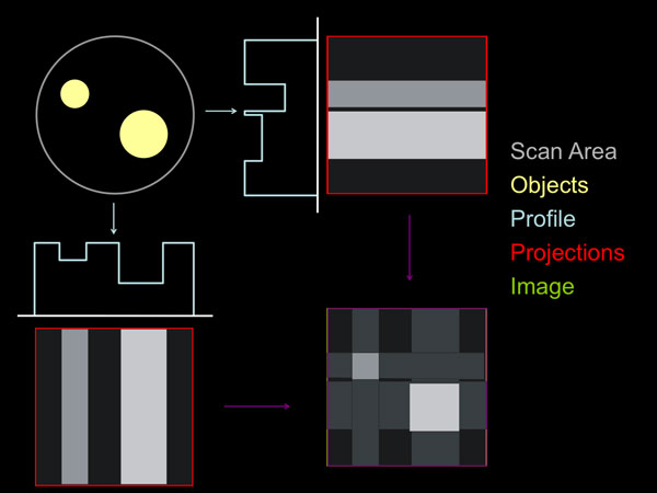
FIGURE 3. Micro-CT grey value frequency distribution plot. The graph reveals three peaks representing air, matrix and fossil. The tooth was calibrated by applying a global threshold at the minima, which separated the fossil peak (vertical red line).
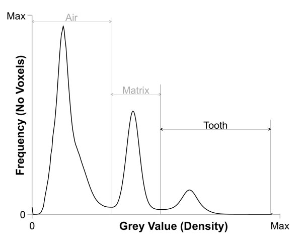
FIGURE 4. Virtual preparation (segmentation) of the fossil was carried out in three steps. (4.1) Surface determination (4.2) ROI growing and (4.3) masking. (4.4) The vascular system was extracted using a combination of masking and region growing.
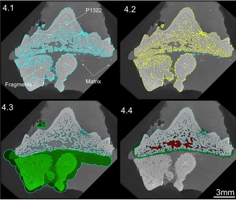
FIGURE 5. Virtually prepared fossil rendered with lights, colour and perspective. (5.1) Fossil and vascular system, (5.2) transparent fossil and vascular system and (5.3) vascular system.
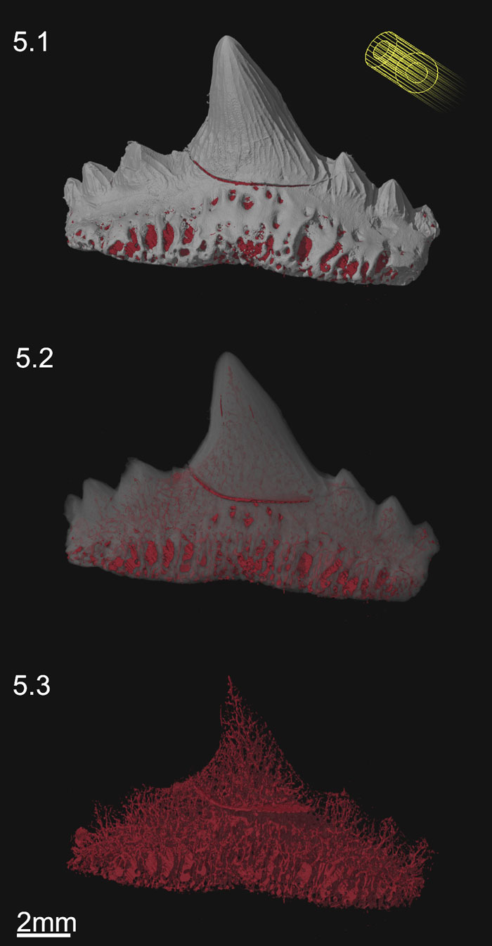
FIGURE 6. Stereo-anaglyph of virtually prepared fossil. Rendered with lights, colour and perspective. The image can be viewed in three-dimensions using red-cyan (or red green) spectacles.
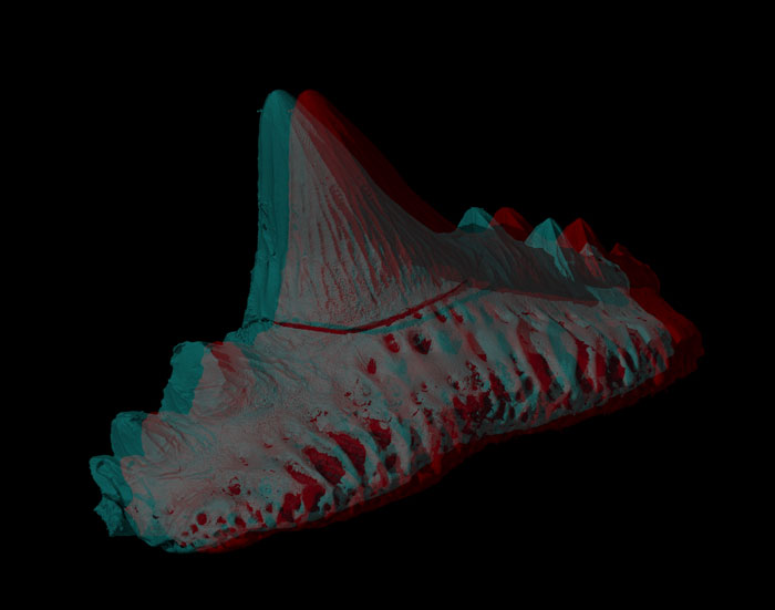
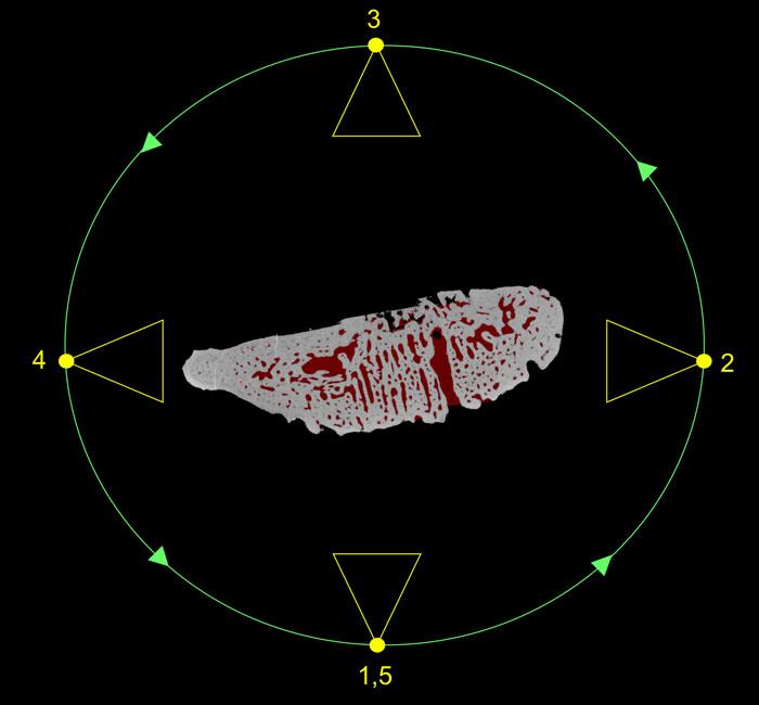
FIGURE 7. Animation keyframer and key frames used to produce supplementary movies 1 and 2. The camera (yellow) follows a circular path (green) around the tooth (shown as a cross section). At each key frame the camera is rotated toward the specimen. At key frames 1 and 2 the tooth is opaque but at 3, 4 and 5 it is transparent. The key frame interpolates these settings as a single camera rotation about the tooth with a transparency developing between key frames 2 to 3. This reveals the underlying vascular structure.
Richard Leslie Abel MSK Laboratory
Department of Surgery and Cancer
Charring Cross Hospital
Imperial College
W6 8RF London
United Kingdom
and
Image and Analysis Centre
Mineralogy Department
Natural History Museum
Cromwell Road
SW7 5BD London
United Kingdom
Richard Abel is a zoologist and micro-tomography specialist. He has collaborated on a wide variety of CT based projects studying: fossils, meteorites, shells, sharks, coral, stone tools, Egyptian mummies, and insect pupae. At present Richie is a lecturer in Musculoskeletal Science at Imperial College (London) researching bone quality and age related disease. Richie is also a research associate at the Natural History Museum (London, UK) and an associate member of Ancient Human Occupation of Great Britain (AHOB3).
![]()
 Carolina Rettondini Laurini
Laboratório de Paleontologia
Departamento de Biologia
FFCLRP - USP
Av. Bandeirantes
3900 - CEP 14040-901
Bairro Monte Alegre
Ribeirão Preto
SP, Brazil
Carolina Rettondini Laurini is researching the taxonomy and systematics of Elasmobranchii fossils from Brazilian basins. Currently a Ph.D. student at the Departamento de Zoologia, Instituto de Biociências of Universidade de São Paulo. Carolina graduated with a BSc in Biology (2008) and an MSc in Comparative Biology (2010). Both from Faculdade de Filosofia, Ciências e Letras de Ribeirão Preto of Universidade de São Paulo.
![]()
 Martha Richter
Department of Palaeontology
Natural History Museum
Cromwell Road
SW7 5BD London
United Kingdom
Dr Martha Richter is a fish palaeontologist whose research focuses mainly on Palaeozoic sharks and actinopterygians from Western Gondwana. She currently manages the Vertebrates, Anthropology and MIcropalaeontology collections at the Natural History Museum in London.
A palaeobiologist's guide to 'virtual' micro-CT preparation
Plain Language Abstract
The aim of this paper is to provide a brief but comprehensive guide to creating, preparing and dissecting a ‘virtual’ fossil, using a worked example to demonstrate some standard data processing techniques. Computed tomography (CT) is a 3D imaging modality for producing ‘virtual’ models of an object on a computer. In the last decade, CT technology has greatly improved, allowing bigger and denser objects to be scanned increasingly rapidly. The technique has now reached a stage where systems can facilitate large-scale, non-destructive comparative studies of extinct fossils and their living relatives. Consequently the main limiting factor in CT-based analyses is no longer scanning, but the hurdles of data processing (see disclaimer). The latter comprises the techniques required to convert a 3D CT volume (stack of digital slices) into a virtual image of the fossil that can be prepared (separated) from the matrix and ‘dissected’ into its anatomical parts. This can be applied to specimens or part of specimens embedded in the rock matrix that until now have been otherwise impossible to visualise. This paper presents a suggested workflow explaining the steps required, using as example a fossil tooth of Sphenacanthus hybodoides (Egerton), a shark from the Late Carboniferous of England. The original NHMUK copyrighted CT slice stack can be downloaded for practice of the described techniques which include segmentation, rendering, movie animation, stereo-anaglyphy, data storage and dissemination. Fragile, rare specimens and type materials in university and museum collections, can therefore be virtually processed for a variety of purposes, including virtual loans, website illustrations, publications and digital collections. Micro CT and other 3D imaging techniques are increasingly been utilized to facilitate data sharing among scientists and on education and outreach projects. Hence there is the potential to usher in a new era of global scientific collaboration, and public communication using specimens in museum collections.
Resumen en Español
Guía para la preparación de mnicrotomografías computarizadas virtuales en paleobiología
En este artículo se proporciona una breve, pero exhaustiva, guía para la creación, preparación y disección de un fósil 'virtual' utilizando un ejemplo práctico para mostrar las técnicas de procesamiento de datos estándar. La tomografía computarizada (TC) es una modalidad de representación en 3D para producir modelos 'virtuales' de un objeto en un ordenador. Durante la última década, la tecnología TC ha mejorado notablemente, permitiendo el escaneo cada vez más rápido de objetos más densos y de mayor tamaño. En la actualidad, la técnica ha alcanzado un estadio en el que los sistemas facilitan los estudios comparativos, a gran escala y no destructivos, entre los organismos fósiles extinguidos y sus parientes actuales. El principal factor limitante en los análisis mediante TC ya no es, por tanto, el escaneo, sino los problemas que presenta el procesamiento de los datos. Este último incluye las técnicas necesarias para convertir un conjunto de imágenes de secciones transversales en una imagen virtual del fósil que pueda ser preparada (separada) de la matriz y 'diseccionada' en sus partes anatómicas. Esta técnica puede ser aplicada a ejemplares, o partes de ejemplares, incluidos en una matriz rocosa y que hasta ahora resultaba imposible visualizar. En este artículo se sugiere un método de trabajo en el que se explican los sucesivos pasos, usando como ejemplo un diente fósil de Sphenacanthus hybodoides (Egerton), un tiburón del Carbonífero superior de Inglaterra. Para practicar las técnicas descritas, que incluyen segmentación, renderización, animación, generación de anaglifos, almacenamiento de datos y diseminación, se puede descargar una serie de imágenes transversales con derechos reservados del Museo de Historia Natural del Reino Unido. Ejemplares raros, frágiles y material tipo conservado en colecciones de museos y universidades pueden, por tanto, ser procesados virtualmente con distintos objetivos, incluyendo préstamos virtuales, ilustraciones de páginas de internet, publicaciones y colecciones digitales. La microtomografía computarizada y otras técnicas de generación de imágenes 3D están siendo cada vez más utilizadas para facilitar la intercomunicación de datos entre científicos y en proyectos educativos y de alcance. Estas técnicas pueden, por tanto, marcar el inicio de una nueva era en la colaboración científica y la comunicación a nivel global con el uso de ejemplares depositados en colecciones museísticas.
Palabras clave: Microtomografía computarizada; Microtomografía de rayos X; diente fósil de tiburón; segmentación y preparación virtual
Traducción: Miguel Company
Résumé en Français
Un guide du paléobiologiste pour la préparation micor-CT virtuelle.
Cet article est un guide court mais complet de la création, préparation et dissection d'un fossile virtuel. L'utilisation d'un exemple de travail permet de mettre en évidence quelques techniques de traitements de données. La tomographie assistée par ordinateur (CT) est une modalité d'imagerie 3D pour produire des modèles virtuels d'un objet sur un ordinateur. Dans la dernière décennie, la technologie CT s'est beaucoup améliorée permettant de scanner de plus en plus rapidement des objets plus grands et plus denses.Cette technique a maintenant atteint un niveau où les systèmes facilitent les études comparatives non destructrices à grande échelle de fossiles disparus et de leur parents actuels. De fait, le principal facteur limitant dans les analyses tomographiques n'est plus le scan lui-même, mais la course d'obstacle des données à traiter (voir "disclaimer"). Elle implique des techniques requises pour convertir un volume 3D CT (pile de coupes digitales) en image virtuelle du fossile qui peut par la suite être préparé (séparé) de sa matrice et disséqué en ses diverses parties anatomiques. Cette technique peut être appliquée à des spécimens entiers ou fragmentaires complètement recouverts de matrice sédimentaire et qui jusqu'à présent n'étaient pas visualisables autrement. Cette étude suggère et explique donc les étapes du travail à réaliser en utilisant comme exemple une dent fossile de Sphenacanthus hybodoides (Egerton), une espèce de requin de la fin du Carbonifère d'Angleterre. Les coupes CT originales appartenant au NHMUK peuvent être téléchargées pour étudier les techniques décrites ici et qui incluent la segmentation, l'apparence finale, l'animation, la stéréo-anaglyphie, le stockage des données et l'accès. Les spécimens rares et fragiles ainsi que les types dans les collections universitaires ou muséologiques peuvent donc être traités virtuellement avec divers objectifs comme les prêts virtuels, les illustrations pour les sites web, les publications et les collections digitales. L'imagerie Micro CT et autres techniques 3D sont utilisées de manière croissante pour faciliter le partage des données entre scientifiques mais aussi pour l'éducation et les projets de valorisation. Il existe donc un potentiel indéniable pour entrer dans une nouvelle ère de collaboration scientifique et de communication publique à l'échelle globale en utilisant les spécimens des collections de Musées.
Mots-cléfs: tomographie assistée par ordinateur; scan micro-CT; microtomographie par rayons X; dent de requin fossile; ségmentation et préparation virtuelle.
Translator: Loïc Costeur
Deutsche Zusammenfassung
Führer eines Paläobiologen zur virtuellen Mikro-CT Präparation
Dieser Artikel präsentiert einen kurzen aber umfassenden Führer für das Erzeugen, Präparieren und Sezieren eines virtuellen „Fossils". Um einige übliche Datenverarbeitungsmethoden aufzuzeigen, wird eine Beispielanwendung verwendet. Computertomographie (CT) ist ein dreidimensionales Bildgebungsverfahren mit dem am Computer virtuelle Modelle von Objekten hergestellt werden können. Während der letzten Dekade machte die CT-Technologie große Fortschritte und die Möglichkeit immer größere und dichtere Objekte zu scannen nahm schnell zu. Die Technologie ist jetzt an einem Punkt angekommen mit solchen Verfahren groß angelegte, zerstörungsfreie vergleichende Untersuchungen an Fossilien und deren lebenden Verwandten erleichtern können. Folglich ist der Hauptlimitationsfaktor für CT-basierte Analysen nicht länger das Scannen sondern die Hürden bei der Datenverarbeitung (siehe Disclaimer). Letzterer enthält die benötigten Techniken um eine 3D CT – Volumenmenge (ein Stapel von digitalen Schnitten) in ein virtuelles Abbild des Fossils zu konvertieren, das dann aus der Matrix heraus präpariert (getrennt) werden kann und in anatomische Einzelteile zerlegt werden kann. Diese Technik kann bei Stücken angewandt werden, die in Gesteinsmatrix eingebettet sind und bei denen bis jetzt eine Visualisierung unmöglich gewesen wäre. Dieser Artikel stellt einen vorgeschlagenen Arbeitsablauf vor und erklärt die erforderlichen Schritte. Als Beispiel wird der fossile Zahn Sphenacanthus hybodoides (Egerton) verwendet, ein Hai aus dem späten Karbon von England. Der original CT-Schnitt, mit einem Copyright des NHMUK kann zur Übung der beschriebenen Techniken wie Segmentierung, Übertragung, Filmanimation, Stereoanaglyphie, Datenspeicherung und Verbreitung, heruntergeladen werden. Fragile, seltene Stücke und Typenmaterial in Univeritäts-und Museumssammlungen können daher für unterschiedliche Verwendungszwecke wie virtueller Ausleihe, Abbildungen auf Webseiten, Publikationen und digitale Sammlungen verarbeitet werden,. Mikro-CT und andere 3D bildgebende Techniken werden zunehmend genutzt, um den Datenaustausch unter Wissenschaftlern zu vereinfachen und bei Bildungs- und Vermittlungsprojekten. Somit besteht Potential eine neue Ära der globalen wissenschaftlichen Zusammenarbeit und öffentlichen Kommunikation einzuleiten, indem Stücke aus Museumssammlungen genutzt werden.
Schlüsselwörter: Computertomographie; Mikro-CT-Scan; Röntgen-Mikrotomographie; fossiler Haizahn; Segmentierung und virtuelle Präparation
Translators: Eva Gebauer and Anke Konietzka
Arabic
Translator: Ashraf M.T. Elewa
Polski Abstrakt
PRZEWODNIK PALEONTOLOGICZNY „WIRTUALNEJ" PREPARACJI W MIKROTOMOGRAFII KOMPUTEROWEJ
Artykuł ten stanowi krótki ale kompletny przewodnik do tworzenia, preparacji oraz sekcji „wirtualnej" skamieniałości na podstawie przerobionego przykładu mającego ilustrować standardowe techniki obróbki danych. Tomografia komputerowa (CT) jest metodą obrazowania trójwymiarowego (3D) służącą do tworzenia „wirtualnych" modeli objektów na komputerze. W ciągu ostatnich dziesięciu lat technologia CT została znacznie ulepszona, co pozowliło na skanowanie bardziej gęstych i większych objektów coraz większą prędkością. Technika ta osiągnęła obecnie fazę, w której ułatwia szeroko zakrojone, niedestrukcyjne badania porównawcze wymarłych organizmów i ich współczesnych krewnych. Tym samym głównym czynnikiem ograniczającym analizy CT nie jest już samo skanowanie ale żmudne przetwarzanie danych (patrz objaśnienia). Ta ostatnia czynność określa metodologię konwersji warstw (zbioru warstw) 3D otrzymanych za pośrednictwem tomografii komputerowej (CT) do wirtualnego obrazu skamieniałości, która może zostać wypreparowana (oddzielona) od otaczającej skały, a jej elementy anatomiczne „poddane sekcji". Metoda ta może zostać zastosowana do wizualizacji okazów lub fragmentów okazów osadzonych w skale, których zobrazowanie było jak do tej pory niemożliwe. Artykuł ten przedstawia sugerowaną metodologię krok po kroku objaśniając poszczególne etapy na podstawie skamieniałego zęba gatunku Sphenacanthus hybodoides (Egerton), rekina z późnego karbonu Anglii. Oryginalny zbiór warstw CT należący do NHMUK można pobrać w celu przećwiczenia opisanych technik, do których należą dzielenie na warstwy, rendering, animacja, stereo-anaglifia, magazynowanie danych oraz rozpowszechnianie. Tym samym delikatne, rzadkie okazy i materiały typowe znajdujące się w kolekcjach uniwersyteckich oraz muzealnych mogą zostać wirtualnie zobrazowane pod kątem różnych zastosowań, takich jak wirtualne wypożyczanie okazu, ilustrowanie na stronach internetowych, publikacje i kolekcje cyfrowe. Mikrotomografia komputerowa i inne metody obrazowania 3D sącoraz częściej stosowane dla ułatwienia dzielenia się wiedzą pomiędzy naukowcami oraz do projektów edukacyjnych i tych o szerokim zakresie dzialania. Tym samym prawdopodobnie możemy rozpocząć nową erę globalnej współpracy pomiędzy naukowcami i komunikacji przy zastosowaniu kolekcji muzealnych.
Słowa kluczowe: tomografia komputerowa, mikrotomografia, mikrotomografia rentgenowska, skamieniały ząb rekina, dzielenie na warstwy i wirtualna preparacja
Translators: Dawid Mazurek, Robert Bronowicz, and Daniel Madzia
-
-
-
Review: The Princeton Field Guide to Mesozoic Sea Reptiles
 The Princeton Field Guide to Mesozoic Sea Reptiles
Article number: 26.1.1R
April 2023

 Poster Winners 2024