

Article Search
Volume 27.1
January–April 2024
Full table of contents
ISSN: 1094-8074, web version;
1935-3952, print version
Recent Research Articles
See all articles in 27.1 January-April 2024
See all articles in 26.3 September-December 2023
See all articles in 26.2 May-August 2023
See all articles in 26.1 January-April 2023
Juan Carlos Fernicola CONICET - División Paleontología
Museo Argentino de Ciencias Naturales “Bernardino Rivadavia”
Av. Ángel Gallardo 470
C1450DJR Ciudad Autónoma de Buenos Aires
Argentina. jctano@yahoo.com, jctano@macn.gov.ar
and
Universidad Nacional de Luján
Departamento de Ciencias Básicas
Ruta Nacional 5 y Av. Constitución
6700 Luján
Argentina
Juan Carlos Fernicola is Professor of Vertebrate Biology at the Universidad Nacional de Luján, Buenos Aires Province, Argentina, and researcher of CONICET (Consejo Nacional de Investigaciones Científicas y Técnicas). He graduated from the Universidad de Buenos Aires, Argentina, and obtained his PhD in Zoology from the Universidad de la República, Uruguay. He is author and co-author of different contributions on systematic, phylogeny, biostratigraphy, paleobiology, and peleoecology of glyptodonts and armadillos. In the last eight years, he has done field work mainly in the Miocene Santacrucian outcrops from the Santa Cruz Province, Patagonia, Argentina.
![]()
Néstor Toledo División Paleontología Vertebrados
Museo de La Plata
Paseo del Bosque s/n
B1900FWA
La Plata
Argentina. CONICET
Néstor Toledo graduated in Palaeontology from the Universidad Nacional de La Plata, Buenos Aires Province, Argentina, and obtained his PhD in functional morphology of Miocene sloths at the same institution. Currently he is performing his postdoctoral studies granted by CONICET (Consejo Nacional de Investigaciones Científicas y Técnicas). He had published several studies in functional morphology and anatomy of fossil xenarthrans. He is teaching assistant of Comparative Anatomy of Vertebrates at the Universidad Nacional de La Plata. In the last six years, he has participated in paleontological expeditions to the Miocene Santacrucian outcrops of the Santa Cruz Province, Patagonia, Argentina.
![]()
M. Susana Bargo División Paleontología Vertebrados
Museo de La Plata
Paseo del Bosque s/n
B1900FWA
La Plata
Argentina. CIC
Susana Bargo is a vertebrate paleontologist at the Museo de la Plata (Argentina), and a researcher of the Comisión de Investigaciones Científicas de la Provincia de Buenos Aires. She graduated and obtained her PhD from the Universidad Nacional de La Plata. Her research to date has focused on the paleobiology of South American fossil mammals, mainly xenarthrans, through the application of functional morphology, biomechanics and morphometrics. She has authored about 50 scientific papers and book chapters, and edited one book. She was the Editor in charge of Vertebrates for Ameghiniana, winning an award in 2008 for a publication in that journal. Since 2003, she has participated in numerous field seasons to Miocene outcrops of Patagonia, Argentina.
![]()
Sergio F. Vizcaíno División Paleontología Vertebrados
Museo de La Plata
Paseo del Bosque s/n
B1900FWA
La Plata
Argentina. CONICET
Sergio Vizcaíno is Professor of Vertebrate Zoology at the Universidad Nacional de La Plata (Argentina) and a researcher of the Consejo Nacional de Investigaciones Científicas y Técnicas working at the Museo de La Plata. He graduated and obtained his PhD from the Universidad Nacional de La Plata. His research focuses on the paleobiology of South American fossil vertebrates, mostly mammals, through the application of functional morphology, biomechanics and morphometrics. He has authored approximately 100 research papers and book chapters, and edited two books and several special volumes. He has participated in numerous fieldwork seasons in Argentina and Antarctica. He was the President of the Asociación Paleontológica Argentina (APA), and in 1996 and 2008 he won awards for his publications in Ameghiniana, the journal of the APA.
![]()
TABLE 1. P values for paired t-tests for interobserver results at different pixel widths (in µm) with constant magnification. Bold results indicate a significant difference between observers (P < 0.05).
|
Pixel width (µm) |
14.81 |
7.41 |
3.70 |
1.85 |
0.93 |
0.74 |
|
NS |
|
|
0.18 |
0.00 |
0.00 |
0.72 |
|
SP |
|
|
0.00 |
0.00 |
0.00 |
0.04 |
|
WS |
0.75 |
0.28 |
0.00 |
0.00 |
0.00 |
0.35 |
|
LP |
0.01 |
0.06 |
0.17 |
0.03 |
0.00 |
0.01 |
|
WS+LP |
0.00 |
0.00 |
0.04 |
1.00 |
0.61 |
0.03 |
TABLE 2. Pearson correlation coefficients (PCC), comparing observer one to observer two at different pixel widths (in µm) with constant magnification. Significant tests (P < 0.05) are in bold. In this case, all PCCs ere significant.
|
Pixel width (µm) |
14.81 |
7.41 |
3.70 |
0.91 |
0.93 |
0.74 |
|
NS |
|
|
0.49 |
0.62 |
0.46 |
0.55 |
|
SP |
|
|
0.61 |
0.69 |
0.62 |
0.56 |
|
WS |
0.49 |
0.59 |
0.82 |
0.69 |
0.74 |
0.48 |
|
LP |
0.56 |
0.64 |
0.91 |
0.83 |
0.86 |
0.85 |
|
WS+LP |
0.67 |
0.68 |
0.81 |
0.71 |
0.71 |
0.58 |
|
Average |
0.57 |
0.64 |
0.73 |
0.71 |
0.68 |
0.60 |
FIGURE 1. Skull of Pleistocene glyptodonts: 1- Neosclerocalyptus paskoensis (MACN-Pv 18107); 2- Neosclerocalyptus sp. (MACN-Pv 8091); 3- Glyptodon reticulatus (MACN-Pv 10153); 4- Panochthus tuberculatus (MLP 16-38); 5- Doedicurus sp. (MACN Pv. 2762). Scale bar equals 5 cm.
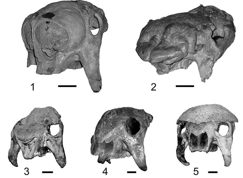
FIGURE 2. Skull of cingulates in dorsal view: 1- Chaetophractus sp.; 2- Glyptodon reticulatus (MACN-Pv 10153); 3- Neosclerocalyptus paskoensis (MACN-Pv 18107). Scale bar equals 5 cm.
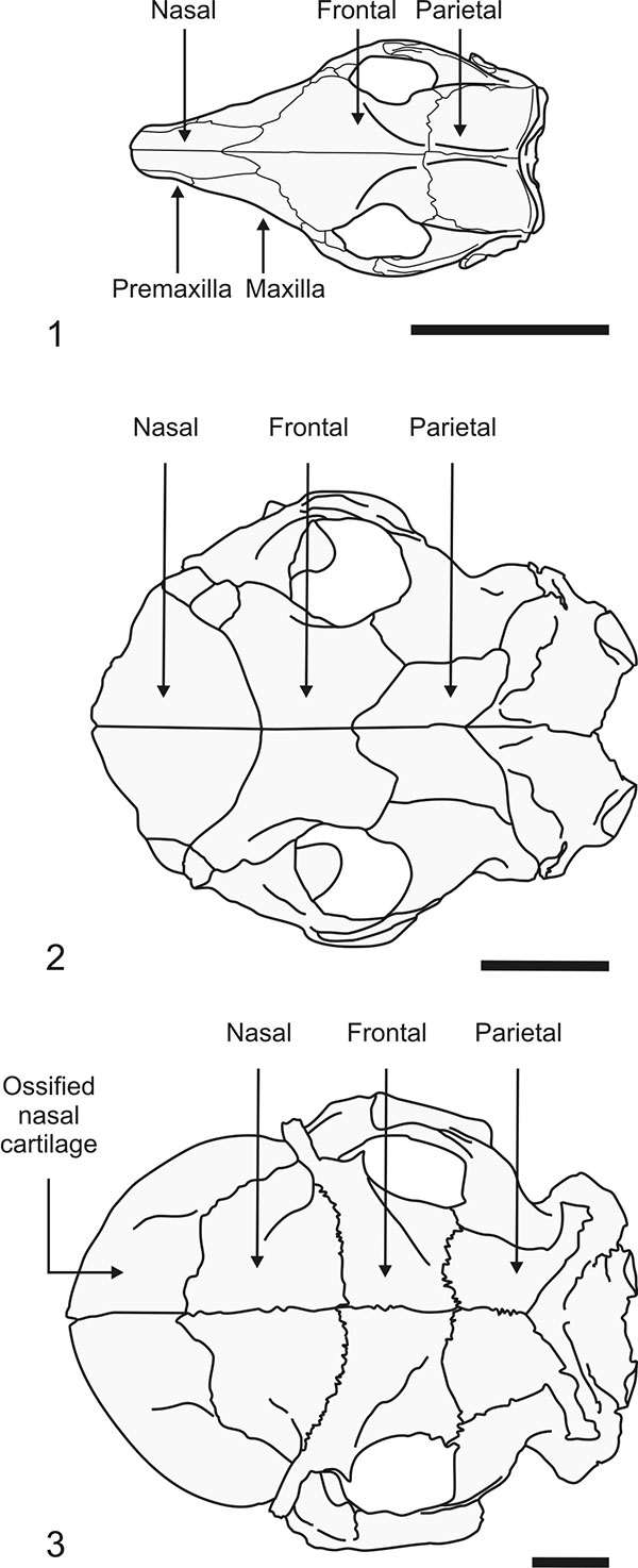
FIGURE 3. Skulls of Neosclerocalyptus in lateral view (nasal region to the left), showing different morphotypes: 1- Neosclerocalyptus paskoensis (MLP 16-384); 2- Neosclerocalyptus pseudornatus (MACN-Pv 8579); 3- Neosclerocalyptus paskoensis (MACN-Pv 18107); 4- Nesoclerocalyptus sp. (MACN-Pv 8091); 5- Neosclerocalyptus ornatus (MLP 16-28); 6- Neosclerocalyptus pseudornatus (MACN-Pv 8773). Scale bar equals 5 cm.
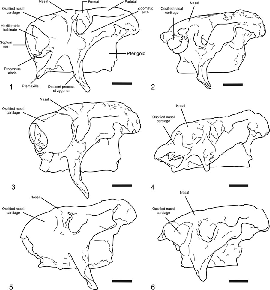
FIGURE 4. Cladogram depicting phylogenetic hypothesis used in this work. Modified from Fernicola (2008), Porpino et al. (2010), and Fernicola and Porpino (2012).
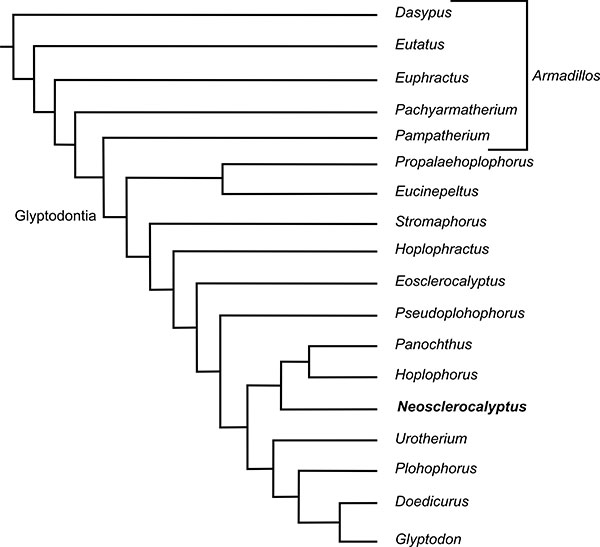
FIGURE 5. Skull of Neosclerocalyptus paskoensis (MACN-Pv 18107) depicting ossified nasal cartilages conforming the rostral most region (light orange) and turbinates (red).
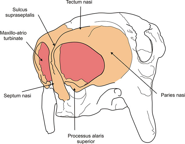
FIGURE 6. Neosclerocalyptus paskoensis (MACN-Pv 18107). Parasagittal CT slice (above) and diagram depicting structures explained in the text (below). Scale bar equals 5 cm.
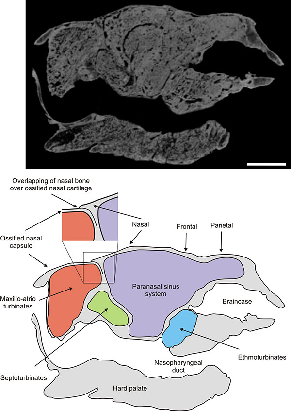
FIGURE 7. Neosclerocalyptus paskoensis (MACN-Pv 18107). Frontal CT slice (above) and diagram depicting paranasal sinuses explained in the text (below). Scale bar equals 5 cm.
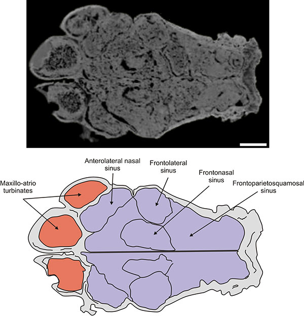
FIGURE 8. Neosclerocalyptus paskoensis (MACN-Pv 18107). Block diagram depicting paranasal sinuses explained in the text.
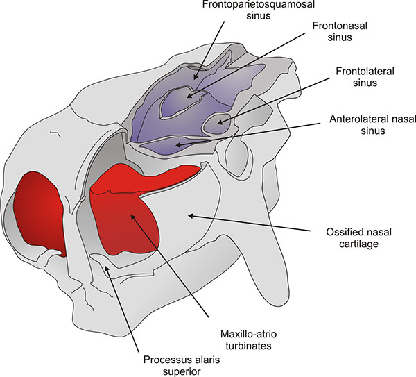
FIGURE 9. Neosclerocalyptus paskoensis (MACN-Pv 18107). Transverse CT slice (above) and diagram depicting structures explained in the text (below). Scale bar equals 5 cm.
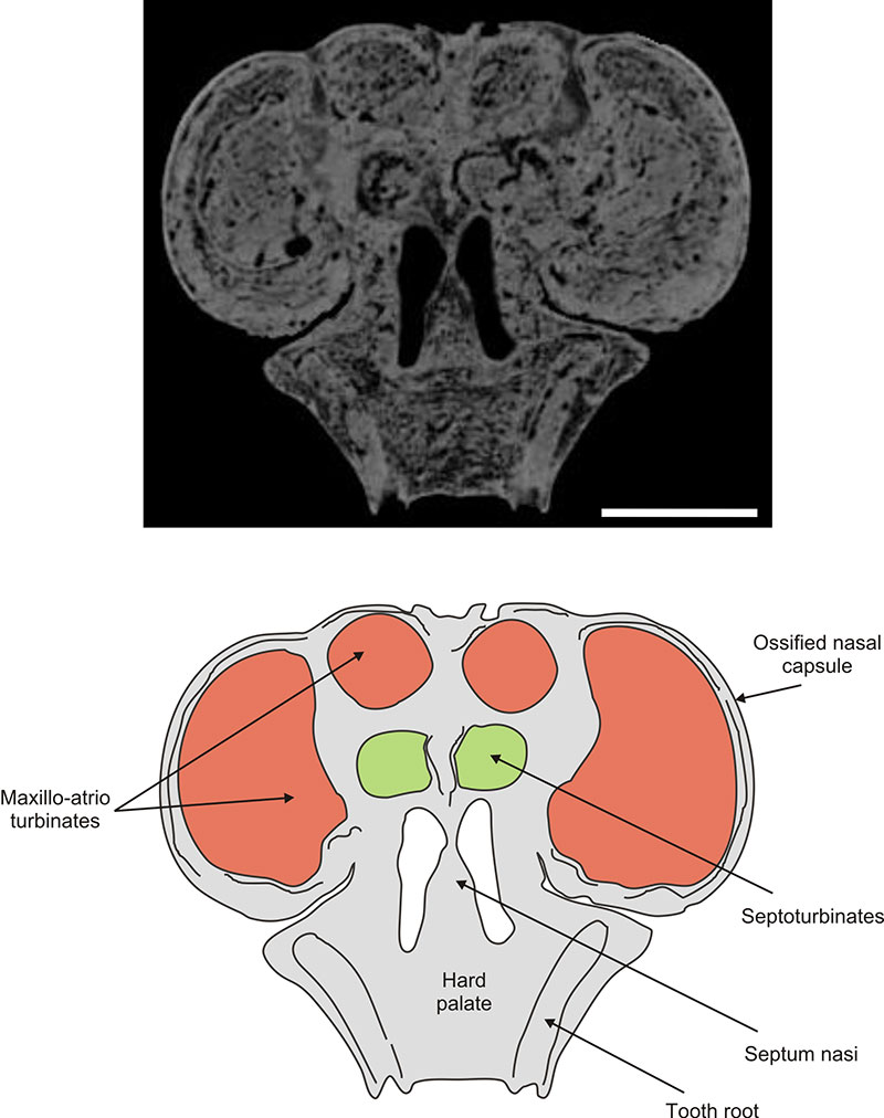
FIGURE 10. Neosclerocalyptus paskoensis (MACN-Pv 18107). Frontal CT slice (above) and diagram depicting structures explained in the text (below). Scale bar equals 5 cm.
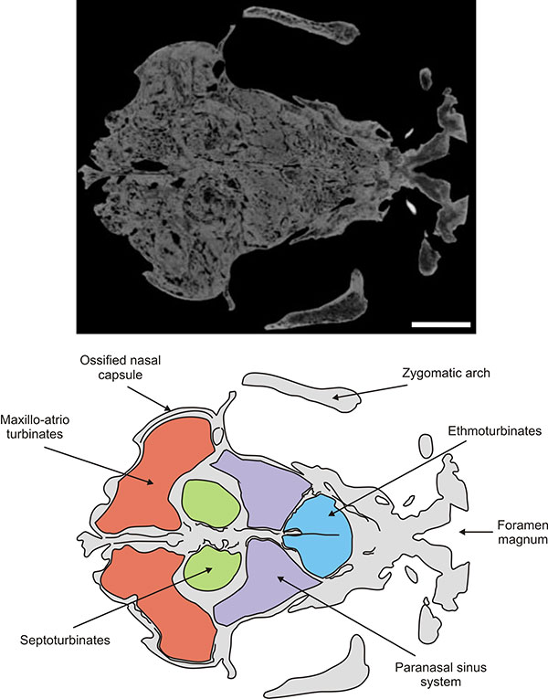
FIGURE 11. Neosclerocalyptus paskoensis (MACN-Pv 18107). Block diagram depicting nasal meatuses explained in the text.
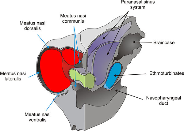
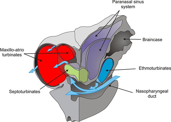
FIGURE 12. Neosclerocalyptus paskoensis (MACN-Pv 18107). Block diagram depicting structures explained in the text and inferred entering airflow (light blue arrows).

FIGURE 13. Neosclerocalyptus paskoensis (MACN-Pv 18107). Block diagram depicting structures explained in the text and inferred leaving airflow (light blue arrows).
A neomorphic ossification of the nasal cartilages and the structure of paranasal sinus system of the glyptodont Neosclerocalyptus Paula Couto 1957 (Mammalia, Xenarthra)
Plain Language Abstract
Glyptodonts are extinct animals belonging to the group of xenarthrans, South American placental mammals also comprising sloths, anteaters and armadillos. The group containing both armadillos and glyptodonts is known as Cingulata, and members of this group have a characteristic bony armor protecting their head, trunk and tail. Among Pleistocene (~ 2.5 Ma) glyptodonts the skull of Neosclerocalyptus is very remarkable due to its bizarre, globular and expanded narial region. In this work we analyzed external and internal anatomy of the skull of Neosclerocalyptus using computed tomography (CT) scanning. We found that the most anterior portion of the narial region is formed by ossification of the nasal cartilages, a feature absent in other mammals and that probably represents a shift in the timing of developmental stages when compared with other cingulates (a phenomenon called “heterochrony”). This neomorphic ossification housed very well developed respiratory turbinates, folded bony structures that warm and humidify the air entering to the nasal cavity during breathing. The other bones of the skull roof (nasals, frontals and parietals) have an expanded system of interconnected inner cavities, called paranasal sinuses. The function of these cavities remains enigmatic. Finally, at the bottom of the nasal cavity there were, as in other mammals, olfactory turbinates (called “ethmoturbinates”), widely separated from the respiratory ones. The nasal cavity as a whole, comprising ossified nasal cartilages and turbinates, probably had similar functions to those of other mammal. Thus, this neomorphic ossification would constitute a morphological novelty, but not necessarily a functional one.
Resumen en Español
Una osificación neomórfica de los cartílagos nasales y la estructura del sistema de senos paranasales del gliptodonte Neosclerocalyptus Paula Couto 1957 (Mammalia, Xenarthra)
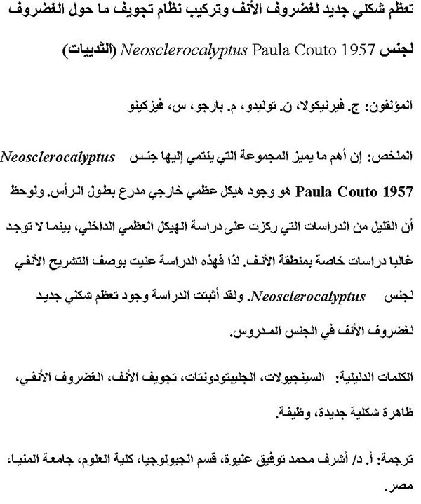
Los gliptodontes, junto con los armadillos, los pampatéridos y los peltefilinos constituyen los Cingulata, uno de los tres clados de los Xenarthra. La característica más notable de este grupo es la presencia de un exoesqueleto acorazado a lo largo de la cabeza, el cuerpo y la cola. Sólo unas pocas contribuciones han descripto el endosqueleto en detalle. En el caso del cráneo, apenas se ha prestado atención a la región nasal. El objetivo de este artículo es proporcionar una descripción de la región nasal del gliptodonte Neosclerocalyptus. Este género tiene la región nasal más expandida y globular entre los gliptodontes, habiendo sido descrita recientemente como parte del sistema de senos fronto-nasales. Nuestro análisis mediante tomografía computarizada muestra que esta región incluye una osificación independiente del cartílago nasal que albergaba los maxilo-atrioturbinales. Esta osificación representaría un carácter neomórfico producido por una adición terminal de un estadio osificado mediante peramorfosis. Otras características anatómicas destacadas son la presencia de un sistema expandido de senos paranasales que involucra a los huesos nasal, frontal, parietal y escamosal, y la amplia separación entre los maxilo-atrioturbinales y los etmoturbinales. Las consecuencias funcionales de esta reorganización son difíciles de predecir o inferir. Por tanto, dicha osificación neomórfica constituiría una novedad morfológica, aunque no necesariamente funcional.
Palabras clave: Cingulados; gliptodontes; cavidad nasal; osificación nasal; neomórfico; función
Traducción: Author and Miguel Company
Résumé en Français
Une ossification néomorphique du cartilage nasal et la structure du système de sinus paranasale chez le glyptodonte Neosclerocalyptus Paula Couto 1957 (Mammalia, Xenarthra)
Les glyphodontes, de même que les tatous, pampathères et peltephilinés constituent les Cingulata, l'un des trois clades de Xenarthra. Le caractère le plus remarquable de ce groupe est la présence d'une armure exo-squelettique le long de tête, du corps et de la queue. Très peu de contributions ont décrit en détail l'endosquelette. Dans le cas du crâne, presque aucune attention n'a été portée à la région des cavités nasales. L'objectif de cette étude est de fournir une description de l'anatomie des narines du glyphodonte Neosclerocalyptus. Ce genre a les narines les plus étendues et les plus globulaires parmi les glyphodontes et a été récemment décrit comme part du système de sinus fronto-nasaux. Notre analyse au CT-scan montre que cette région inclus une ossification indépendante du cartilage nasal qui loge les maxillo-atrioturbinates. Cette ossification pourrait représenter un caractère néomorphique produit par l'ajout final d'un stade d'ossification par péramorphose. D'autres caractères anatomiques remarquables sont la présence d'un système de sinus paranasal étendu qui inclue les os nasal, frontal, parietal et squamosal, et la large séparation entre les maxillo-atrioturbinates et les ethmoturbinates. Les conséquences fonctionnelles de ce réarrangement ne sont pas facilement prédictibles ou déductibles. Donc, cette ossification néomorphique pourrait constituer une innovation morphologique, mais pas nécessairement une innovation fonctionnelle.
Mots clés : Cingulates; Glyptodontes; cavité nasale; ossification des narines; néomorphique; fonctionnement
Translator: Olivier Maridet
Deutsche Zusammenfassung
Eine neomorphe Ossifikation des Nasenknorpel und die Struktur des paranasalen Sinus-Systems des Glyptodonten Neosclerocalyptus Paula Couto 1957 (Mammalia, Xenarthra)
Glyptodonten bilden zusammen mit den Gürteltieren, Pampatheren und Peltephilinen die Cingulata, einer der drei Kladen der Xenarthra. Das auffälligste Merkmal dieser Gruppe ist das gepanzerte Exoskelett am Kopf, Körper und Schwanz. Nur einige wenige Arbeiten befassten sich im Detail mit dem Endoskelett. Beim Schädel wurde der Nasenregion kaum Aufmerksamkeit gewidmet. Das Ziel dieser Studie ist es, eine Beschreibung der Nasenanatomie des Glyptodonten Neosclerocalyptus anzufertigen. Diese Gattung hat die weiteste und kugelförmigste Nasenregion unter den Glyptodonten und war kürzlich als Teil der Fronto-Nasal Sinussysteme beschrieben worden. Unsere auf Computertomographie beruhende Analyse zeigt, dass diese Region eine eigene Nasenknorpel-Ossifikation beinhaltet, die die Maxillo-Atrio-Nasenmuscheln beherbergt. Diese Verknöcherung würde ein neomorphes Merkmal darstellen, das durch eine terminale Ergänzung einer verknöcherten Phase über Peramorphose hervorgerufen wurde. Weitere bemerkenswerte anatomische Merkmale sind das Vorhandensein eines erweiterten paranasalen Sinussystems das das Nasal, Frontale, Parietale und Squamosum umfasst und die breite Trennung zwischen den Maxillo-Atrio-Nasenmuscheln und den ethmoidalen Nasenmuscheln. Die funktionalen Konsequenzen dieser Anordnung sind noch nicht bekannt oder erschlossen. So könnte diese neomorphe Ossifikation zwar ein morhphologisches, jedoch nicht zwingend ein funktionales Novum darstellen.
Schlüsselwörter: Cingulata; Glyptodonten; Nasenkavität; Nariale Verknöcherung; Neomorph; Funktion
Translators: Eva Gebauer
Arabic
Translator: Ashraf M.T. Elewa
-
-
-
Review: The Princeton Field Guide to Mesozoic Sea Reptiles
 The Princeton Field Guide to Mesozoic Sea Reptiles
Article number: 26.1.1R
April 2023

 Poster Winners 2024