

Article Search
Volume 27.1
January–April 2024
Full table of contents
ISSN: 1094-8074, web version;
1935-3952, print version
Recent Research Articles
See all articles in 27.1 January-April 2024
See all articles in 26.3 September-December 2023
See all articles in 26.2 May-August 2023
See all articles in 26.1 January-April 2023
TABLE 1. Table 1 - Spatial data of study sites.
TABLE 2. Absolute and relative frequencies of living foraminifera in winter samples. In gray - samples with less than 48 living individuals (presented in PDF format).
TABLE 3. Absolute and relative frequencies of living foraminifera in summer samples. In gray - samples with less than 48 living individuals (presented in PDF format).
FIGURE 1. Location of the study area; 1) Geographical context of the Guadiana River basin in the Iberian Peninsula (Europe). Adapted from chguadiana.es (2012). Coordinate system: Datum ETRS89 UTM Zone 30N; 2) Study area: Map of the Guadiana Estuary with site locations.
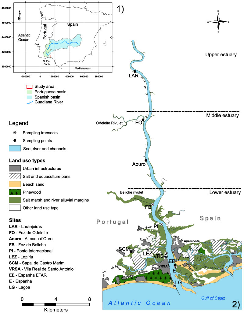
FIGURE 2. Scanning electron micrographs of the foraminifera specimens. Scale bar equals 100 µm. 1-3- Three different specimens of Polysaccammina ipohalina Scott, 1976b, illustrating the differences in size and form. In all specimens it is possible to see attached organic matter; 4-5- Polysaccammina hyperhalina Medioli, Scott, and Petrucci, 1983. 4- complete specimen of P. hyperhalina ; 5- aperture view; 6- specimen with several side branches; 7-10- different sized specimens of Ammovertellina sp.; 11-14- various specimens of Reophax nana Rhumbler, 1913; 15-17- Leptohalysis scottii (Chaster, 1892); 15 and 16- side view of two complete specimens; 17- detail on the agglutination of the last chamber; 18- complete specimen of Ammobaculites exiguus Cushman and Brönnimann, 1948b; 19- Ammobaculites sp. with the uncoiled portion broken; 20-22- Ammotium salsum (Cushman and Brönnimann, 1948a); 20- best specimen; 21- smaller specimen; 22- aperture detail; 23- Ammotium sp.; 24-26- different specimens of Miliammina fusca (Brady, 1870); 27-28- Miliammina obliqua Heron-Allen and Earland, 1930; 27- view of the interio-marginal arch of the aperture; 29-30- Arenoparrella mexicana (Kornfeld, 1931); 29- ventral side with view to main aperture and supplementary apertures; 30- dorsal side with view to supplementary apertures; 31-32- Deuterammina eddystonensis Brönnimann and Whittaker, 1990; 31- dorsal view; 32- ventral view; 33-35- Jadammina macrescens (Brady, 1870); 33- dorsal view; 34- ventral view; 35- dorsal view of a deformed test.
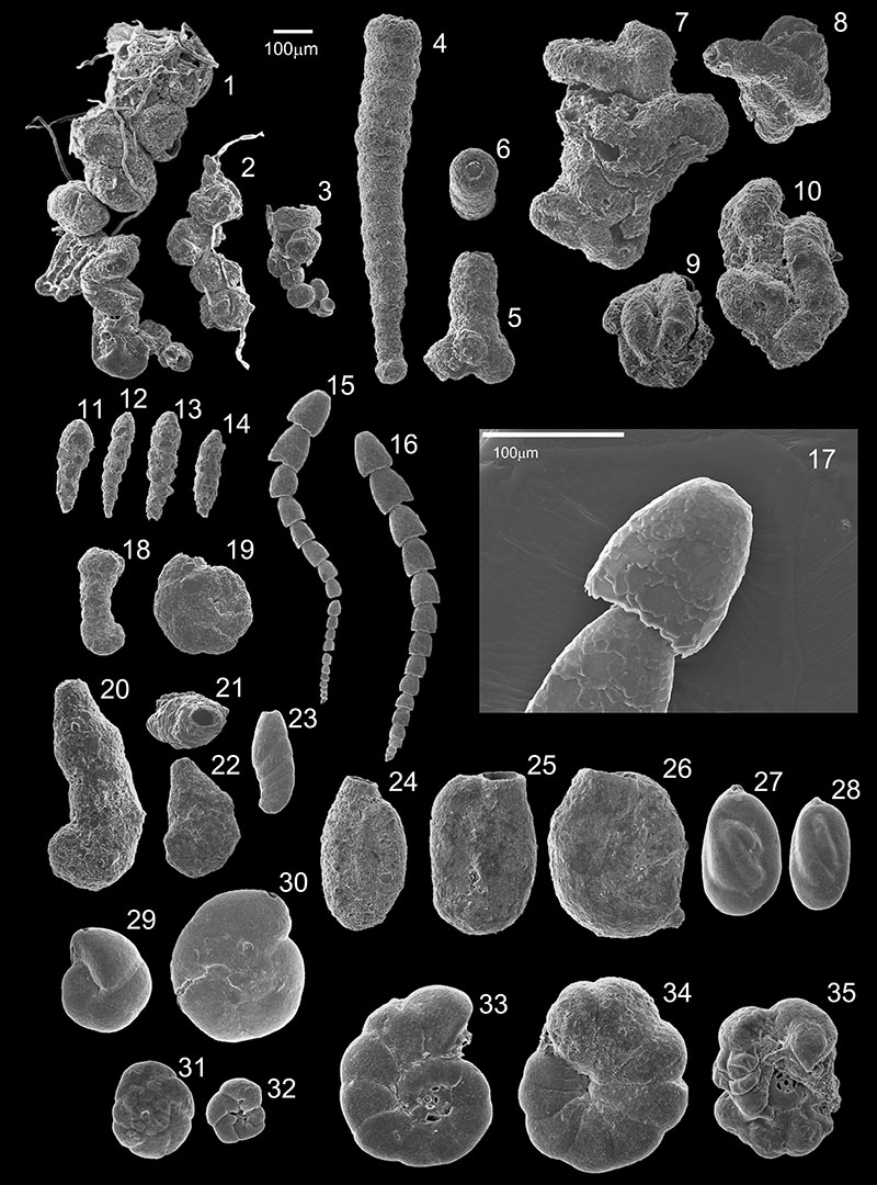
FIGURE 3. Scanning electron and light microscope micrographs of the foraminifera specimens. Scale bar equals 100 µm except where noted otherwhise; 1-2- Jadammina macrescens (Brady, 1870); 1- supplementary apertures view; 2- detail of supplementary apertures (scale bar = 50 µm); 3-4- Lepidodeuterammina plymouthensis Brönnimann and Whittaker, 1990; 3- dorsal view; 4- ventral view; 5-8- Lepidodeuterammina ochracea (Williamson, 1858); 5- dorsal view; 6- ventral view; 7- dorsal view of a smaller specimen; 8- ventral view of a smaller specimen; 9-10- Portatrochammina sp.; 9- dorsal view; 10- ventral view; 11-13- Siphotrochammina sp.; 11- dorsal side with inter-cameral foramen view; 12- dorsal view of a smaller specimen, also with inter-cameral foramen; 13- ventral view of a smaller specimen; 14-16- Tiphotrocha comprimata Saunders, 1957; 14- dorsal view; 15- ventral view; 16- individual strongly attached to a sea-grass leaf; detail of a Pinus pollen grain at the center of the leaf; 17-21- Trochammina inflata (Montagu, 1808); 17- dorsal view; 18- ventral view; 19- ventral view with umbilical tube detail; 20- microspheric form dorsal view; 21- microspheric form ventral view; 22- Eggerelloides scaber (Williamson, 1858); 23-25- Textularia earlandi Parker, 1952; 23- apertural view; 24- lateral view; 25- profile view with aperture in detail; 26-29- Discorinopsis aguayoi (Bermúdez, 1935); 26- scanning electron dorsal view; 27- scanning electron ventral view; 28- light microscope dorsal view; 29- light microscope ventral view.
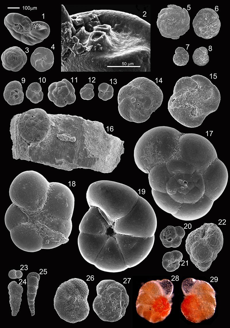
FIGURE 4. Scanning electron micrographs of the foraminifera specimens. Scale bar equals 100 µm. 1-4- different sized specimens of Bolivina ordinaria Phleger and Parker, 1952, new name; 5- Buliminella elegantissima (d’Orbigny, 1839b); 6 - 7- Cornuspira involvens (Reuss 1850); 8-10- Miliolid sp1; 8- apertural view; 9- front view; 10- back view; 11-13- Miliolid sp2; 11- apertural view; 12- front view; 13- back view; 14-16- Miliolid sp3; 14- front view; 15- apertural view; 16- back view; 17-18- Miliolid sp4; 17- apertural view; 18- front view; 19-21- Miliolid sp5; 19- apertural view; 20- front view; 21- back view; 22-23- Miliolid sp6; 22- front and apertural view; 23- back view; 24-26- Miliolid sp7; 24- front view; 25- apertural view; 26- front and apertural view; 27-29- Miliolid sp8; 27- front view; 28- apertural view; 29- front view of a smaller specimen.
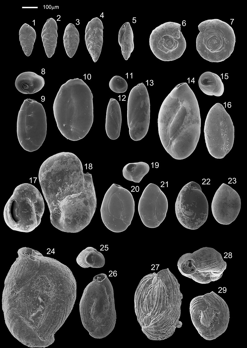
FIGURE 5. Scanning electron and light microscope micrographs of the foraminifera specimens. Scale bar equals 100 µm. 1- Lamarckina haliotidea (Heron-Allen and Earland, 1911) ventral view; 2-3- Asterigerinata mamilla (Williamson, 1858); 2- dorsal view; 3- ventral view; 4-5- Discorbis sp.; 4- dorsal view; 5- ventral view; 6-7- Helenina anderseni (Warren, 1957); 6- dorsal view; 7- ventral view; 8- Haynesina depressula (Walker and Jacob, 1798) side view; 9- Haynesina germanica (Ehrenberg, 1840) side view; 10- Elphidium advenum (Cushman, 1922) side view; 11- Elphidium excavatum (Terquem, 1875) side view; 12-15- Elphidium wiliamsoni Haynes, 1973; 12- side view in light microscope image; 13- side view in scanning electron image; 14- profile view in scanning electron image; 15- side view of a smaller specimen in scanning electron image; 16-18- side view of different size Elphidium gerthi Van Voorthuysen, 1957; 19-21- Elphidium oceanensis (d’Orbigny, 1826); 19- side view in scanning electron image; 20- profile view in scanning electron image; 21- side view in light microscope image; 22- Elphidium poeyanum (d’Orbigny, 1826) side view; 24-27- Ammonia sp1; 24- dorsal view; 25- profile view; 26- ventral view; 27- dorsal view in light microscope image; 28-31- Ammonia sp2 ( Ammonia aberdoveyensis Haynes, 1973); 28- dorsal view; 29- profile view; 30- ventral view; 31- dorsal view in light microscope image; 32-35- Ammonia sp3 ( Ammonia aberdoveyensis Haynes, 1973); 32- dorsal view; 33- profile view; 34- ventral view; 35- dorsal view in light microscope image.
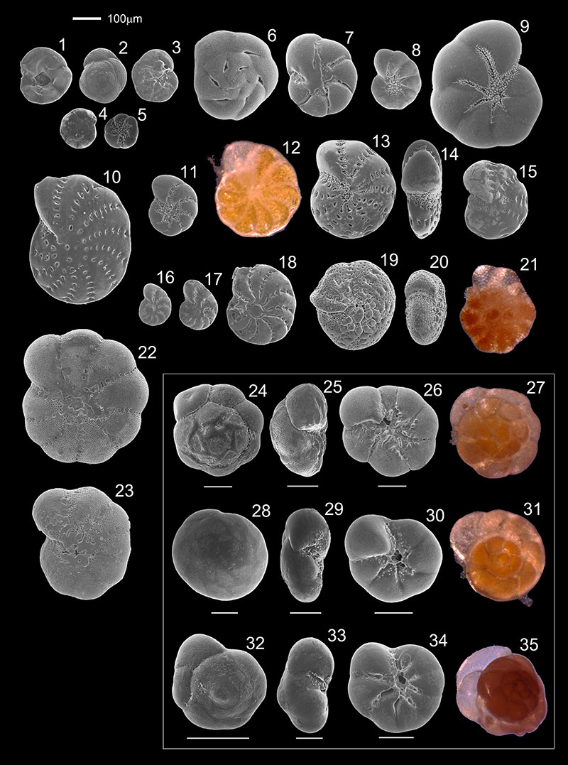
FIGURE 6. RDA attribute plot representing the distribution and abundance of the dominant species in Guadiana Estuary according to elevation and distance-to-sea variables.

FIGURE 7. Distribution patterns of the common to dominant species in the samples collected in winter along a distance-to-sea and elevation gradients (in relation to MSL).
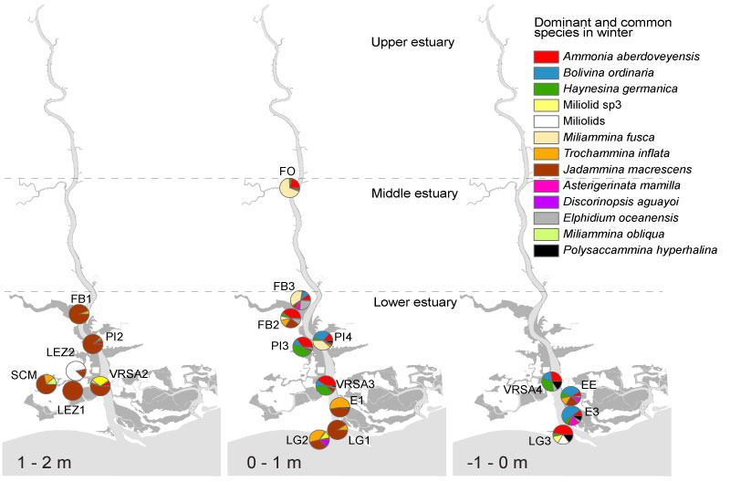
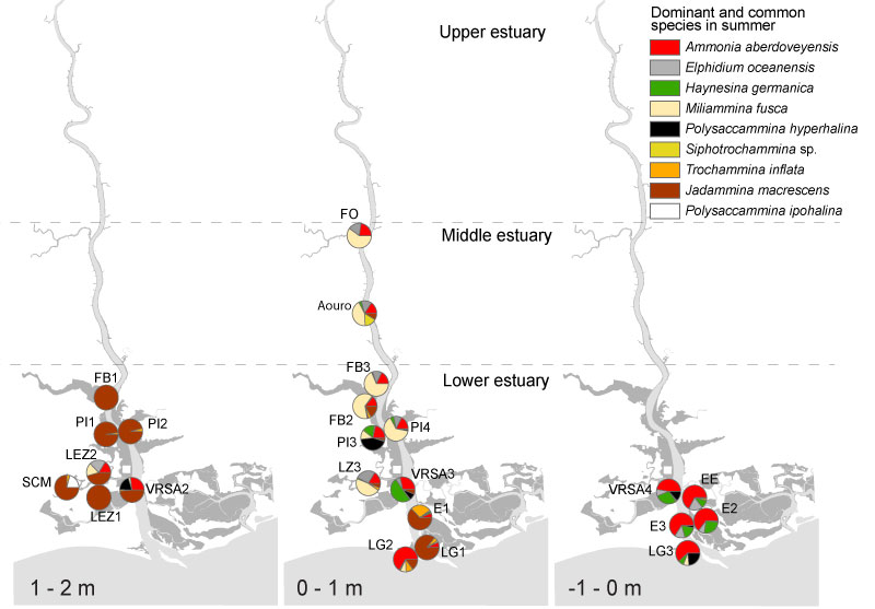
FIGURE 8. Distribution patterns of the common to dominant species in the samples collected in summer along a distance-to-sea and elevation gradients (in relation to MSL).
 Sarita Graça Camacho
Center of Environmental and Marine Research (CIMA)
Faculdade de Ciências e Tecnologia
Universidade do Algarve
Campus de Gambelas
8005-139 Faro
Portugal
scamacho@ualg.pt
Sarita Camacho was born in 1974 in Luanda, Angola. She holds a degree in Marine Biology and Fisheries from Algarve University, Portugal. Her first steps in micropaleontology were in 1998 at Dalhousie University, Halifax, under the supervision of Prof. David Scott, who taught her about the foraminifera and thecamoebians of transitional environments. This experience led her to join the Centre for Marine and Environmental Research (CIMA), where she has participated in several multidisciplinary projects on paleoenvironmental reconstruction of estuarine systems along the Portuguese south coast. Currently, her main interest is in knowing more about the potential of estuarine foraminifera and thecamoebians as environmental stress indicators.

 Delminda Maria de Jesus Moura
CIMA, Faculdade de Ciências e Tecnologia
Universidade do Algarve, Campus de Gambelas
8005-139 Faro
Portugal
dmoura@ualg.pt
Delminda Moura is a lecturer in morphology and coastal morphodynamics for 1st and 2nd study cycles in the Algarve University. Currently she is a member of the commission of the 1st cycle on Marine Sciences. Her scientific activity, developed in the Centre for Marine and Environmental Research (CIMA), is on the evolution of coastal environments along the Quaternary with particular emphasis on estuaries and rocky shores.
 Simon Connor
School of Earth, Atmosphere and Environment
Faculty of Science
Monash University
Clayton 3800
Australia
Simon.Connor@monash.edu
Simon Connor is a lecturer in palaeoecology and biogeography at Monash University. He has a PhD from the University of Melbourne and was a research fellow in environmental dynamics at the Centre for Marine and Environmental Research (CIMA), Portugal, from 2008-2012. Simon has research interests in the history of vegetation and human impacts on the environment in the Caucasus, Balkans, Iberia, Macaronesia and Australasia.
 David B. Scott
Centre for Environmental and Marine Geology
Department of Earth Sciences
Dalhousie University
Halifax, Nova Scotia
Canada B3H 4J1
dbscott@dal.ca
David B. Scott was born in 1947 in California. He has been Professor of Earth Sciences, Dalhousie University, Halifax, for more than 35 years, with more than 40 grad students and over 130 papers. His long career as micropaleontologist as taken him to almost all parts of the world to study recent and ancient foraminifera, ostracoda and thecamoebians and their use as paleoenvironmental indicators and coastal zone management tools. His main contribution to micropaleontology research is related to his pioneering studies that use salt-marsh foraminiferal biozonation to infer sea-level changes in the past. Currently his main interest is related to global climate changes, most recently using deep-sea corals as a climate archive.
 Tomasz Boski
CIMA, Faculdade de Ciências e Tecnologia
Universidade do Algarve
Campus de Gambelas
8005-139 Faro
Portugal
tboski@ualg.pt
Tomasz Boski was born in Lodz, Poland, in 1950. He holds a Degree in Petrology and Geochemistry from University of Warsaw, Poland, a Master in Quaternary Geology and a doctorate in geology, from University of Brussels, and aggregation in Marine & Environmental Science, in the University of Algarve, Portugal. His research is dedicated to studies of climate and sea level change during the Quaternary, the geochemistry of sedimentary processes and integration of environmental sciences in land territorial/coast. He is president of the Portuguese Group for Quaternary Studies and Scientific Coordinator of the Center for Marine and Environmental Research (Algarve University) where he manages 24 research projects, promoted 25 master's degrees, 7 doctorates and 5 postdocs.
Taxonomy, ecology and biogeographical trends of dominant benthic foraminifera species from an Atlantic-Mediterranean estuary (the Guadiana, southeast Portugal)
Plain Language Abstract
Foraminifera are single celled marine organisms that possess a hard shell which often remains preserved in the sediments after death. In estuaries, foraminifera have a high ecological value, as they are distributed according to environmental conditions. Once preserved in the estuarine sediments, their fossils can provide relevant information about the evolution of the estuary’s condition and tell us more about the reasons for environmental change, both natural and anthropogenic.
The Guadiana Estuary belongs to one of the biggest river systems on the Iberian Peninsula. The estuary is controlled by two countries (Portugal and Spain) and has a Mediterranean climate, which means that extreme seasonal environmental variations occur. In our study, the foraminiferal assemblages and their occurrence within the estuary were analyzed in order to improve their value as environmental indicators. Allied to the study of the ecological distribution of the most important estuarine species, a photographic and systematic inventory was created in order to serve as a basis for future studies of environmental management and reconstructions of past environments.
Resumen en Español
Taxonomía, ecología y tendencias biogeográficas de las especies de foraminíferos bentónicos dominantes de un estuario del Atlántico-Mediterráneo (Guadiana, sureste de Portugal)
Este estudio analiza la taxonomía, ecología y biogeografía de las especies de foraminíferos bentónicos que viven en los márgenes intermareales del estuario del Guadiana (SE de Portugal, SW de España). De los 54 taxones identificados durante las campañas de muestreo de invierno y verano, 49 son listados sistemáticamente e ilustrados mediante fotografías de microscopio electrónico de barrido (SEM). Ammonia spp. fueron los taxones calcáreos más ubicuos en ambas temporadas. El análisis morfológico y las imágenes de SEM sugirieron tres morfotipos distintos del género Ammonia, dos de los cuales resultaron ser Ammonia aberdoveyensis sobre la base de los análisis de rRNA parcial. Jadammina macrescens y Miliammina fusca fueron los taxones aglutinados más ubicuos en el estuario. Jadammina macrescens domina las zonas de humedal superior casi exclusivamente, con muy altas densidades. Ammonia spp. son los taxones más abundantes en las zonas de humedal inferior y de plataforma de marea del curso inferior del estuario del Guadiana, aunque están distribuidas por todo el estuario, especialmente durante el verano cuando las condiciones ambientales favorecen su proliferación. Miliammina fusca domina las zonas de humedal inferior y de plataforma de marea con escasa vegetación del curso superior, donde se asocia con especies calcáreas. Debido a su posición geográfica, el sistema del Guadiana comparte características tanto de estuarios del Atlántico como del Mediterráneo. Esto se refleja en las asociaciones de foraminíferos, con un predominio de especies termófilas y una zonación ecológica típica de la zona climática mediterránea.
Palabras clave: foraminíferos bentónicos; taxonomía; biogeografía; zonación ecológica; análisis genético; estuario del Guadiana
Traducción: Enrique Peñalver
Résumé en Français
Taxonomie, écologie et tendances biogéographiques des espèces de foraminifères benthiques dominants de l'estuaire de l'Atlantique-Méditerranée (le Guadiana, au sud du Portugal)
Cette étude analyse la taxonomie, l'écologie et la biogéographie des espèces de foraminifères benthiques vivant sur les marges intertidales de l'estuaire du Guadiana (SE Portugal, SW Espagne). Parmi les 54 taxons identifiés pendant l'échantillonnage des campagnes en hiver et en été, 49 sont systématiquement répertoriés et illustrés par des photographies de microscope électronique à balayage (MEB). Ammonia spp. étaient les taxons calcaires les plus omniprésent dans les deux saisons. L'analyse morphologique et les images MEB suggéré trois morphotypes distincts du genre Ammonia, dont deux se sont avérés d'être Ammonia aberdoveyensis sur la base des analyses d'ARNr partielle. Jadammina macrescens et Miliammina fusca étaient les taxons agglutiné les plus omniprésent dans l'estuaire. Jadammina macrescens domine les zones supérieures de marais presque exclusivement, se produisant à des densités très élevées. Ammonia spp. sont les plus abondants dans les bas et replats de marais de la partie inférieure de l'estuaire du Guadiana, mais sont très répandus dans l'estuaire, surtout pendant l'été lorsque les conditions environnementales favorisent leur prolifération. Miliammina fusca domine la végétation clairsemée des bas de marais et les zones de replats de marée de la partie supérieure, où il est associé avec des espèces calcaires. En raison de sa position géographique, le système de Guadiana partage des caractéristiques avec les estuaires de l'Atlantique et de la Méditerranée. Cela se reflète dans les foraminifères, avec une dominance des espèces thermophiles et d'un zonage écologique typique de la zone climatique méditerranéenne.
Mots-clés: foraminifères benthiques; taxonomie; biogéographie; zonation écologique; analyse génétique; estuaire du Guadiana
Translator: Kenny J. Travouillon
Deutsche Zusammenfassung
Taxonomie, Ökologie und biogegrafische Tendenzen von domonanten benthischen Foraminiferenaten aus einem Atlantisch-Mediterranen Ästuar (Guadiana, Südost-Portugal)
Diese Studie analysiert die Taxonomie, Ökologie und Biogeografie der Arten von benthischen Foraminiferen, die an den intertidalen Rändern des Guardiana Ästuars (Südost-Portugal, Südwest-Spanien) leben. Von den 54 Taxa, die während der Sammlungskampagnen im Sommer und Winter identifiziert wurden, sind 49 systematisch gelistet und via Rasterelektronen-mikroskopie-Fotos (REM) illustriert. Ammonia spp. waren die am meist verbreiteten kalzitischen Taxa in beiden Jahreszeiten. Morphologische Analysen und REM-Aufnahmen deuteten auf drei distinkte Morphotypen der Gattung Ammonia hin, von denen zwei sich auf der Basis von partiellen RRNA-Analysen als Ammonia aberdoveyensis herausstellten. Jadammina macrescens und Miliammina fusca waren die am meisten verbreiteten agglutinierten Taxa im Ästuar. Jadammina macrescens dominiert fast alleine die oberen Marschzonen und tritt bei hoher Dichte auf. Ammonia spp. kommen am häufigsten in der niederen Marsch und der Gezeitenzone der Unterläufe des Guadiana Ästuars vor, sind aber auch weit verbreitet im gesamten Ästuar, besonders während des Sommers wenn Umweltbedingungen die Ausbreitung begünstigen. Miliammina fusca dominiert die spärlich bewachsene niedere Marsch und Gezeitenzonen der Oberläufe wo sie mit kalzitischen Arten assoziiert ist. Wegen seiner geografischen Position teilt das Guardiana-System Merkmale mit sowohl atlantischen als auch mediterranen Ästuaren. Dies spiegelt sich in den Foraminiferen-Assemblagen wieder mit einer Dominanz von thermophilen Arten und einer ökologischen Zonierung die typisch für die mediterrane Klimazone ist.
Schlüsselwörter: benthische Foraminifera; Taxonomie; Biogeographie; ökologische Zonierung; genetische Analyse; Guadiana Ästuar
Translator: Eva Gebauer
Arabic
Translator: Ashraf M.T. Elewa
-
-
-
Review: The Princeton Field Guide to Mesozoic Sea Reptiles
 The Princeton Field Guide to Mesozoic Sea Reptiles
Article number: 26.1.1R
April 2023

 Poster Winners 2024