

Article Search
Volume 27.1
January–April 2024
Full table of contents
ISSN: 1094-8074, web version;
1935-3952, print version
Recent Research Articles
See all articles in 27.1 January-April 2024
See all articles in 26.3 September-December 2023
See all articles in 26.2 May-August 2023
See all articles in 26.1 January-April 2023
APPENDIX
Principle of fringe projection
In a fringe projection profilometry system (FPP), a series of straight, vertical or horizontal and equally spaced fringes are generated by computer and projected onto the surface of an object using a digital video projector. At the same time, a CCD (charged-coupled device) camera captures the intensity of the fringes from the surface of the object for later processing. By using the phase shifting (PS) method, “N” fringe patterns of frames that were captured by the camera, each nth intensity pattern I n( x , y ) can be commonly described as:
I n ( x , y ) = a ( x , y ) + b ( x , y ) cos [ (2 π / p ) x + φ ( x , y ) + θ ∙ n ]; n=1...N
where N is the total number of images acquired; n is the number of the phase-shifting steps, ( x , y ) denotes the coordinates of an arbitrary point in the object being analyzed; p is the period of the equally spaced fringes on the reference plane; φ ( x , y ) is the phase map related to the object profile, also called wrapped phase ; and θ is the assigned phase shift value that is usually equal to 2π/N (Ma et al., 2012).
The numerical process to recover the wrapped phase from the set of captured images is performed with a phase shift algorithm (PSA). The correct design of this algorithm, in accordance with a specific setup, determines the complexity and resolution of the results that, briefly, uses the intensity values obtained by shifting the fringes on the object to calculate the phase (Rathjen, 1995). Next, an unwrapping procedure is needed to make this phase map continuous, which is done by removing the artificial discontinuities added by the FPP technique. This phase’s unwrapping is a complex process and the techniques used to perform it assume that the neighboring pixel differences of the unwrapped phase can be estimated by adding an integral multiple of 2π, when these differences are less than π. Using some mathematical methods it is possible to obtain a cloud of data points that is proportional to the analyzed object (Ghiglia and Pritt, 1998). After this, the temporal carrier is calculated and subtracted (Li et al., 1998) and the topography or profile is calculated by:
h ( x, y ) = l 0 ∙ φ ( x, y )/[ 2 π f 0 d + φ ( x, y ) ]
where l 0 is the distance between the CCD camera and the reference plane, and d is the distance between the camera and the projector (Takeda and Mutoh, 1983).
FIGURE 1. Optical set up specifications for fringe projection profilometry (FPP) used in this study for the recovery of a 3-D image of a hemimandible sample.
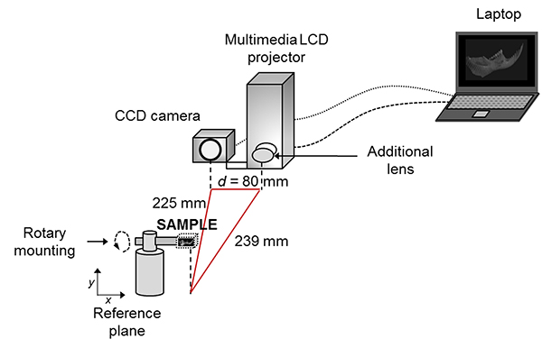
FIGURE 2. Flowchart of the 'OTY' procedure employed in this study, where: α = angle (60º in this case), * = MBE algorithm (Gutiérrez-García et al., 2013), and ** = Goldstein algorithm. OTY: name given to the white light system together with the phase shifting algorithm filter, based on the fact that it was developed for use on Ototylomys samples (see main text).
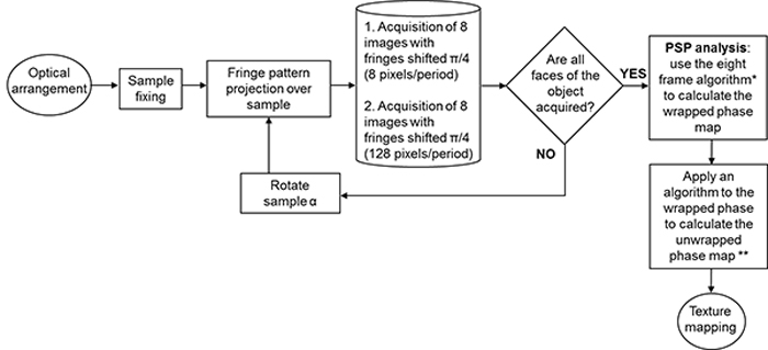
FIGURE 3. Magnitude maps of the sample obtained every 60º for 8 and 128 pixels/period. The images show the resolution and detail levels given the number of fringes projected over the sample. The measurements’ accuracy of surface and depth depends on the number of projected fringes, which include as many as the system can display (8 pixels/period for each fringe in this case). When the acquisition of details is difficult, a wider fringe is required (based on our sample size, we used 128 pixels).
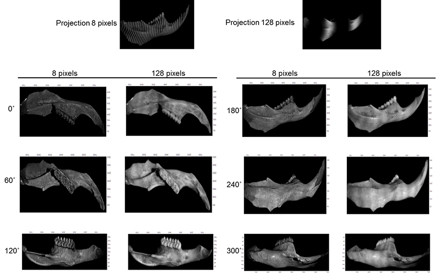
FIGURE 4. Process applied to recover the topography of the fossil sample. Where: 1) image captured by the CCD of the fringe projection on the sample, 2) wrapped phase obtained of the 8 frames after applying the MBE filter, 3) unwrapped phase map, 4) phase carrier compensation, and 5) surface of one of the views of the fossil recovered.
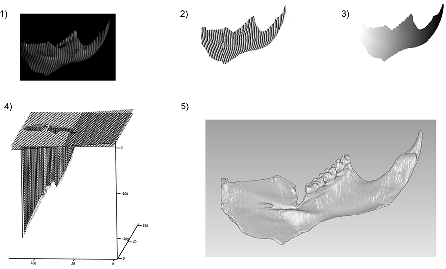
FIGURE 5. Processed views of a fossil sample acquired by the optical FPP system OTY. Each view is re-oriented 60º degrees with respect to the prior. Where: “a” indicates a zone that has no information at 60º in this stage of the process and will be corrected with the information of the next view, as is shown in “b”.
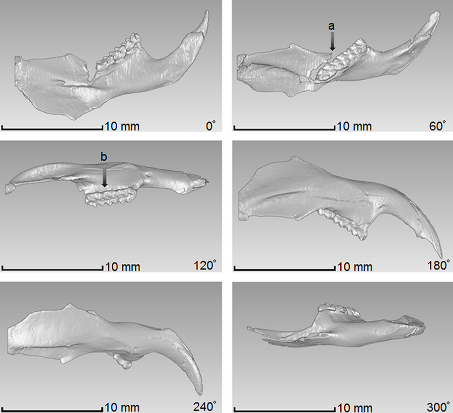
FIGURE 6. Full 3-D image of the reconstructed fossil after merging all the six views. 1) Cloud of points, 2) Final mesh.
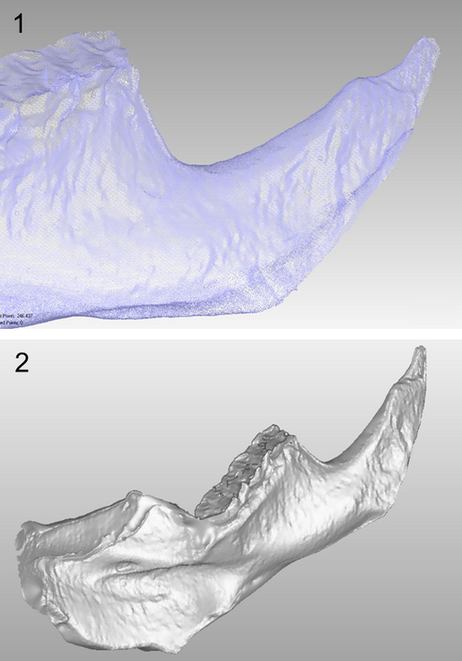
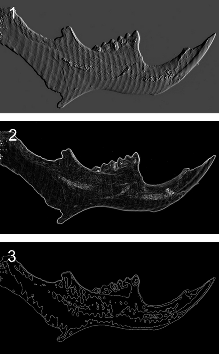
FIGURE 7. Examples of analyses that can be performed with the obtained data from the fossils: 1) denoting the relief (emboss filter), 2) detecting edges and transitions (sobel filter), 3) study of the roughness and waviness of a sample (topography filter).
 Juan C. Gutiérrez-García
Centro de Investigaciones en Óptica
Loma del Bosque 115, A.P. 1-948
León, Guanajuato, 37150
México
ing.juancarlosg@gmail.com
Juan Carlos Gutiérrez-García was graduated in 2006 as Mechatronic Engineer at Centro de Enseñanza Técnica Industrial (CETI), in Guadalajara, México, and obtained his MS degree in Optical Sciences at the Centro de Investigaciones en Óptica (CIO) in León, México. He is Ph. D. candidate in Optical Sciences at CIO. His research interest includes the signal and image processing, optical metrology, models of light-matter interaction, polarization in optical media, bioinformatics and morphometrics.

 Tania A. Gutiérrez-García
Departamento de Ciencias Computacionales
Centro Universitario de Ciencias Exactas e Ingenierías
Universidad de Guadalajara
Blvd. Marcelino García Barragán 1421
Guadalajara, Jalisco, 44430
México
taniagutierrezgarcia@gmail.com
Tania A. Gutiérrez-García, Ph. D. is a research scientist at the Department of Computational Sciences, Universidad de Guadalajara. She is dedicated to the study of the genetic patterns of biodiversity, including the geographical and historical perspectives. Her main research lines are phylogeography, ancient DNA, metagenomics and bioinformatics.
 J.F. Mosiño
Tecnológico Nacional de México - Instituto Tecnológico de León
Avenida Tecnológico S/N
León, Guanajuato, 37290
México
jfmosino@gmail.com
Dr. Mosiño was graduated in 1995 as Bachelor in Electronic Engineering from University of Guanuajuato and received also the Master Degree in Electrical engineering in 1997. In 2001 he obtained the Ph. D. in Optical Sciences at the Optical Research Centre CIO in León Gto. México. Before he joined the Instituto Tecnológico de León as professor he spend time at CIO as researcher from 2001-2009. Since 2002 is Member of the National Research System (SNI level 2) by the Mexican Council of Science and Technology (CONACYT). His research interest includes: Signal and image processing, optical metrology, fringe analysis, energy transfer in solids and propagation of polarized light in anisotropic media.
 Ella Vázquez-Domínguez
Departamento de Ecología de la Biodiversidad
Instituto de Ecología
Universidad Nacional Autónoma de México
Apartado Postal 70-275, Ciudad Universitaria
México Distrito Federal, 04510
México
evazquez@ecologia.unam.mx
Works on research within molecular ecology, evolution and population genetics of vertebrates, with emphasis on questions regarding phylogeography and historical diversification and distribution of natural populations, extant and extinct.
 Amalia Martínez
Centro de Investigaciones en Óptica
Loma del Bosque 115, A.P. 1-948
León, Guanajuato, 37150
México
amalia@cio.mx
Amalia Martínez-García received her BS degree in physics from Facultad de Ciencias Físico-Matemáticas, UANL, Monterrey, her MS degree from the Division of Applied Physics, Centro de Investigación Científica y de Educación Superior de Ensenada (CICESE), and her PhD in Optics at Centro de Investigaciones en Óptica (CIO), León, Mexico. She was research scientist at CICESE. Currently, she is researcher at CIO, member of the National System of Researchers, Mexico, Mexican Academy of Optics, SPIE and OSA. She is the President of the Mexican Academy of Optics (AMO), 2015-2016. Her research interests are optical metrology with holography, speckle and moiré techniques, images correlation and stereo vision systems.
 Joaquín Arroyo-Cabrales
Laboratorio de Arqueozoología “M. en C. Ticul Alvarez Solórzano”, Subdirección de Laboratorios y Apoyo Académico, Instituto Nacional de Antropología e Historia
Moneda #16, Colonia Centro
06060 Distrito Federal
México
arromatu5@yahoo.com.mx
Joaquín Arroyo-Cabrales, Ph. D. is a Senior Scientist at the Archaeozoology Lab, National Institute of Anthropology and History, the Mexican federal agency that takes care of the Historical, Archaeological, and Paleontological Heritages, and currently is in charge of the Paleontological Collection. His research focuses on Late Quaternary vertebrates and their contribution for understanding the paleoenvironments in which human survived in the Americas.
A novel application of the white light/fringe projection duo: recovering high precision three-dimensional images from fossils for the digital preservation of morphology
The fringe projection profilometry technique (FPP) is a widely used optical technique in which several frames with contrast fringes are projected onto a three-dimensional object, in order to acquire information of its topography in form of an array of data (cloud of points). This technique is useful to obtain three-dimensional (3-D) images that, when used in combination with white light, is an ideal non-contact option to digitalize a variety of objects. Despite its advantages, this technique has been scarcely used for imaging biological materials, mainly because of the difficulties involved in the optical setup and the algorithms needed for image processing. By using the latest advances in both fringe projection and white light, we designed a technique to acquire 3-D images from biological objects. We successfully obtained an exceptionally high quality 3-D image from a fossil jawbone of a tropical wild rodent. The digital preservation of the jawbone was crucial because it had to be destroyed to perform DNA extraction. The optical technique we describe adequately solves the setup problems and allows obtaining a 3-D exact model of rather small, delicate samples.
Resumen en Español
Una nueva aplicación del dúo luz blanca/proyección de franjas: recuperando imágenes 3-D de alta precisión en fósiles para la preservación digital de la morfología
Existe un interés histórico en la preservación digital de características morfológicas de materiales biológicos, especialmente porque a menudo la preservación de moléculas sensibles es crítica para estudios evolutivos. Con este fin, hemos aplicado exitosamente los recientes avances en la técnica de perfilometría de proyección de franjas en conjunción con luz blanca y un nuevo algoritmo de fase para digitalizar la forma de una hemimandíbula de un roedor fósil. Generamos una nube de puntos en un arreglo de datos que nos permitió plotear una restauración digital tridimensional (3-D) de la muestra fósil completa. La resolución máxima de este sistema puede utilizarse con objetos del rango 1–30 mm, minimizando los errores sistemáticos inducidos por pequeñas vibraciones o fluctuaciones de luz y, como consecuencia, mejorando la relación señal-a-ruido de la nube de datos recuperada. Se trata de una herramienta útil para preservar imágenes 3-D de fósiles u otros objetos biológicos para los cuales se necesita información morfológica bastante detallada, como por ejemplo en investigaciones biológicas y paleontológicas o, como en este caso, cuando los investigadores necesitan una réplica morfológica porque la muestra será destruida para extracción de ADN.
Palabras clave: morfometría; luz blanca; roedor; imágenes 3-D; proyección de franjas
Traducción: Diana Elizabeth Fernández
Résumé en Français
Une nouvelle application du duo lumière blanche/projection de franges : obtention d'images 3D de haute précision pour la conservation digitale de la morphologie
Un intérêt historique pour la conservation digitale des caractères morphologiques des matériels biologiques existe de longue date, en particulier car la préservation de molécules sensible est souvent cruciale pour les études en sciences évolutives. À cette fin, nous avons appliqué avec succès les avancées récentes de la technique de profilométrie par projection de franges combinée avec la lumière blanche et un nouvel algorithme de phase pour digitaliser la forme d'une hémi-mandibule de rongeur fossile. Nous avons pu générer un nuage de points dans un tableau de données qui nous a permis d'effectuer une restauration digitale tridimensionnelle (3D) de l'échantillon fossile complet. La résolution maximale de ce système est donnée par la limite de diffraction (de l'ordre du micron), et nous montrons que ce système perfectionné peut être utilisé sur des objets se situant dans la gamme de tailles de 1 mm à 30 mm. Cela minimise les erreurs systématiquement causées par de petites vibrations ou fluctuations de lumière et, par conséquent, améliore le rapport signal/bruit du nuage de données obtenu. Cela est un outil utile pour conserver des images 3D de fossiles et d'autres objets biologiques pour lesquels des informations morphologiques relativement détaillées sont nécessaires, comme dans les recherches des biologistes et paléontologues ou, comme le cas présent, quand des chercheurs ont besoin d'une réplique morphologique car l'échantillon va être détruit pour en extraire de l'ADN ancien.
Mots-clés : morphométrie ; lumière blanche ; rongeur ; imagerie 3D ; projection de franges
Translator: Antoine Souron
Deutsche Zusammenfassung
Eine neue Anwendung des weißen/Fringe-Projektions Duos: Gewinnung hochpräziser 3D-Abbildungen von Fossilien zur digitalen Morphologie-Erhaltung
Es besteht ein langes historisches Interesse an der digitalen Erhaltung morphologischer Merkmale von biologischem Material, vor allem weil die Erhaltung von sensiblen Molekülen oft kritisch für Evolutionsstudien ist. Zu diesem Zweck haben wir erfolgreich die neuesten Fortschritte der Fringe-Projektions-Profilometrie Technik angewandt, in Kombination mit weißem Licht und einem neuen Phasenalgorithmus um die Form einer fossilen Nager-Hemimandibel zu digitalisieren. Wir waren in der Lage eine Punktwolke in einer Reihe von Daten zu generieren, die es uns erlaubte eine dreidimensionale (3D) Rekonstruktion der gesamten Fossilprobe zu plotten. Die maximale Auflösung dieses Systems wird durch die Beugungsgrenze bestimmt (im Mikrometerbereich) und wir zeigen, dass das weiterentwickelte System für Objekte von 1-30mm genutzt werden kann. Die systematischen Fehler, hervorgerufen durch leichte Vibrationen oder Lichtfluktuationen, werden minimiert und demzufolge wird der Rauschabstand der gewonnenen Datenwolke verbessert. Dies ist ein nützliches Werkzeug um 3D-Abbildungen von Fossilien und anderen biologische Objekten zu bewahren, für die detaillierte morphologische Informationen gewünscht sind wie bei biologischen oder paläontologischen Forschungsstudien. Oder wie im vorliegenden Fall, wenn Forscher eine morphologische Replik benötigen weil die Probe zwecks Gewinnung von aDNA zerstört wird.
Schlüsselwörter: Morphometrie; weißes Licht; Nagetier; 3D-Abbildung; Fringe-Projektion
Translator: Eva Gebauer
Arabic
Translator: Ashraf M.T. Elewa
-
-
-
Review: The Princeton Field Guide to Mesozoic Sea Reptiles
 The Princeton Field Guide to Mesozoic Sea Reptiles
Article number: 26.1.1R
April 2023

 Poster Winners 2024