

Article Search
Volume 27.1
January–April 2024
Full table of contents
ISSN: 1094-8074, web version;
1935-3952, print version
Recent Research Articles
See all articles in 27.1 January-April 2024
See all articles in 26.3 September-December 2023
See all articles in 26.2 May-August 2023
See all articles in 26.1 January-April 2023
 Yusuke Takeda. Department of Earth and Planetary Science, The University of Tokyo, 7-3-1 Hongo, Bunkyo-ku, Tokyo, 113-0033, Japan. ytakeda@um.u-tokyo.ac.jp
Yusuke Takeda is a PhD candidate at the University of Tokyo and belongs to the Department of Paleontology and Historical Geology at the University Museum, The University of Tokyo. He has worked on palaeoecology and evolution of cephalopods. Especially he has focused on their biotic interactions, such as predator-prey relationships. His research targets include cephalopod fossils from his hometown Hokkaido, Japan, Western Interior, USA and modern nautilids from the Pacific.

 Kazushige Tanabe. Department of Earth and Planetary Science, The University of Tokyo, 7-3-1 Hongo, Bunkyo-ku, Tokyo, 113-0033, Japan. tanabe@um.u-tokyo.ac.jp
Kazushige Tanabe, Ph.D., an Emeritus Professor of the University of Tokyo, is now working as a research fellow at the Department of Earth and Planetary Science and University Museum, the University of Tokyo. His research interests include all aspects of paleobiology and biology of modern and fossil cephalopod mollusks, and marine ecology of bivalve mollusks. For more details of his academic records, see the following site: researchmap.jp/read0007821/?lang=english.
 Takenori Sasaki. The University Museum, The University of Tokyo, 7-3-1 Hongo, Bunkyo-ku, Tokyo, 113-0033, Japan. sasaki@um.u-tokyo.ac.jp
Takenori Sasaki is an associate professor in charge of the Department of Paleontology and Historical Geology at The University Museum, The University of Tokyo. His research has focused on comparative morphology, anatomy and systematics of molluscs, covering both neontology and paleontology.
 Kentaro Uesugi. Japan Synchrotron Radiation Research Institute (JASRI), 1-1-1, Kouto, Sayo-cho, Sayo-gun, Hyogo 679-5198 Japan. ueken@spring8.or.jp
Kentaro Uesugi, Ph.D., a researcher of JASRI, is now working as a beamline scientist at SPring-8, Japan. His research interest is development of x-ray micro-tomography system using synchrotron radiation.
 Masato Hoshino. Japan Synchrotron Radiation Research Institute (JASRI), 1-1-1, Kouto, Sayo-cho, Sayo-gun, Hyogo 679-5198 Japan. hoshino@spring8.or.jp
Masato Hoshino, Ph.D., a researcher of JASRI, is now working as a beamline scientist at SPring-8, Japan. His research interest is development of a novel imaging and measurement system using synchrotron X-rays.
FIGURE 1. Left lateral (1), dorsal (2) and ventral (3) views of Phyllopachyceras ezoensis with preserved upper and lower jaws in situ within the body chamber. UMUT MM 27831 (modified from Tanabe et al., 2013).
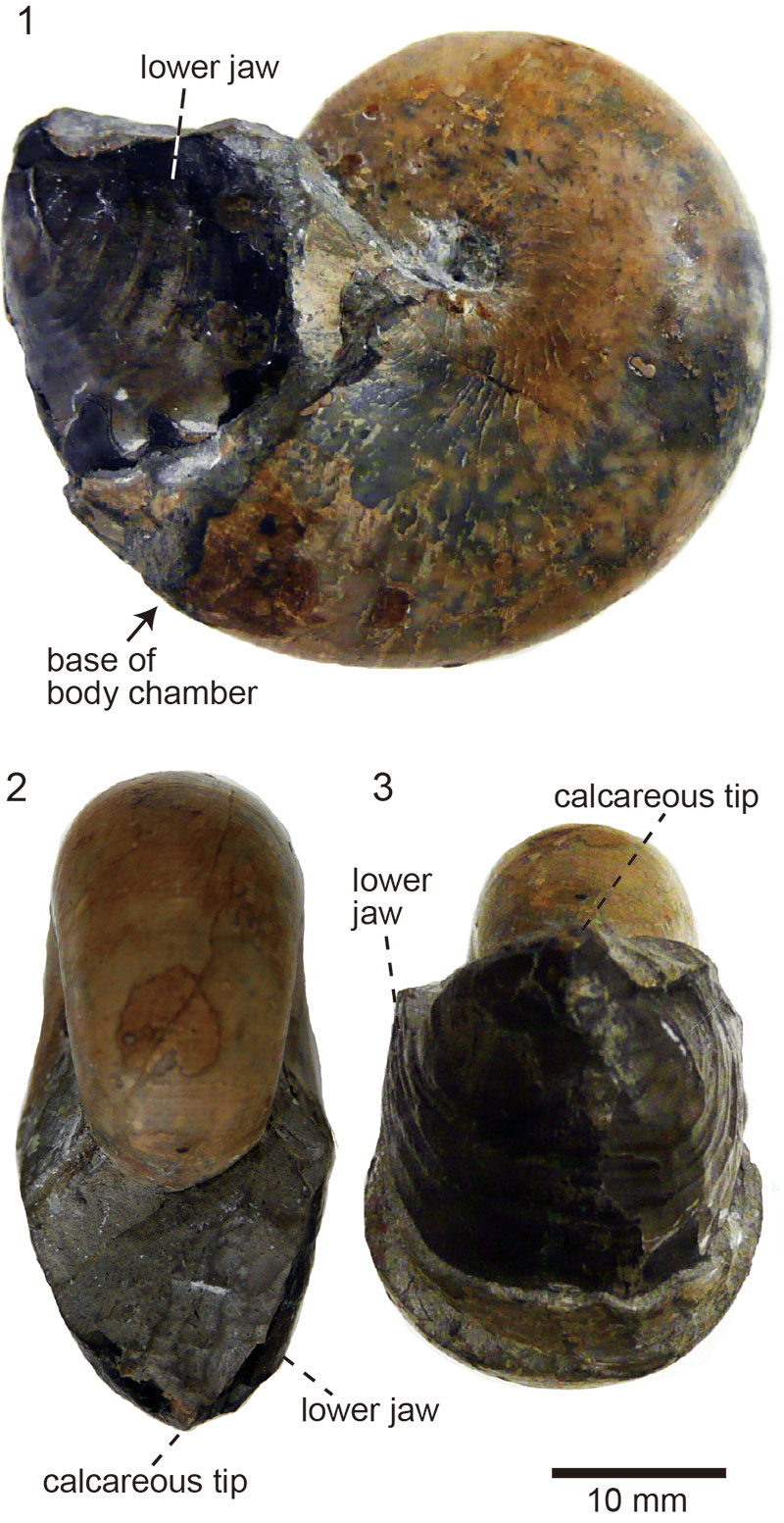
FIGURE 2.Reconstructed tomographic images of the specimen (1) and its internal structure in median section (2). The lower and upper jaws are enlarged in (3) and (4), respectively.
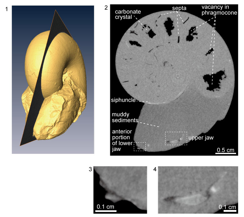
FIGURE 3. Serial cross-sections of the body chamber portion of the specimen cut from the venter (1) to the dorsum (4), in which sectioned images of the upper jaw are shown. Note that the vertical stripes are due to the separated scanning.
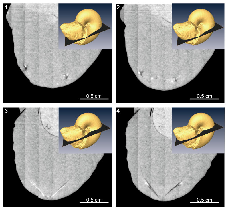
FIGURE 4. Linear absorption coefficient (LAC) of the internal portions of the specimen estimated by their mean luminance values in the tomographic images. The numbers (1)-(10) correspond to the materials in Table 1. The dashed lines indicate the known values for the materials (Chantler et al., 2005) that could be expected to be observed in the specimen. Note that glycine is the most dominant amino acid in jaws of Octopus vulgaris (Hunt and Nixon, 1981). The relationship between LAC values and luminance values is based on the assumption that the LAC values for the surrounding air are zero and that the crystals precipitated in the phragmocone are calcite.
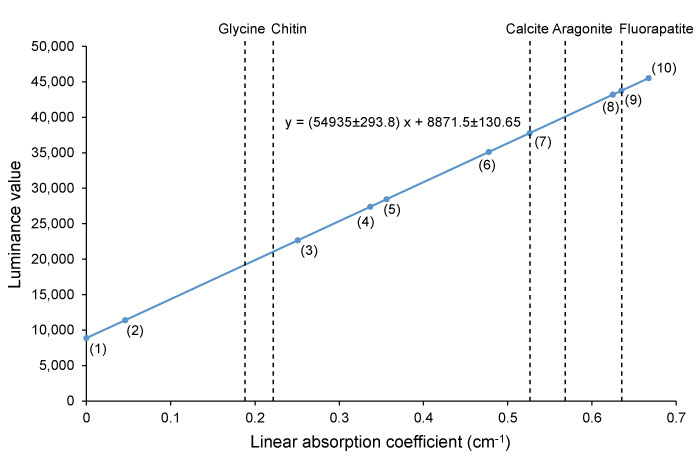
FIGURE 5. Three-dimensional reconstruction of the upper and lower jaws preserved in the body chamber of the specimen. The reconstructed parts are inside the specimen (1). The jaws are preserved close to each other (2).
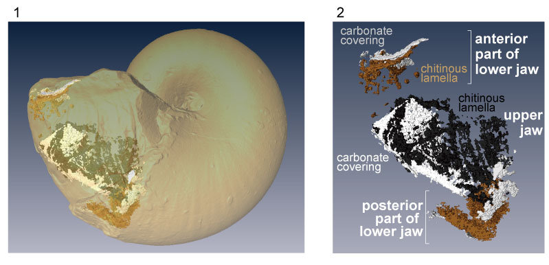
FIGURE 6. Result of segmentation of the lower jaw of the specimen, from lateral view which is restricted to its anterior and posterior portion (1). Three-dimensional reconstruction (2) suggests a wide distribution of calcareous material. The outer calcareous layer on the outer “chitinous” layer is partly taken off in (2). The transverse section of the area indicated as a square in (1) shows that the calcareous covering of the lower jaw also covers the internal surface of the “chitinous” lamella (3). The abbreviation is indicated in (2).
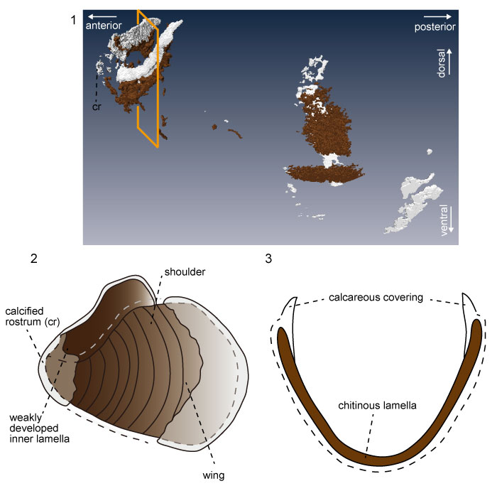
FIGURE 7. Result of segmentation of the upper jaw of the specimen, from frontal (1), rear (2), left-lateral (3) views and the transverse section of the area (4) indicated as a square in (3). The three-dimensional reconstruction (5) shows areal distributions of the “chitinous” lamellae and the calcareous covering. The reconstruction of the transverse section (6), which corresponds to (4), shows the architecture of the outer lamella. The abbreviations are indicated in (5).
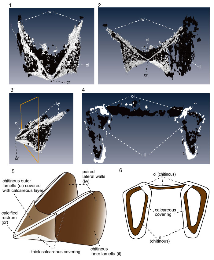
TABLE 1. Linear absorption coefficient (LAC) in the tomographic images of the Phyllopachyceras ezoensis. The numbers (1)-(10) correspond to plots in Figure 4.
| mean | minimum | maximum | standard deviation | |
| air surronding the specimen (1) | 0.000 | -0.001 | 0.003 | 0.0015 |
| phragmocone vacancy (2) | 0.046 | 0.027 | 0.068 | 0.0123 |
| septa (3) | 0.251 | 0.199 | 0.343 | 0.0467 |
| lower jaw (dark-colored part) (4) | 0.337 | 0.317 | 0.351 | 0.0103 |
| upper jaw (dark-colored part) (5) | 0.356 | 0.333 | 0.384 | 0.0149 |
| sediments in body chamber (6) | 0.478 | 0.473 | 0.483 | 0.0040 |
| carbonate crystal in phragmocone (7) | 0.526 | 0.516 | 0.538 | 0.0067 |
| siphuncle (8) | 0.625 | 0.610 | 0.645 | 0.0136 |
| upper jaw (light-colored part) (9) | 0.635 | 0.581 | 0.708 | 0.0525 |
| lower jaw (light-colored part) (10) | 0.667 | 0.633 | 0.706 | 0.0262 |
Non-destructive analysis of in situ ammonoid jaws by synchrotron radiation X-ray micro-computed tomography
Virtually all modern cephalopod mollusks possess a well-developed jaw, consisting of upper and lower elements, and a radula as their primary feeding organs. These structures are housed in a muscular organ called the buccal mass within the digestive tract and allow the mollusk to bite and shear their prey. Fossilized remains of jaws and radulae are occasionally preserved within the body chambers of ammonoid conchs, but complete excavation of them is difficult as typically they are embedded in a consolidated sedimentary matrix. This study introduces for the first time a three-dimensional (3D) reconstruction of the jaw of the Late Cretaceous phylloceratid ammonoid, Phyllopachyceras ezoensis, created using high-resolution synchrotron radiation X-ray tomography. Our analysis suggests that both the upper and lower jaws of the species were originally made of a chitin-protein complex similar to that of modern cephalopods, but their outer surfaces are wholly covered by a calcareous material. The overall jaw architecture resembles that of other Mesozoic ammonoids, except for the development of a calcareous covering on both the upper and lower jaws, which appears to reflect the predatory-scavenging feeding habits of the species. This and previous work suggest that the overall morphology and composition of the jaw in Mesozoic Ammonoidea have been developed under genetic and functional factors.
Resumen en Español
Análisis no destructivo de mandíbulas de ammonoideos in situ por microtomografía computarizada de rayos-X con radiación de sincrotrón
Presentamos la tomografía de rayos X de alta resolución con radiación de sincrotrón para la reconstrucción tridimensional no destructiva del aparato mandibular preservado en la cámara que alberga el cuerpo del ammonoideo Phylloceratidae del Cretácico Tardío Phyllopachyceras ezoensis. El análisis de las imágenes de rayos X usando la estimación del coeficiente de absorción lineal revela que la mandíbula superior consiste principalmente de láminas internas y externas compuestas de carbonato apatito, que originalmente pudo haber sido un complejo de quitina-proteína, con bordes angulosos de material calcítico grueso. Las características morfológicas indican que el aparato mandibular de esta especie es del tipo rhynchaptychus. La arquitectura tridimensional del aparato mandibular de estos especímenes es similar a la de otros ammonoideos, excepto por el desarrollo de un grueso depósito calcificado tanto en la mandíbula superior como en la inferior, lo que puede ser considerado como una evidencia de hábitos alimenticios depredadores para la especie. Las características de la mandíbula de esta especie parecen haber tenido constricciones de factores filogenéticos y morfofuncionales.
Palabras clave: radiación de sincrotrón; tomografía computarizada de rayos X; Ammonoidea; Cretácico; aparato mandibular; Phylloceratina
Traducción: Enrique Peñalver (Sociedad Española de Paleontología)
Résumé en Français
text
Translator: Kenny J. Travouillon or Antoine Souron
Deutsche Zusammenfassung
Zerstörungsfreie Untersuchung von in situ Ammonitenkiefern mit Synchotron Mikrocomuptertompgrafie
Wir präsentieren erstmals hochauflösende Synchotron-Röntgentomografie für eine zerstörungsfreie, dreidimensionale Rekonstruktion des Kieferapparates - erhalten innerhalb der Wohnkammer - des spätkretazischen phylloceratiden Ammonoiden Phyllopachyceras ezoensis. Untersuchungen der Röntgenbilder mit einem linearen Absorptionskoeffizienten zeigen, dass der Oberkiefer hauptsächlich aus inneren und äußeren Lamellen aus Karbonat-Apatit besteht, der möglicherweise ursprünglich ein Chitin-Protein Komplex mit abgewinkelten Rändern aus dickem kalzitischem Material war. Die morphologischen Merkmale weisen darauf hin, dass der Kieferapparat dieser Art vom Rhynchaptychus-Typ war. Die dreidimensionale Architektur des Kieferapparats dieser Stücke gleicht dem anderer Ammonoiden, bis auf eine dicke kalzifizierte Ablagerung in Ober-und Unterkiefer, die wahrscheinlich die räuberisch-aasfresserischen Fressgewohnheiten dieser Art unterstützten. Die Kiefermerkmale dieser Art scheinen sowohl durch phylogenetische als auch funktional-morphologische Faktoren begrenzt zu sein.
Schlüsselwörter: Synchotronstrahlung; Röntgencomputertomografie; Ammonoidea; Kreide; Kieferapparat; Phylloceratina
Translator: Eva Gebauer
Arabic
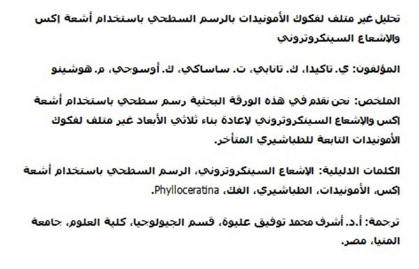
Translator: Ashraf M.T. Elewa
-
-
-
Review: The Princeton Field Guide to Mesozoic Sea Reptiles
 The Princeton Field Guide to Mesozoic Sea Reptiles
Article number: 26.1.1R
April 2023

 Poster Winners 2024