FIGURE 1. Ultrastenos huberi, QM F31060, holotype, maxillary rostrum. A, dorsal view. B, ventral view. C, left lateral view. Abbreviations: appal, antepenultimate premaxillary alveolus; cl, canthus lacrimalis; fpal, first premaxillary alveolus; fr, frontal; idp, interdental reception pit; ju, jugal; lpal, last premaxillary alveolus; la, lacrimal; lac, lacrimal canal; lr, lateral ridge; mal, maxillary alveolus; mt, maxillary tooth; mr, medial ridge; mx, maxilla; na, nasal; nar, naris; om, orbital margin; ppt, penultimate premaxillary tooth; pm, premaxilla; pmf, premaxillary fenestra; pf, prefrontal; rn, reception notch for dentary tooth 4; rp, reception pit; rt, replacement tooth; sof, suborbital fenestra. Note that the premaxillary teeth are not numbered because the loss of a tooth position renders numbering ambiguous. Scale bar equals 50 mm.
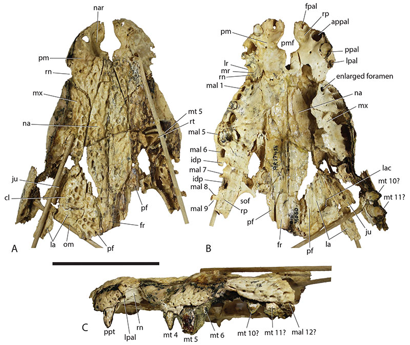
FIGURE 2. Ultrastenos huberi, QM F42665, significant fragments of the holotype of U. willisi Stein et al. A, B, basicranial fragment with displaced adherent left pterygoid in right lateral (A) and occipital views (B). C, D, right temporal fragment in lateral (C) and dorsal views (D). E, G, I, posterior end of left mandibular ramus in lateral (E), medial (G) and dorsal (I) views. F, H, disarticulated right suranguar and angular in medial (F) and lateral (H) views. J, right surangular in dorsal view (anterior to the left). Scale bar equals 50 mm.
FIGURE 3. Ultrastenos huberi, QM F31075, White Hunter Cranial Form 1, posterior cranial fragment. A, B, dorsal view as a photograph (A), and as an annotated photograph with interpretive overlay (B). C, D, occipital view as a photograph (C), and an annotated photograph with interpretive overlay (D). Abbreviations: bo, basioccipital; fa, foramen aereum; fm, foramen magnum; fr, frontal; mf, metotic foramen; mptf, median pharyngeal tube foramen; ncr, nuchal crest; oc, occipital condyle; oto, otoccipital; pa, parietal; parp, paroccipital process; pbs, parabasisphenoid; pcf, posterior carotid foramen; po, postorbital; pop, postoccipital process of the supraoccipital; ptf, posttemporal fenestra; ptyf, pharyngotympanic foramen; q, quadrate; qc, quadrate condyle; qjss, sutural surface for articulation with the quadratougal; so, supraoccipital; sq, squamosal; stf, supratemporal fenestra; XIIp, posterior foramen for cranial nerve XII (posterior hypoglossal foramen). Scale bar equals 50 mm.
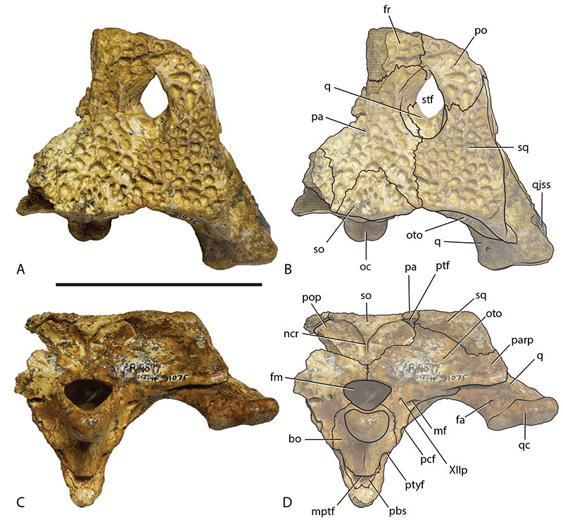
FIGURE 4. Ultrastenos huberi, QM F31075, White Hunter Cranial Form 1, posterior cranial fragment. A, B, right lateral view as a photograph (A), and as an annotated photograph with interpretive overlay (B). C, D, right ventrolateral view as a photograph (C), and an annotated photograph with interpretive overlay (D). Abbreviations: bo, basioccipital; boa, bony otic aperture; cb, caudal bridge of the laterosphenoid; cp, capitate process of the laterosphenoid; lb, lateral bridge of the laterosphenoid; ls, laterosphenoid; mf, metotic foramen; npd, posterior wall of the nasopharyngeal duct; oc, occipital condyle; oto, otoccipital; pa, parietal; pbs, parabasisphenoid; pcf, posterior carotid foramen; po, postorbital; pro, prootic; psr, base of parasphenoid rostrum; pt, pterygoid; ptyf, pharyngotympanic foramen; q, quadrate; qc, quadrate condyle; qjss, sutural surface for articulation with the quadratougal; sq, squamosal; ss, squamosal sulcus; stf, supratemporal fenestra; styf, subtympanic foramen; suof, supraorbital foramen; V, foramen for cranial nerve V (trigeminal foramen); XIIp, posterior foramen for cranial nerve XII (posterior hypoglossal foramen). Scale bar equals 50 mm.
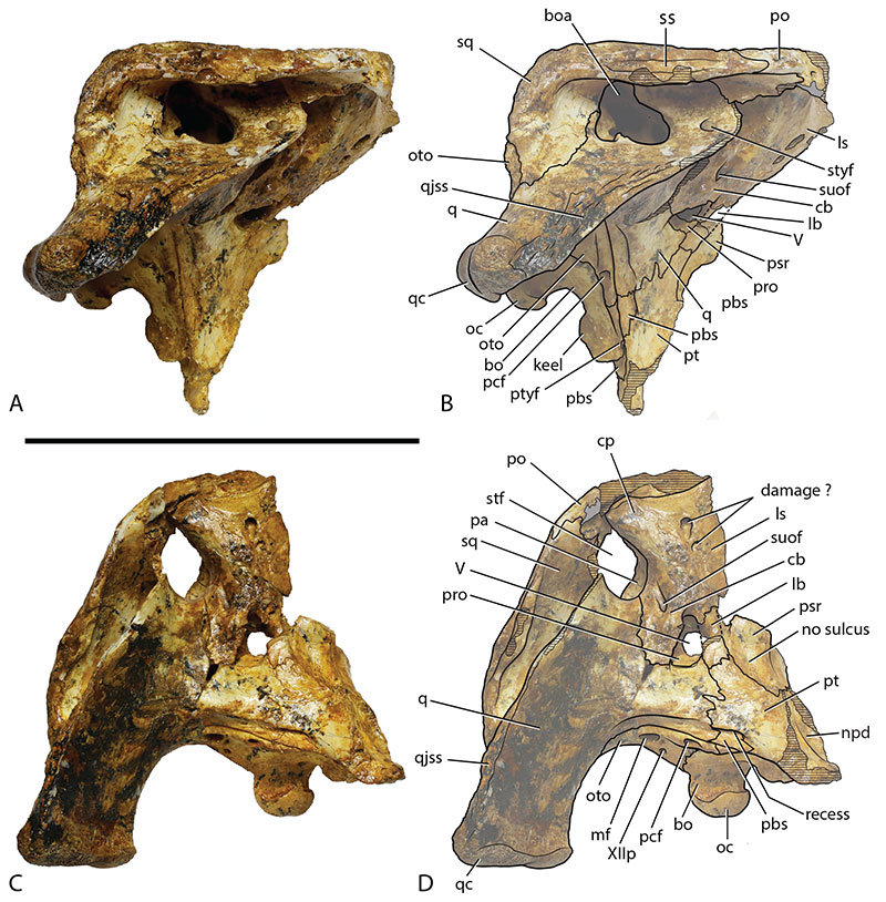
FIGURE 5. Ultrastenos huberi QM F31076, White Hunter Cranial Form 1, posterior cranial fragment. A, dorsal view. B, ventral view. C, occipital view. D, right lateral view. Abbreviations: bo, basioccipital; boa, bony otic aperture; cb, caudal bridge of the laterosphenoid; cp, capitate process of the laterosphenoid; cqp, cranioquadrate passage; fa, foramen aereum; fm, foramen magnum; fr, frontal; I, foramen for cranial nerve I (olfactory foramen); II, foramen for cranial nerve II (optic foramen); III+orb, common foramen for cranial nerve III (oculomotor nerve) and the orbital artery and vein; ioc, incisure of the cranioquadrate passage in the otic aperture; IV, foramen for cranial nerve IV (trochlear foramen); lb, lateral bridge of the laterosphenoid; ls, laterosphenoid; mf, metotic foramen; ncr, nuchal crest; oto, otoccipital; ott, olfactory tract trough; pa, parietal; parp, paroccipital process; pbs, parabasisphenoid; pcf, posterior carotid foramen; pfss, sutural surface for articulation with the prefrontal; plp, posterolateral process of the sqamosal; po, postorbital; pob, postorbital bar; pop, postoccipital process of the supraoccipital; ptf, posttemporal fenestra; q, quadrate; qjss, sutural surface for articulation with the quadratougal; so, supraoccipital; sq, squamosal; ss, squamosal sulcus; stf, supratemporal fenestra; V, foramen for cranial nerve V (trigeminal foramen); XIIa, anterior foramen for cranial nerve XII (anterior hypoglossal foramen); XIIp, posterior foramen for cranial nerve XII (posterior hypoglossal foramen). Scale bar equals 50 mm.
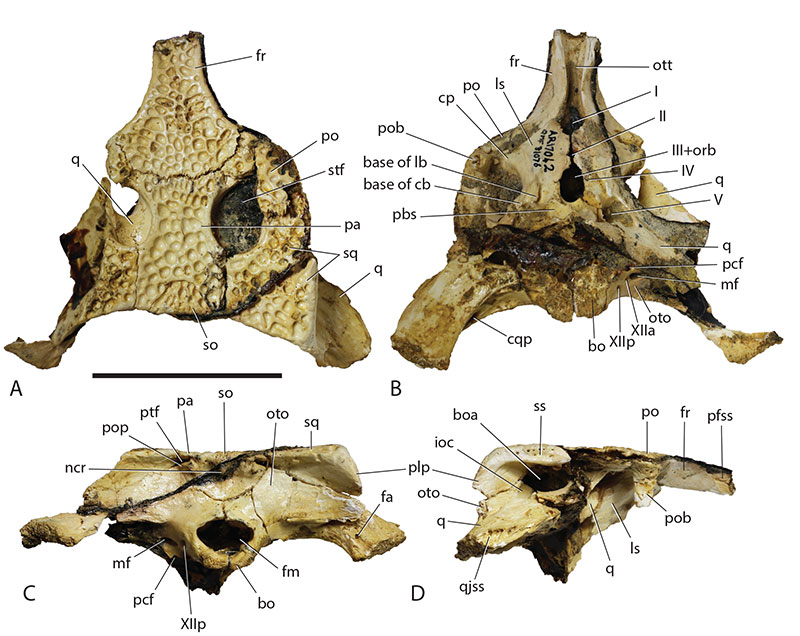
FIGURE 6. Comparison of the occipital proportions of various crocodylians. A: Osteolaemus tetraspis NTM unregistered cast, a brevirostrine form. B: Ultrastenos huberi, QM F31075. C. Tomistoma schlegelii, NTM P3142 (cast), a slender longirostrine form. Images are not to scale.
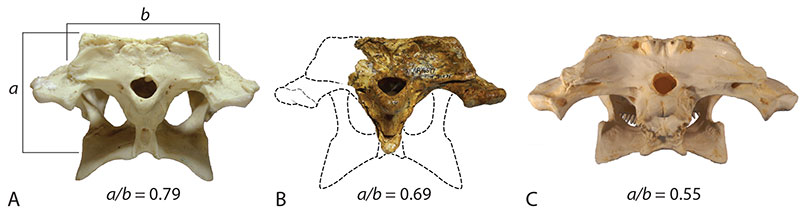
FIGURE 7. Ultrastenos huberi, QM F31060 (holotype, maxillary rostrum) and QM F31076 (posterior cranial table) placed in approximate life position, relative to each other, in dorsal view. Scale bar equals 50 mm.

FIGURE 8. Dermal bone ornamentation of Baru wickeni and Ultrastenos huberi compared. A: Explanatory drawing of the skull of Baru wickeni in dorsal view with frontal and right squamosal marked in red and the areas shown in (B) and (C) marked by boxes. B: Baru wickeni (NTM P91171-1), frontal in dorsal view showing the ornamentation. C: Baru wickeni (NTM P902-4), right squamosal in dorsal view showing the ornamentation. D: Explanatory drawing of the skull of Ultrastenos huberi in dorsal view with frontal and right squamosal marked in red and the areas shown in (E) and (F) marked by boxes. E: Ultrastenos huberi (QM F F31076), frontal in dorsal view showing the ornamentation. F: Ultrastenos huberi (QM F F31075), right squamosal in dorsal view showing the ornamentation. Scale bars in (B, C, E, F) equal 10 mm. (A, D) are not to scale.
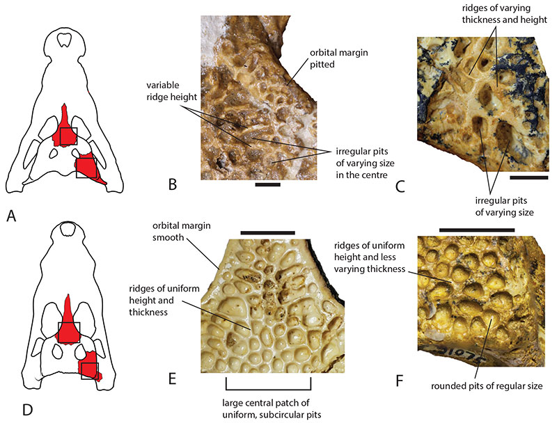
FIGURE 9. Comparison of Quinkana timara to White Hunter Cranial Form 2, QM F31079, here referred to Q. meboldi Willis. A: Q. timara (NTM P8695-114), right squamosal. B: Q. timara (NTM P9464-177), parietal margin of the right supratemporal fenestra. C, D: Q. meboldi (QM F31079), posterior cranial fragment. B, right postorbital. C, parietal margin of the right supratemporal fenestra. Scale bars equal 10 mm.
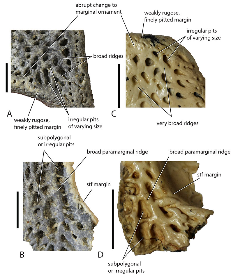
FIGURE 10. Ultrastenos sp. “Bullock Creek Taxon”, isolated specimens from the Middle Miocene, Bullock Creek Local Fauna, Northern Territory. A, B: NTM P87115-3, posterior cranial fragment. A, right lateral view. B dorsal view. C: NTM P9660, right maxilla in ventral view. D: NTM P895-15, left mandibular ramus in lateral view. Derived character states shared with U. huberi Willis: 1, posterolateral process of the squamosal descends at a steep angle (74⁰); 2, medial, posterior and lateral margins of the supratemporal fenestra rounded and curve gradually into one another; 3, enlarged anterior maxillary foramen; 4, transverse palatal premaxillomaxillary suture lying anterior to the level of the first maxillary alveolus; 5, absence of a posterior lamina of the maxilla separating the anterior tip of the ectopterygoid from the margin of the suborbital fenestra; 6, dermal ornament consisting of regular, subcircular pits separated by walls of even height; 7, posterior extent of the dentary posterior to the posterior margin of the external mandibular fenestra; 8, length of the reduced external mandibular fenestra is less than 25% of the distance between the dentary toothrow and the anterior margin of the mandibular glenoid. Scale bars equal 50 mm.
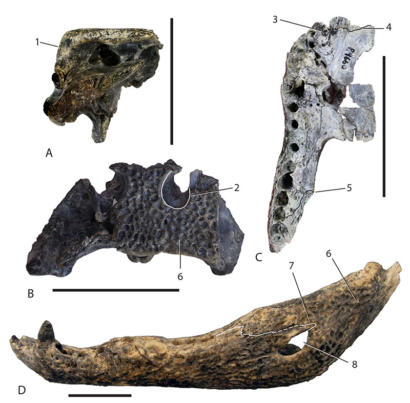
FIGURE 11. Comparison of the squamosal in right lateral view of Ultrastenos huberi with various species of Baru, showing the angle of descent of the posterolateral process and sculptural elements of the lateral surface. A: Ultrastenos huberi, QM F31075. Note that the lateral squamosal sulcus is both broad and extensive and the posterolateral process descends at a steep angle. B: Baru wickeni, NTM P9464-10 (image reversed for comparison). Note that the lateral surface is largely flat with a short, narrow sulcus and the posterolateral process descends at a shallow angle. C: Juvenile Baru iylwenpeny (NTM P6478). Note the flat lateral surface with a short, narrow sulcus and the shallow angle of descent. D: Adult Baru iylwenpeny (NTM P6515) Note the flat lateral surface with a short, narrow sulcus and the shallow angle of descent shared with other Baru specimens. Note also the presence of a posterolateral boss shared with (B) but not (C). Dashed lines demarcate each posterolateral boss. Hatched areas demarcate lateral sulci. Scale bars equal 20 mm.
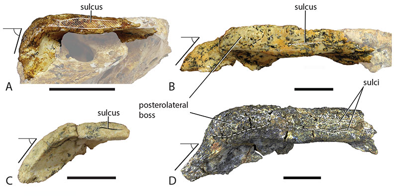
FIGURE 12. Ultrastenos huberi, shared derived character states. A-D: QM F42665, holotype of Ultrastenos willisi. A, B, Right temporal fragment in dorsal view (A) and lateral view (B). C, D, ventral portion of braincase (with attached, displaced fragment of right pterygoid) in right lateral view (C) and occipital view (D). E-H: QM F31075, ‘WH Cranial Form 1’. E, F, right temporal region in dorsal view (E) and lateral view (F). G, H, ventral portion of braincase in right lateral view (G) and occipital view (H). Numbered character states: 1, distance from the ventrolateral tip of the paroccipital process to the dorsal margin of the quadrate condyle is less than the transverse width of the quadrate condyle (shown by rectangular bracket); 2, short posterolateral process of the squamosal descends at an angle steeper than 65⁰; 3, parabasisphenoid-pterygoid suture recessed within a sulcus; 4, enlarged sagittal, ventral keel of the basioccipital prominent in lateral view; 5, otoccipital-basioccipital suture (marked with red line) fails to cross the synovial surface of the occipital condyle. Scale bars equal 20 mm.
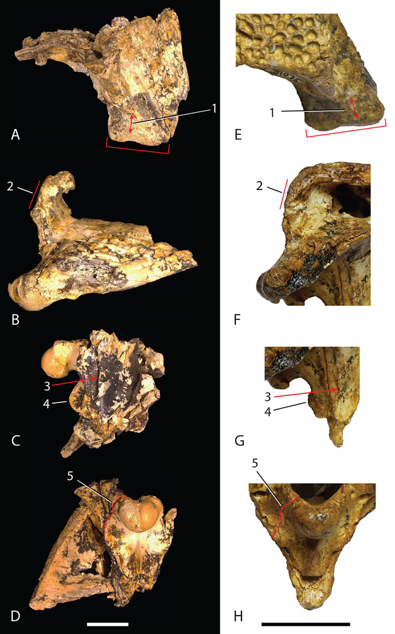
FIGURE 13. Single most parsimonious tree found by Ristevski et al. (2023) in a traditional search of their dataset using implied weights (k = 25), showing U. huberi sharing a more recent common ancestry with mekosuchine genera such as Mekosuchus than with Baru. Large, polyspecific, named clades that do not include Mekosuchinae have been collapsed into single branches (names in bold type) to simplify the diagram. Abbreviations: CI, consistency index; MPT, most parsimonious tree; #OTUs, number of operational taxonomic units; RI, retention index. Redrawn from figure 24 in Ristevsi et al. (2023).
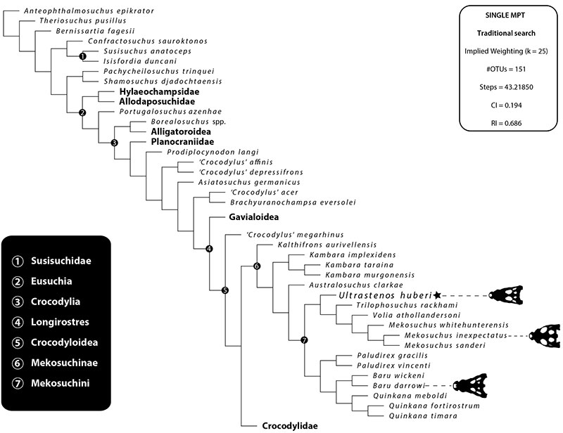
FIGURE 14. Reconstruction of Ultrastenos huberi based largely upon QM F31060 and QM F31076 and scaled to the size of these two specimens. A, occipital view. B, dorsal view. C, ventral view. D, right lateral view. Abbreviations: bo, basioccipital; boa, bony otic aperture; cl, canthus lacrimalis; ect, ectopterygoid; fa, foramen aëreum; fr, frontal; itf, infratemporal fenestra; ju, jugal; la, lacrimal; mx, maxilla; mal, maxillary alveolus; na, nasal; nar, naris; oc c, occipital condyle; oto, otoccipital; pa, parietal; pal, palatine; pbs, parabasisphenoid; pm, premaxilla; pmf, premaxillary fenestra; po, postorbital; pf, prefrontal; pt, pterygoid; ptf, posttemporal fenestra; q, quadrate; q co, quadrate condyle; qj, quadratojugal; rp, reception pit; so, supraoccipital; sof, suborbital fenestra; sq, squamosal; stf, supratemporal fenestra. Scale bar equals 100 mm.
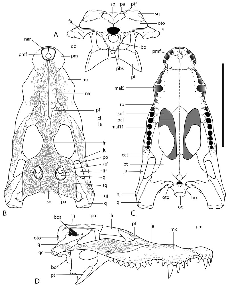
FIGURE 15. Ultrastenos huberi, rostral fragments. A-B, QM F31064, fragment of right maxilla. A, ventral view. B, lateral view. C-F, QM F31061, rostral fragment, including right premaxilla and fragment of right maxilla. C, ventral view. D, dorsal view. E, lateral view. F, medial view. Abbreviations: appa, antepenultimate premaxillary alveolus; ect sut, sutural surface for articulation with the ectopterygoid; idp, interdental reception pit; lpal, last premaxillary alveolus; lr, lateral ridge; mal, maxillary alveolus; mn, margin of the naris; mr, medial ridge; msy, articular surface for maxillary symphysis; nar, naris; nc, nasal cavity; nsy, articular surface for nasal symphysis; pm, premaxilla; pmf, premaxillary fenestra; ppal, penultimate premaxillary alveolus; psy, articular surface for premaxillary symphysis; rp, reception pit; sofm, margin of the suborbital fenestra; Vpal, maxillary foramen for palatine ramus of cranial nerve V2 (maxillary division of the trigeminal nerve), note that the margins of this foramen are broken, thus enlarging the apparent size of the foramen. Scale bar equals 20 mm.
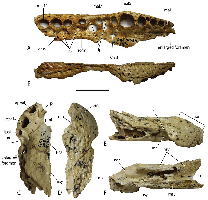
FIGURE 16. Ultrastenos huberi, QM F61097, cranial fragment. A, dorsal view. B, ventral view. C, left lateral view. Abbreviations: cl, canthus lacrimalis; ect, ectopterygoid; fr, frontal; ju, jugal; la, lacrimal; mal, maxillary alveolus; mx, maxilla; na, nasal; or, orbit; pf, prefrontal; rp, reception pit; sof, suborbital fenestra; Vpal, foramen for palatine ramus of cranial nerve V2 (maxillary division of the trigeminal nerve). Scale bar equals 50 mm.
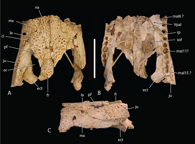
FIGURE 17. Ultrastenos huberi, pterygoid fragments. A: QM F42665, right pterygoid in articulation with right ectopterygoid in ventral view. B: QM F31076, pterygoid pair in ventral view. Abbreviations: ect, ectopterygoid; ecss, sutural surface for articulation with the ectopterygoid; palss, sutural surface for articulation with the palatine; sofm, margin of the suborbital fenestra; vptr, ventral pterygoid ridge. Scale bar equals 10 mm.
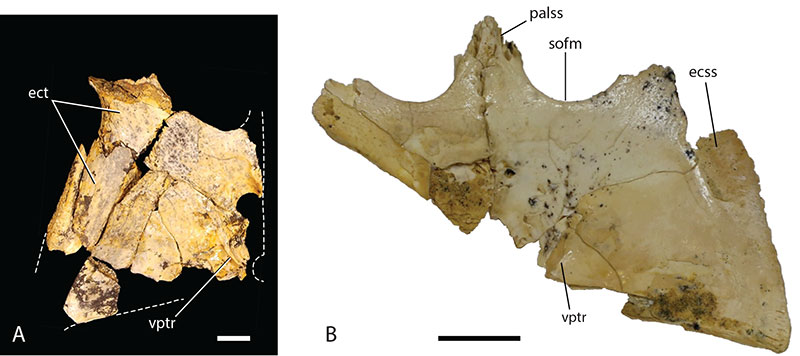
FIGURE 18. Quadrate condyles of Ultrastenos huberi and Baru wickeni compared. A: Ultrastenos huberi, QM F31075, right quadrate condyle, photograph taken normal to the articular plane of the quadrate condyle (dorsal is towards the top of the page). The flattened dorsomedial margin is marked by a rectangular bracket and the dorsal indentation between the medial and lateral hemicondyles is marked by triangular arrow. B: Baru wickeni, NTM P911, right quadrate condyle, photograph taken normal to the articular plane of the quadrate condyle (dorsal is towards the top of the page). Note that the flattened dorsomedial margin, and the dorsal indentation are absent. The small notch visible on the medial margin is due to damage. Scale bar equals 10 mm.
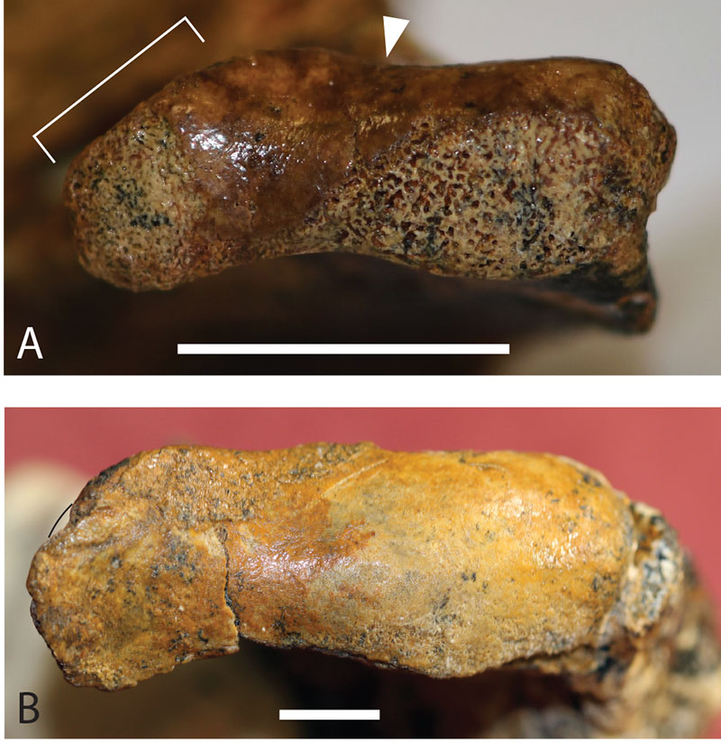
FIGURE 19. Ultrastenos huberi, reconstructed left mandibular ramus based largely upon QM F61096 and QM F42665, scaled to the size of the former. A, dorsal view. B, medial view (reversed for comparison). C, lateral view. Abbreviations: an, angular; ar, articular; c, coronoid; d, dentary; dal, dentary alveolus; dsy, symphyseal surface of the dentary; emf, external mandibular fenestra; fa, foramen aëreum; fic, foramen intermandibularis caudalis; gl, mandibular glenoid; ms, Meckelian sulcus; rap, retroarticular process; sa, suraqngular; sp, splenial. Scale bar equals 50 mm.
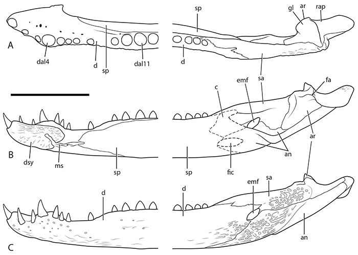
FIGURE 20. Ultrastenos huberi, anterior mandibular fragments. A-C: QM F61096, articulated left and right dentaries. A, dorsal view. B, right lateral view. C, posterior view. Dashed line in (C) represents the anterior margin of the splenial sutural scar. D, E: QM F31068, left dentary fragment. D, dorsal view. E, medial view. White arrows in (E) indicate the anterior tip of the dorsal and ventral anterior splenial processes. F: QM F31069, symphyseal fragment of mandible in posteriomedial view. Abbreviations: avp, anterior ventral process of the splenial; d, dentary; da, dentary alveolus; dldsy, dorsal lobe of the dentary symphyseal surface; dsys, symphyseal surface of the dentary; dt, dentary tooth; ms, Meckelian sulcus; msy, mandibular symphysis; sp, splenial; spss, sutural surface for articulation with the splenial. Scale bar equals 20 mm.
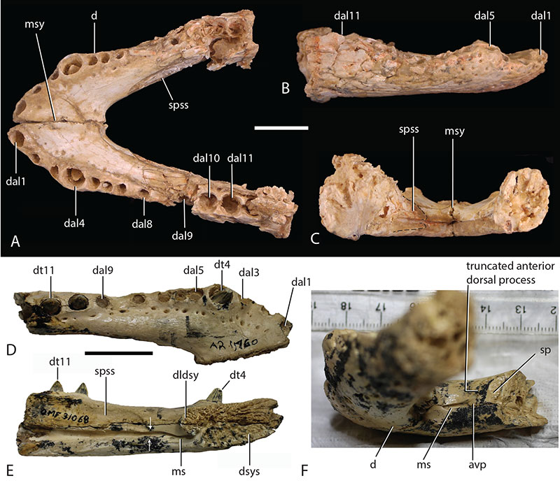
FIGURE 21. Baru wickeni, bones from the maxillary rostrum of small juveniles. A-C: QM F31062, right premaxilla. A, ventral view. B, dorsal view. C, lateral view. D, E: QM F31063, posterior fragment of right maxilla. D, ventral view. E, lateral view. The dashed line in (D) represents margins of the ectopterygoid sutural scar. Abbreviations: jss, sutural surface for articulation with the jugal; ma, maxillary alveolus; mss, sutural surface for articulation with the maxilla; mt, maxillary tooth; nar, naris; nm, narial margin; pa, premaxillary alveolus; pdp, posterior dorsal process of the premaxilla; pml, posterior medial lamina of the maxilla; rp, reception pit; sofm, margin of the suborbital fenestra. Scale bar equals 20 mm.
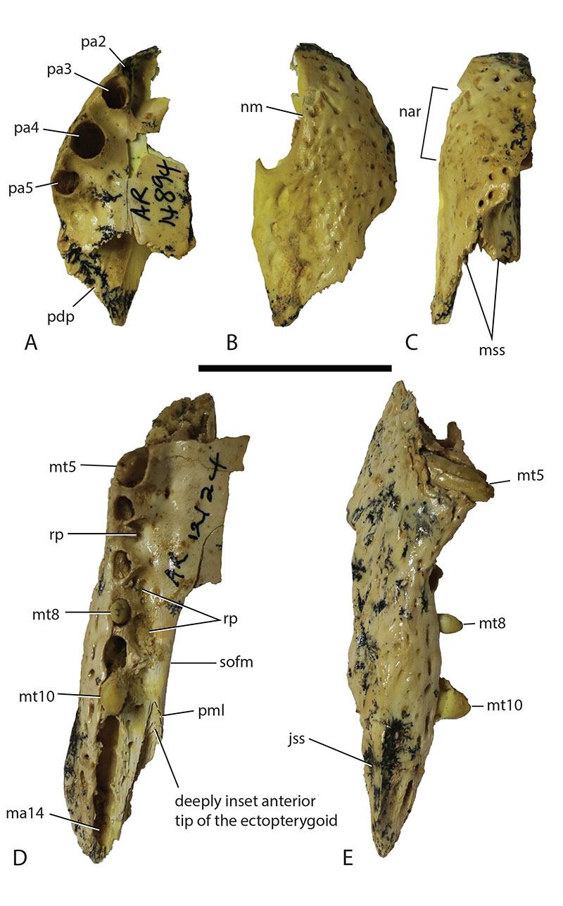
FIGURE 22. Reconstruction of the posterior skull of Ultrastenos huberi. A: Composite reconstruction showing marked anterior narrowing congruent with the slender longirostrine morphotype. Redrawn from Stein et al. (2016). B, C: QM F42665, temporal fragment from the holotype of U. willisi, mirror imaged to create reconstructions. B, temporal fragment arranged to match the reconstruction in (A). C, Alternative arrangement of the temporal fragment to match the brevirostrine reconstruction of U. huberi supported here. Abbreviations: ju, jugal; oto, otoccipital; q, quadrate; qj, quadratojugal; sq, squamosal. Hatched areas represent broken bone surfaces. Scale bar equals 50 mm.
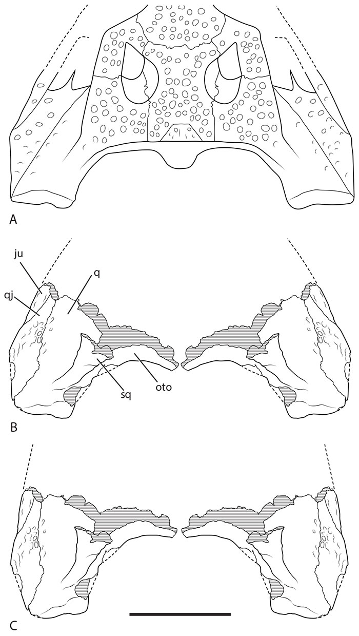
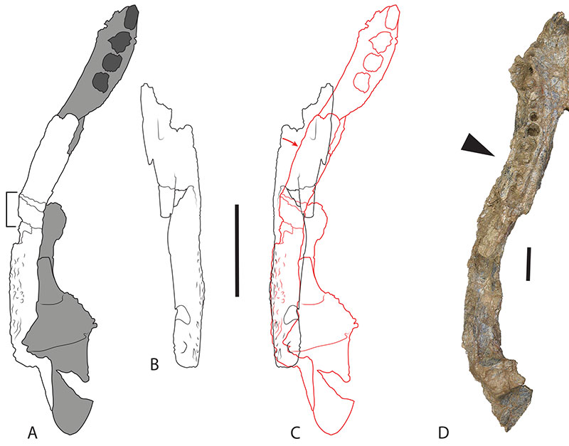
FIGURE 23. Curvature of the mandible of Ultrastenos huberi compared to Baru iylwenpeny Yates et al. A-C: Ultrastenos huberi, QM F42665, mandibular fragments. A, posterior fragment of the left mandibular ramus. B, right surangular. C, mirror image of the right surangular overlain onto the left mandibular ramus fragment (drawn in red). The left surangular in (A) is filled with white, the other bones are shaded grey. The rectangular bracket in (A) indicates a highly fractured and re-glued zone. Note the offset of the end anterior of the left ramus in (C), which has rotated medially relative to the right surangular (marked with red arrow). D: Baru iylwenpeny, NTM P2787, right mandibular ramus (image reversed). The triangle indicates an inflection point similar to that exhibited in QM F42665. Scale bar equals 50 mm.