FIGURE 1. Cranial anatomy of Indohyus indirae. A, skull, dorsal view (RR 601). B, skull, ventral view (RR 601). C, skull, dorsal view (RR 207). D, skull, ventral view (RR 207).
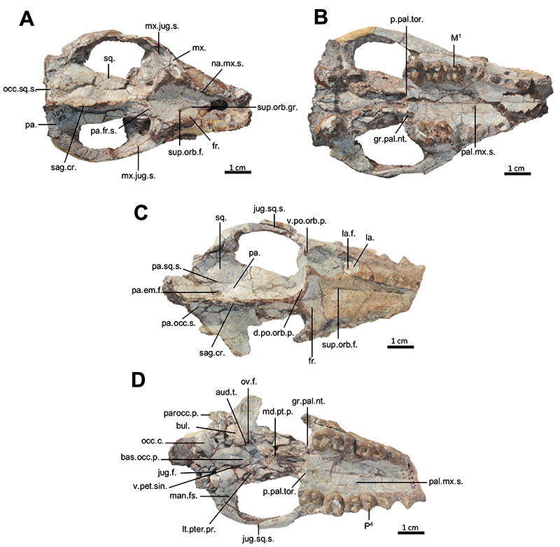
FIGURE 2. Cranial anatomy of Indohyus indirae. A, skull, left lateral view (RR 208). B, skull, right lateral view (RR 208). C, Oblique, ventral view (RR 209). For abbreviations see text.
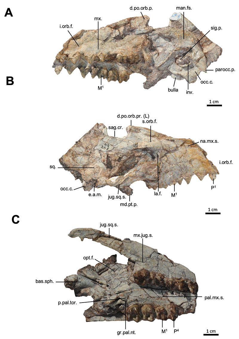
FIGURE 3. Cranial anatomy of Indohyus indirae. A, rostrum, dorsal view (RR 602). B, rostrum, ventral view (RR 602). C, Basicranium (RR 210). D, Juvenile skull with unassociated lower jaw (RR 262).
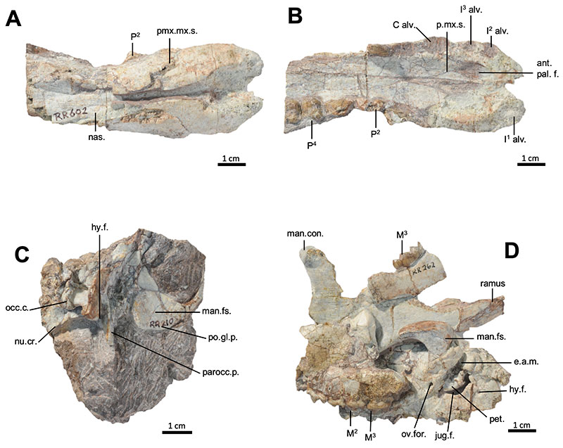
FIGURE 4. Annotated CT slices highlighting the cranial anatomy of Indohyus indirae (RR207), featuring detailed transverse sections of the skull. A, slice 403. B, slice 817. C, slice 1043. D, slice 1180.
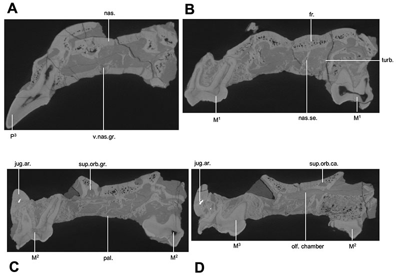
FIGURE 5. Annotated CT slices highlighting the cranial anatomy of Indohyus indirae (RR207), featuring detailed transverse sections of the skull. A, slice 1303. B, slice 2142. C, slice 2230. D, slice 2302.
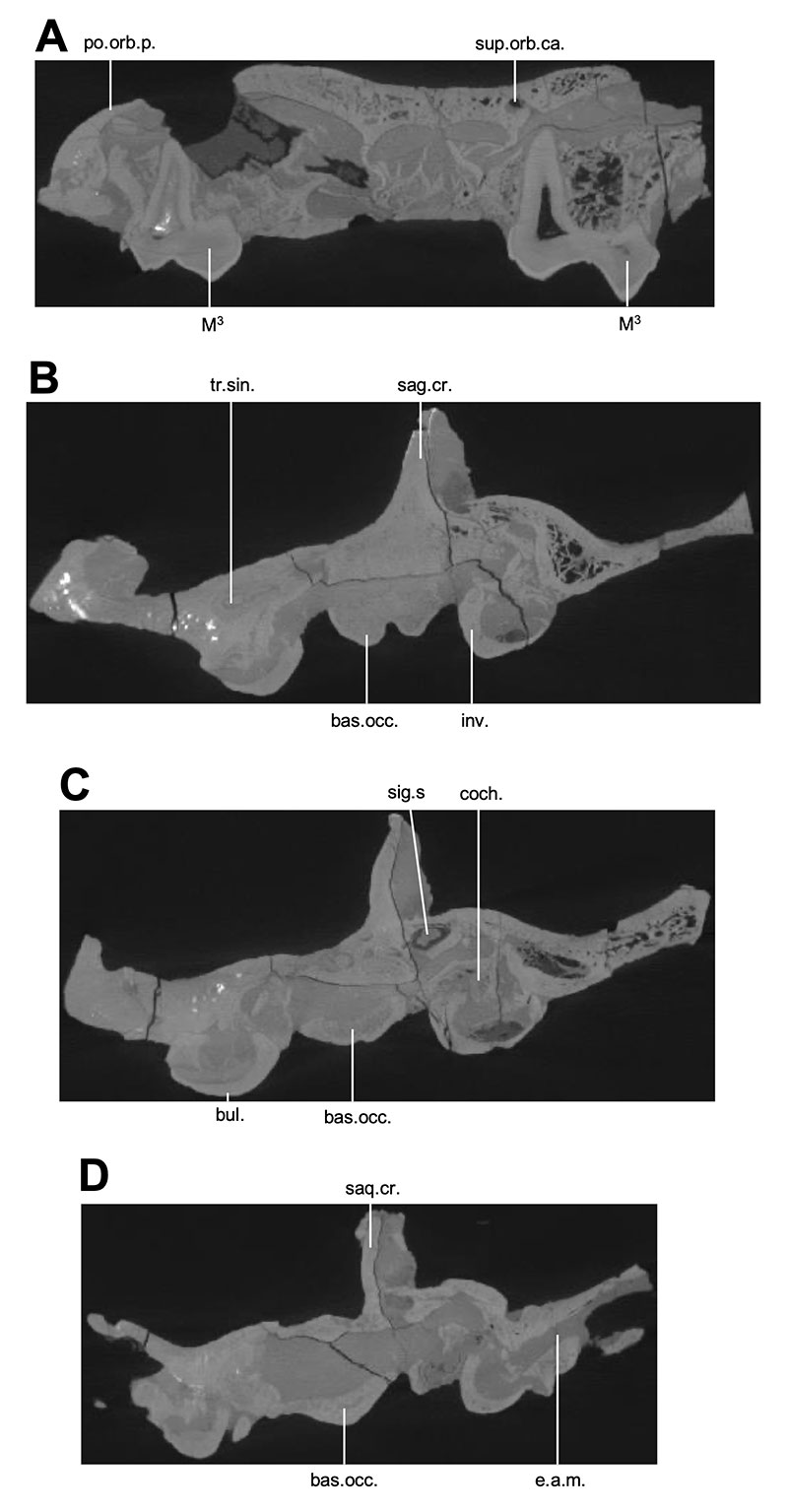
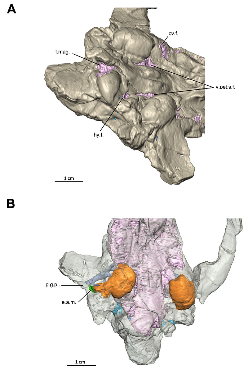
FIGURE 6. 3D rendering of the basicranium of Indohyus indirae in oblique (A) and vental (B) views. In A, the foramina opening into the endocast are highlighted in pink. In B, the bullae are depicted in orange.