FIGURE 1. Locality map and stratigraphic sections of the Otekaikea huata type locality. Arrow shows type locality. Foraminiferal zonation and geochronology are based on Graham et al. (2000).
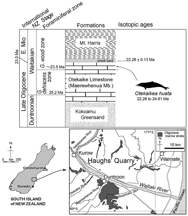
FIGURE 2. Dorsal views of the type skull, OU 22306, Otekaikea huata ( 1 and 2). 3, dorsolateral view, figure is not to scale.
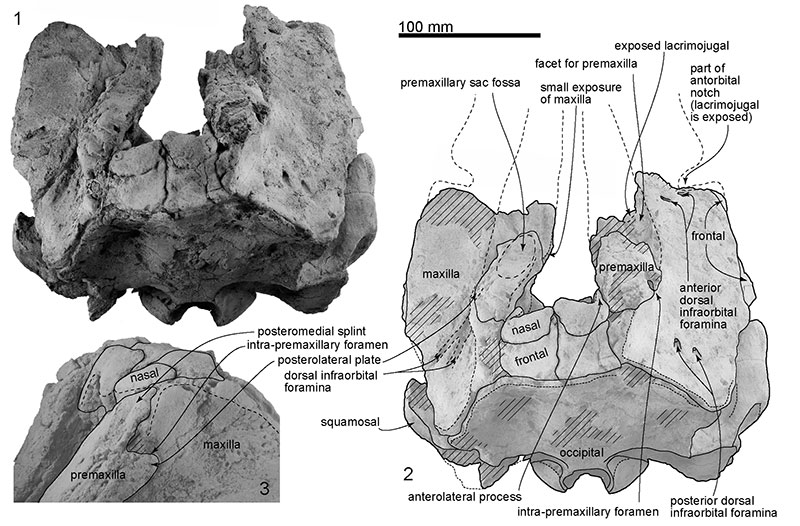
FIGURE 3. Ventral views of the type skull, OU 22306, Otekaikea huata. Pa, the parietal.
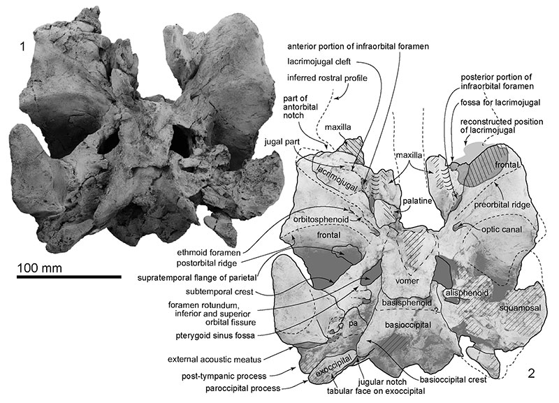
FIGURE 4. Anterior views of the type skull, OU 22306, Otekaikea huata.
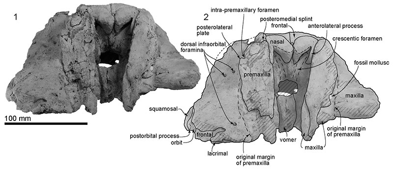
FIGURE 5. Posterior views of the type skull, OU 22306, Otekaikea huata.
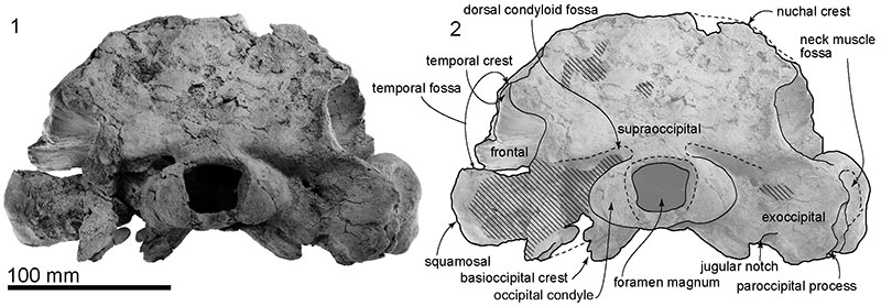
FIGURE 6. Right lateral views of the type skull, OU 22306, Otekaikea huata.
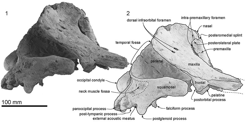
FIGURE 7. Disarticulated rostral elements, OU 22306, Otekaikea huata. 1, dorsal view. 2, left premaxilla lateral view. 3, right premaxilla lateral view. 4, ventral view. 5 and 6, detailed photos of the premaxilla.
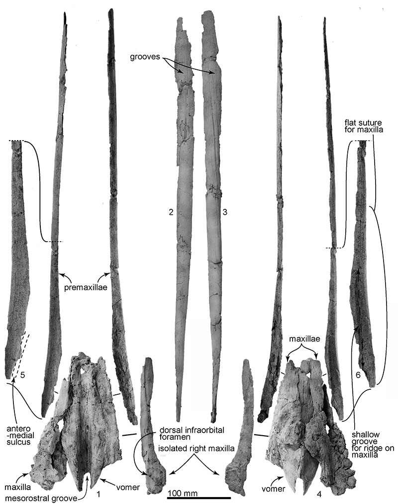
FIGURE 8. Skull of Otekaikea huata, OU 22306. Right 1, lateral view and 2, dorsal view.
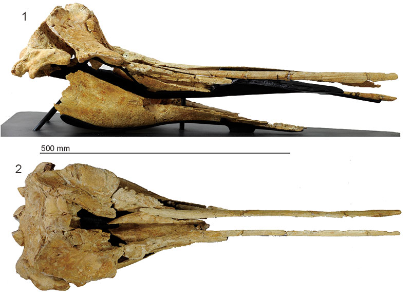
FIGURE 9. Details of right basicranium part, OU 22306, Otekaikea huata. 1 and 2, ventral view. 3 and 4, ventrolateral view. Ali, alisphenoid. Bo, basioccipital. Bs, basisphenoid. Pa, parietal. Vo, vomer.
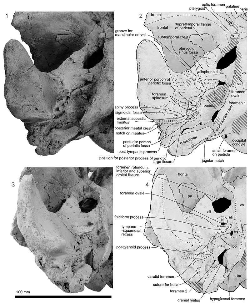
FIGURE 10. The type teeth, OU 22306, Otekaikea huata. Scales vary as indicated, depending on specimen size. 1 and 2, largest incisor. 1, worn surface view. 2, lateral view. 3 and 4, two long tusks, from worn surface. 5 and 6, two medium teeth. 7-9, single rooted smaller medium teeth. 10-12, buccolingually flattened teeth. 13 and 14, tooth with small denticles. 13, buccal view. 14, lingual view.
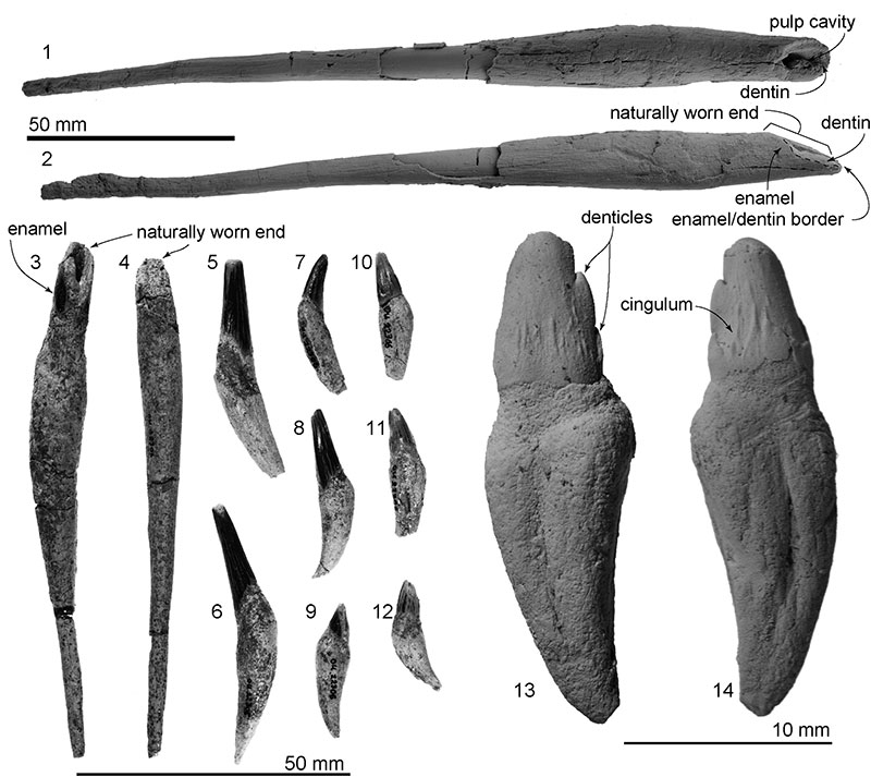
FIGURE 11. Periotic in original position in the right squamosal, ventral view, OU 22306, Otekaikea huata.
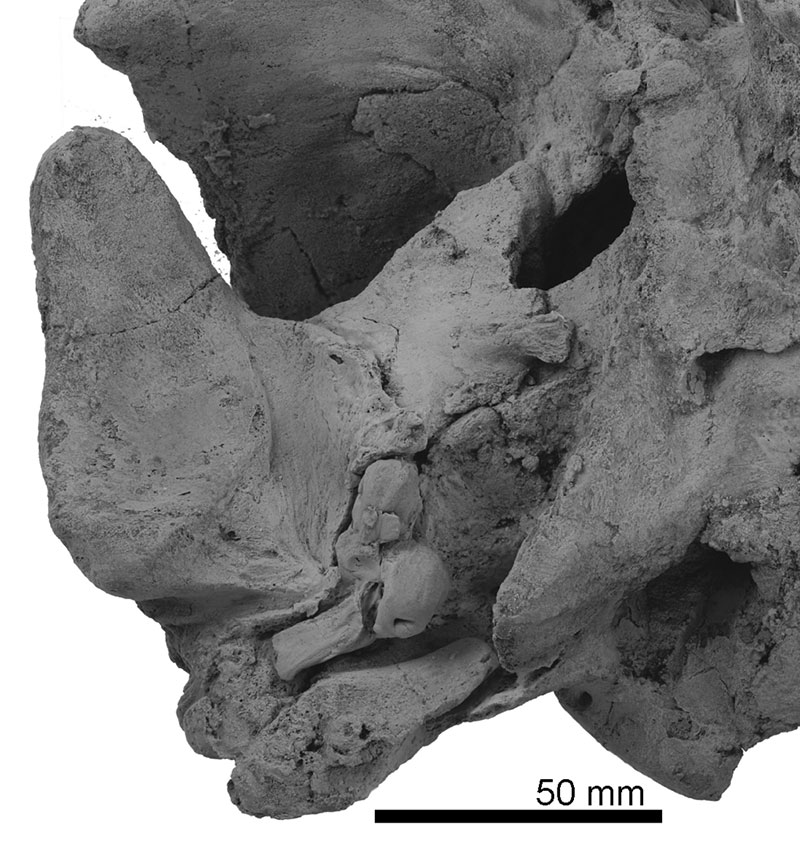
FIGURE 12. The type tympanoperiotics. OU 22306, Otekaikea huata. 1-6, right periotic. 7 and 8, sigmoid process of left bulla. 1, medial view. 2, lateral view. 3, ventral view. 4, dorsal view. 5, posterior view. 6, anterior view. 7, ventral view. 8, posterior view.
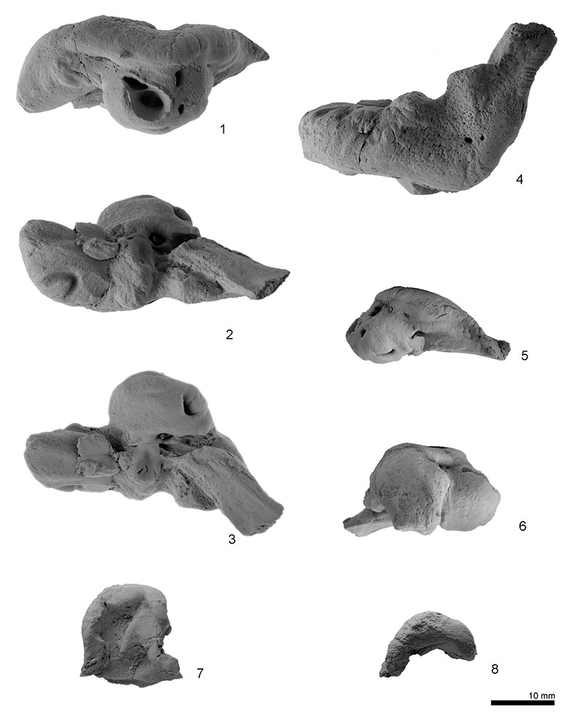
FIGURE 13. Key features of the type tympanoperiotics. OU 22306, Otekaikea huata. 1-6, right periotic. 7 and 8, sigmoid process of left bulla. 1, medial view. 2, lateral view. 3, ventral view. 4, dorsal view. 5, posterior view. 6, anterior view. 7, ventral view. 8, posterior view.
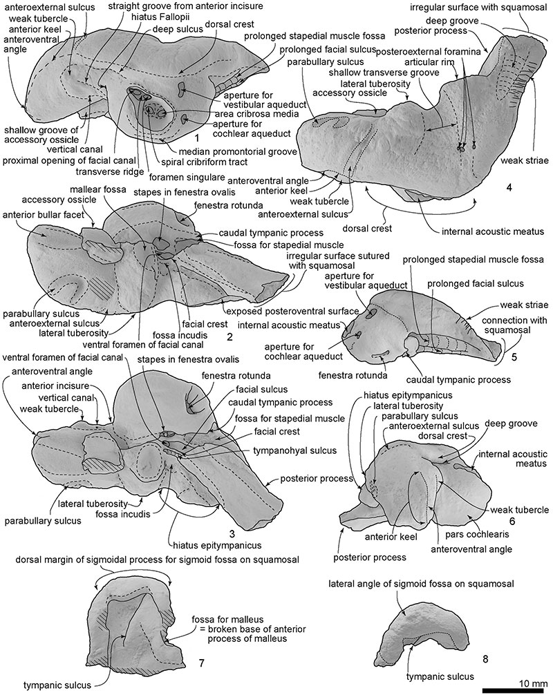
FIGURE 14. The type mandible, OU 22306, Otekaikea huata. 1, anterior view. 2, left lateral view. 3, dorsal view. 4, mandibular symphysis in dorsal view. 5, posterior view. 6, ventral view. 7, anterior end in ventral view. 8, right lateral view.
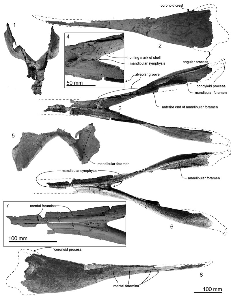
FIGURE 15. The type cervical vertebrae, OU 22306, Otekaikea huata. 1-5, anterior views. 6-8, lateral views. 9-10, dorsal views. 1, atlas. 2 and 8, axis, 3, fourth cervical vertebra. 4, fifth cervical vertebra. 5, sixth cervical vertebra. 6 and 9, atlas and axis. 7 and 10, third to sixth cervical vertebrae. 7, mirrored image.
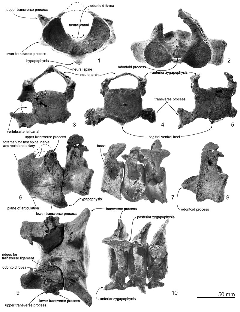
FIGURE 16. The type thoracic and lumbar vertebrae, OU 22306, Otekaikea huata. 1-3, anterior view of three thoracic vertebrae. 4 and 5, a posterior thoracic vertebra, anterior and left lateral views. 6, lumbar vertebrae from anterior view.
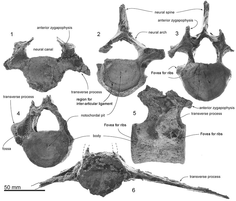
FIGURE 17. The type ribs. OU 22306, Otekaikea huata, from anterior view.
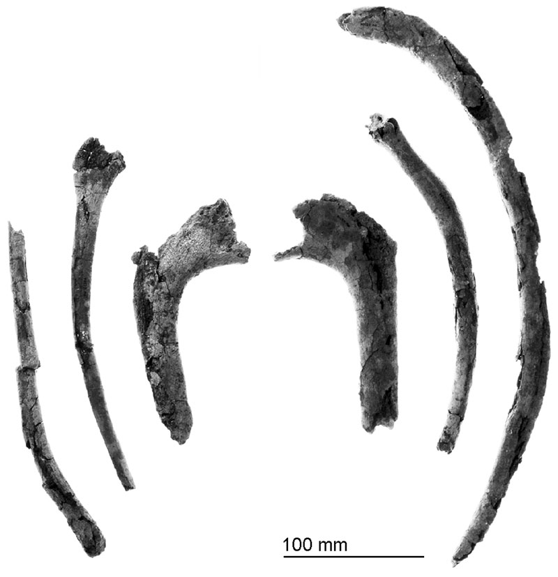
FIGURE 18. The type sternum. OU 22306, Otekaikea huata. 1, ventral. 2, left lateral. 3, dorsal.
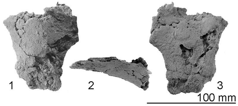
FIGURE 19. The type right forelimb bones in lateral view, OU 22306, Otekaikea huata. 1, right scapula. 2, humerus. 3, ulna. 4, radius.
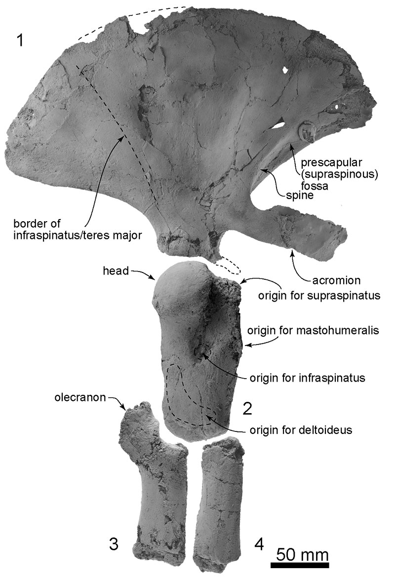
FIGURE 20. The type forelimb bones, OU 22306, Otekaikea huata. 1, right scapula in medial view. 2, right scapula in distal view. 3, left scapula in lateral view. 4, left scapula in distal view. 5-8, right humerus. 5, posterior view. 6, medial view. 7, anterior view. 8, proximal view. 9, carpus. 10, ulna in posterior view.
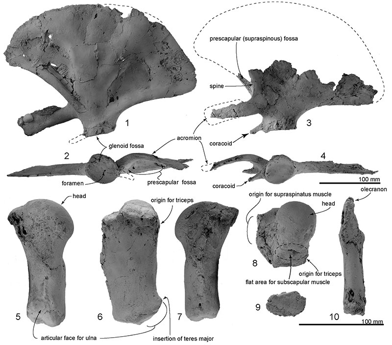
FIGURE 21. Phylogenetic analysis of Otekaikea huata and the Odontoceti. Top, strict consensus tree of equally weighted analysis 1 with decay index values shown. This figure shows simplified cladograms, with species in the more-diverse families crown-ward of Papahu merged for ease of illustrating. Complete trees are provided in Appendices 7 and 8. Bottom, strict consensus tree of implied weighted analysis 2 with decay index labeled. Synapomorphies for some nodes are: Platanistoidea ( sensu lato), Characters 59, 169, 186, 195, 196, 212; Platanistoidea (sensu stricto), Characters 166, 167, 175, 288, 291.
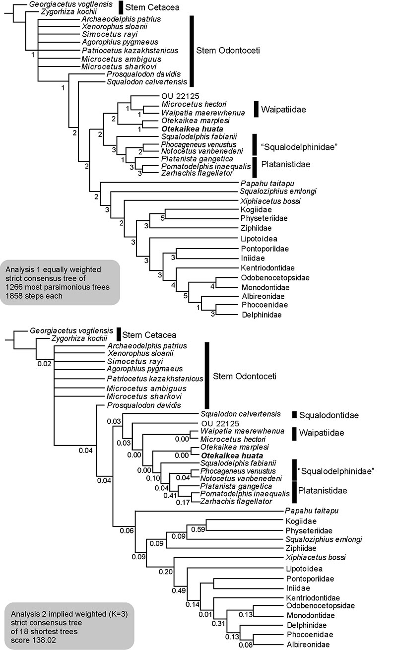
FIGURE 22. Close-up photo of the intra-premaxillary foramen of 1, Otekaikea huata and the posterior accessory foramen of 2, Monodon monoceros and 3, Delphinapterus leucas. Figures are not to scale. Fr, frontal. Ma, maxilla. Na, nasal. Pmax, premaxilla.
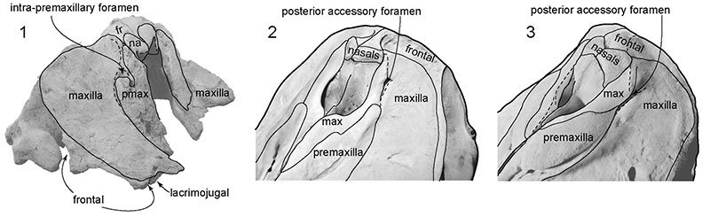
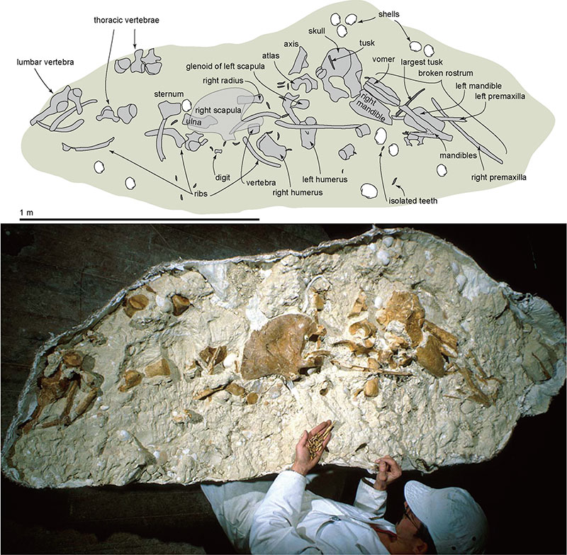
FIGURE 23. The original distribution of type elements, OU 22306, Otekaikea huata. Top, line art based on tracing of bone outlines as uncovered during preparation. Bottom, partly prepared field jacket with bones partly exposed. Andrew Grebneff is holding loose teeth. Photo © R. Ewan Fordyce.