FIGURE 1. Position of the sampling area and tectonic setting of the Tibetan Plateau and Southern China (simplified after Dai et al., 2011). GCT: Great Counter thrust; ZGT: Zhongba-Gyangze thrust.
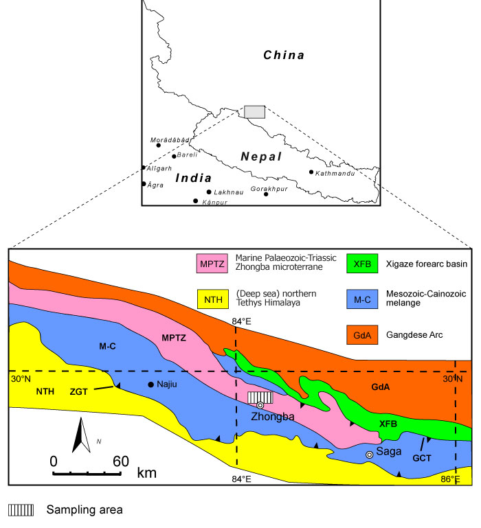
FIGURE 2. Lithological characteristics of the limestones from the Zhongba Formation. 1, limestone sample with abundant bryozoans (SMF 23.262); 2, rudstone containing crinoids, cystoporate (Fistulipora enodata Gorjunova, 1970) and fenestrate bryozoans (SMF 23.263); 3, bindstones with Fistulipora guttata Trizna and Klautzan, 1961 (SMF 23.264); and 4, grain- to packstones with bryozoan and crinoid fragments (SMF 23.265).
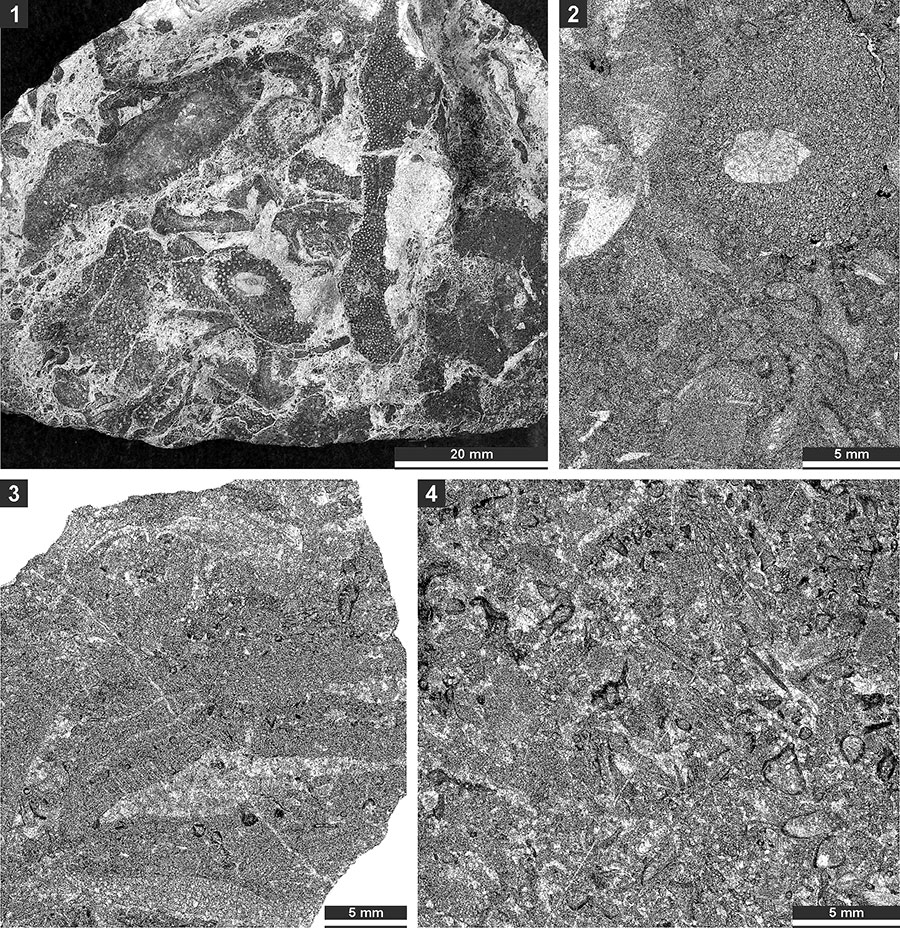
FIGURE 3. Thin section photographs of Fistulipora enodata Gorjunova, 1970, SMF 23.014 (1-4); Fistulipora guttata Trizna and Klautzan, 1961, SMF 23.025 (5-8); and Fistulipora sakagamii n. sp., holotype SMF 23.028 (9 and 10). 1 and 5, longitudinal section showing autozooecial chambers and vesicular skeleton; and 2-4, 6-8, 9, and 10, tangential sections showing autozooecial apertures and vesicles.
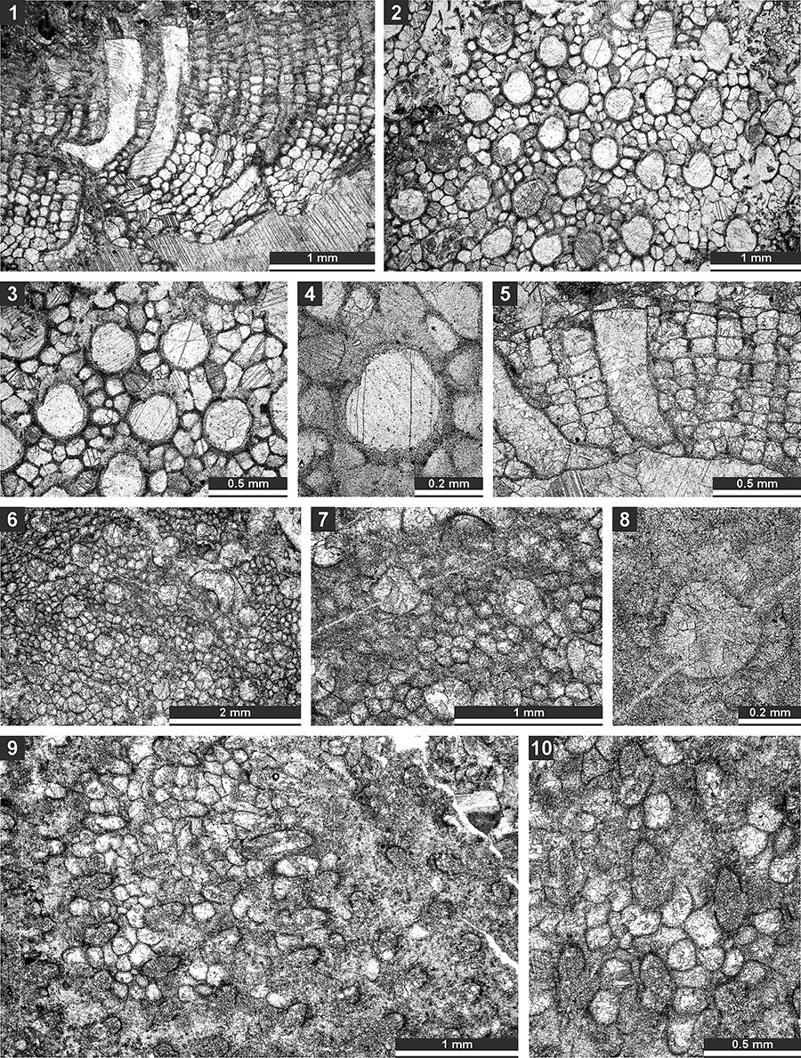
FIGURE 4. Thin section photographs of Fistulipora sakagamii n. sp., holotype SMF 23.028 (1 and 2); Dybowskiella hupehensiformis n. sp., holotype SMF 23.030 and paratype SMF 23.032 (3-7); and Fistuliramus xianzaensis Liu and Wang, 1987, SMF 23.037 (8). 1, 2, 5-7, tangential section showing autozooecial apertures and vesicles; 3 and 4, longitudinal section showing autozooecial chambers and vesicular skeleton; and 8, branch transverse section.
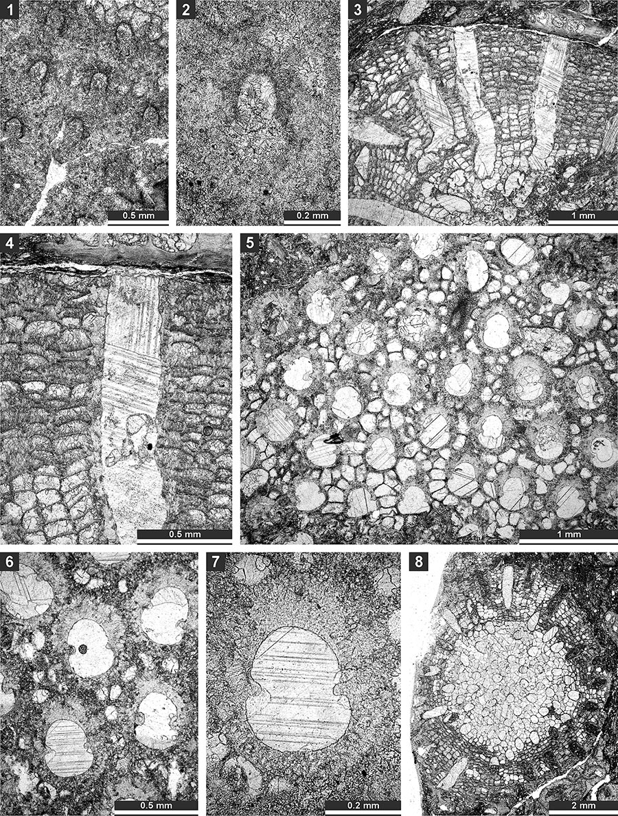
FIGURE 5. Thin section photographs of Fistuliramus xianzaensis Liu and Wang, 1987, SMF 23.042 (1) and SMF 23.038 (2 and 3); and Eridopora uncata Yang and Lu, 1983, SMF 23.052 (4 and 6) and SMF 23.051 (5). 1, longitudinal section; and 2-6, tangential section showing autozooecial apertures and vesicles.
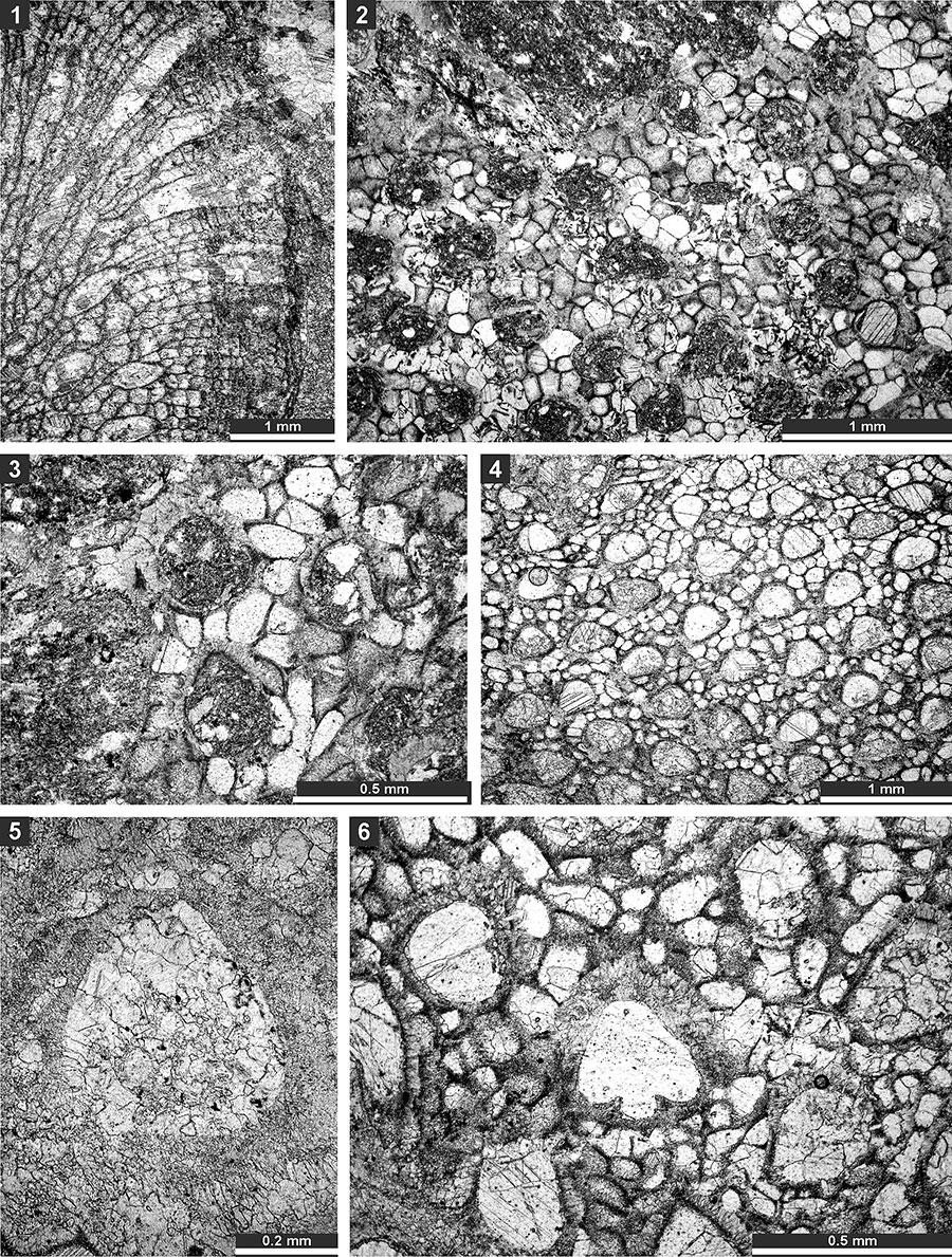
FIGURE 6. Thin section photographs of Eridopora uncata Yang and Lu, 1983, SMF 23.052 (1-3); Cyclotrypa alexanderi Sakagami, 1963 SMF 23.056 (4-5), SMF 23.061 (6), and SMF 23.056 (7); and Hexagonella kobayashii Sakagami, 1968, SMF 23.069 (8-9) and SMF 23.070 (10 and 11). 1-3, tangential section showing autozooecial apertures with lunarial ends indenting into the aperture (arrow); 7, tangential section showing autozooecia and macrozooecia (arrow); 8 and 9, branch transverse section showing autozooecial chambers and vesicular skeleton; and 10 and 11, longitudinal section showing autozooecial chambers and vesicular skeleton.
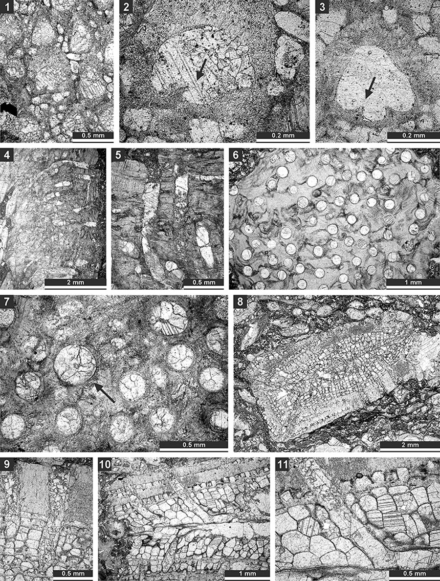
FIGURE 7. Thin section photographs of Hexagonella kobayashii Sakagami, 1968, SMF 23.067 (1-3) and SMF 23.072 (4); and Goniocladia aff. indica Waagen and Pichl, 1885, SMF 23.266 (5), SMF 23.077 (6), and SMF 23.073 (7). 1-3 and 6, tangential section; 7, mid-tangential section showing autozooecial chamber; 4, transverse section showing mesotheca with median tubules (arrow); and 5, external view of the colony form the reverse side.
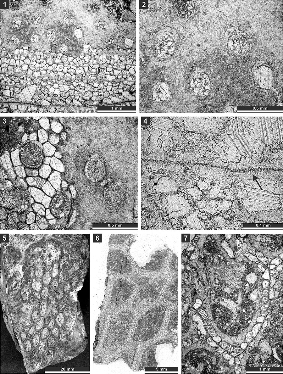
FIGURE 8. Thin section photographs of Goniocladia aff. indica Waagen and Pichl, 1885, SMF 23.076 (1 and 2), SMF 23.077 (3-5), and SMF 23.073 (6 and 7); and Liguloclema meridianus (Etheridge, 1926), SMF 23.093 (8). 1-2, and 6, branch transverse sections showing autozooecia chambers, mesotheca and hemisepta (arrow); 3-5, tangential sections showing autozooecial apertures; 7, longitudinal section showing autozooecial chamber with superior hemisepta (arrow); and 8, branch oblique section showing mesotheca, autozooecial chambers with hemisepta and vesicular skeleton.
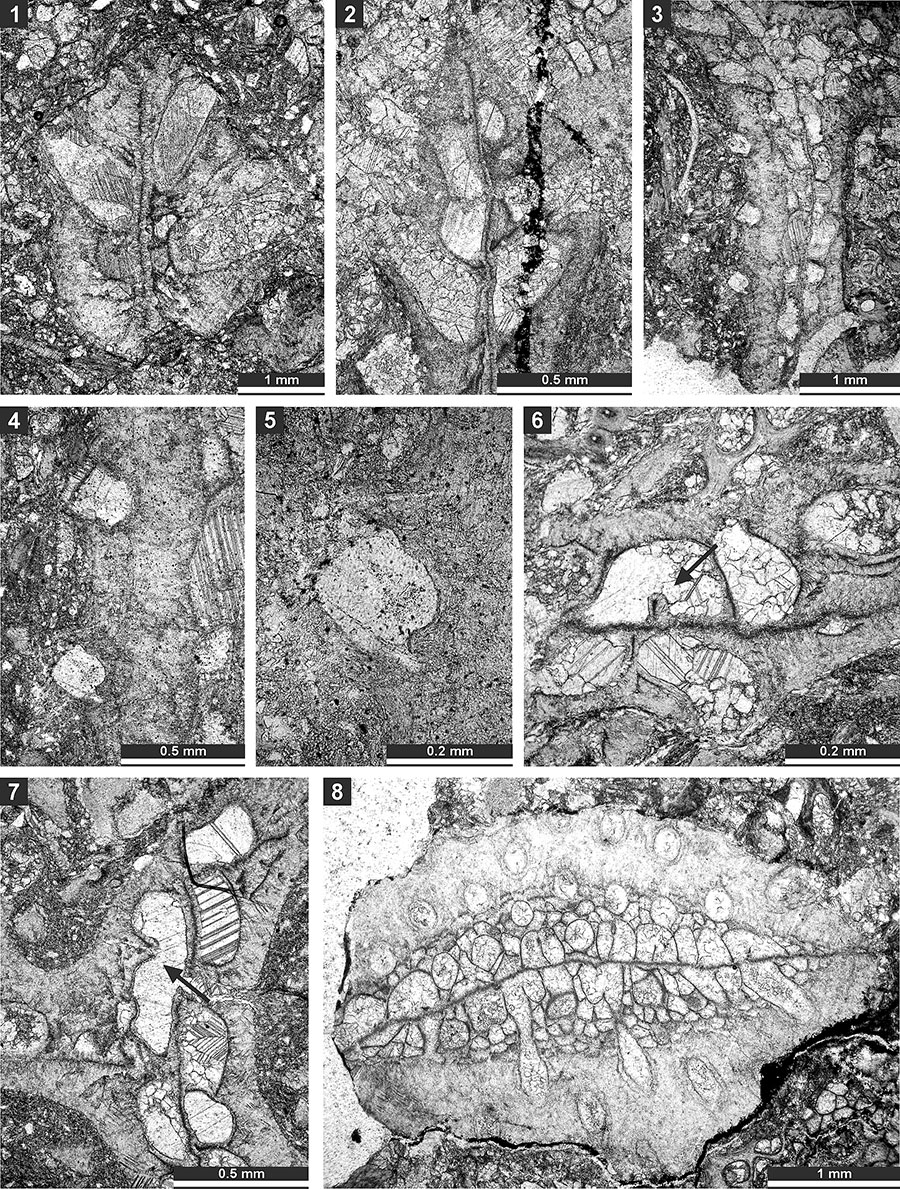
FIGURE 9. Thin section photographs of Liguloclema meridianus (Etheridge, 1926), SMF 23.093 (1), SMF 23.085 (2), and SMF 23.083 (3-5); and Etherella tibetensis n. sp., paratype SMF 23.217 (6). 1 and 2, branch oblique section showing mesotheca, autozooecial chambers with hemisepta and vesicular skeleton; 3 and 4, mid-tangential section showing autozooecial chambers with hemisepta; 5, tangential section showing autozooecial apertures; and 6, transverse section showing extrazooecial skeleton and sparse vesicles.
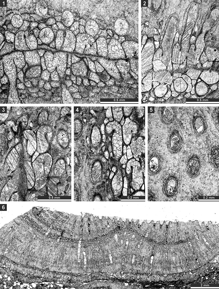
FIGURE 10. Thin section photographs of Etherella tibetensis n. sp., paratype SMF 23.267 (1), paratype SMF 23.216 (2), paratype SMF 23.219 (3 and 4), holotype SMF 23.214 (5-7), and paratype SMF 23.230 (8 and 9). 1, external view of the colony (split along mesotheca); 2-4, tangential section showing arrangement of autozooecia and wall microstructure; 5-7, oblique section showing hemisepta and mesotheca (arrows); and 8 and 9, branch transverse section showing autozooecial chambers and extrazooecial skeleton.
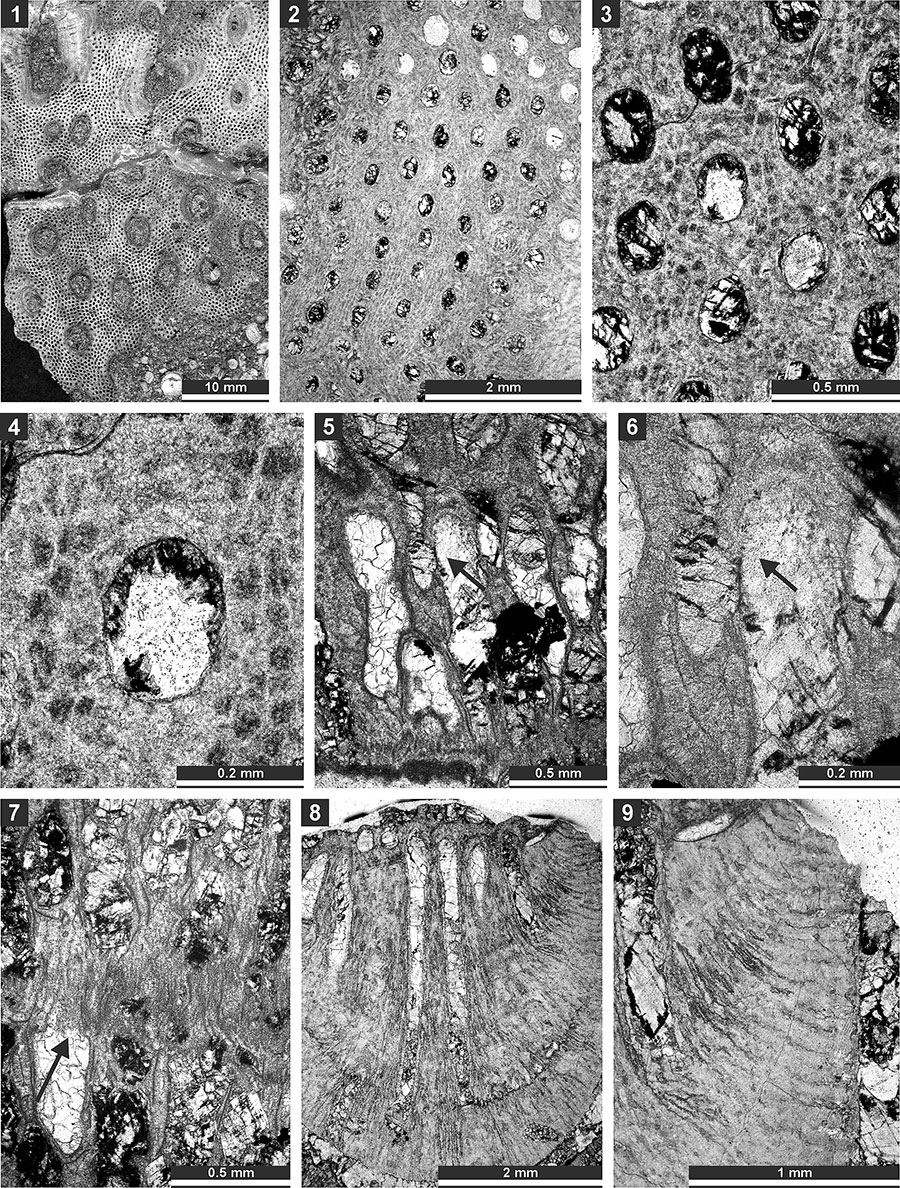
FIGURE 11. Thin section photographs of Tabulipora xinjiangensis Yang and Lu, 1983, SMF 23.233 (1 and 2); Dyscritella lii n. sp., holotype SMF 23.235 (3, 4, and 6) and paratype SMF 23.236 (5); and Ulrichotrypella omanica Ernst et al., 2008, SMF 23.242 (7 and 8). 1, longitudinal section showing ring septa; 2, oblique section showing macroacanthostyles and tubules; 3 and 4, oblique section through the colony; 5 and 6, tangential section showing autozooecial apertures and acanthostyles; and 7 and 8, oblique section.
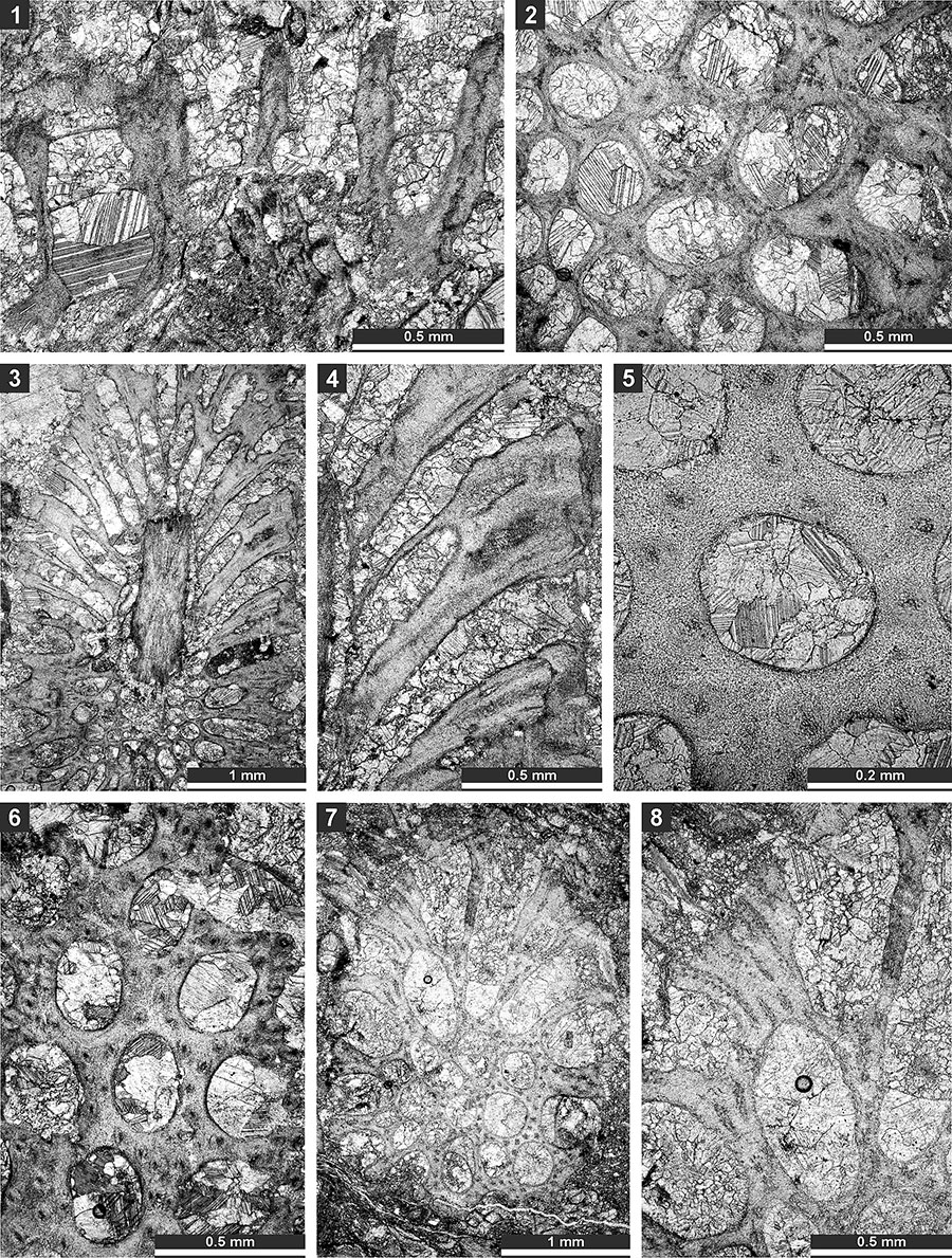
FIGURE 12. Thin section photographs of Ulrichotrypella omanica Ernst et al., 2008, SMF 23.242 (1); Neoeridotrypella astrica (Linskaya, 1951), SMF 23.245 (2 and 3), SMF 23.246 (4-6), and SMF 23.249 (7); and Streblotrypa (Streblotrypa) parviformis n. sp., holotype SMF 23.251 (8-10). 1, 6, 7, and 10, tangential section; 2-5, oblique branch section; and 8 and 9, longitudinal section.
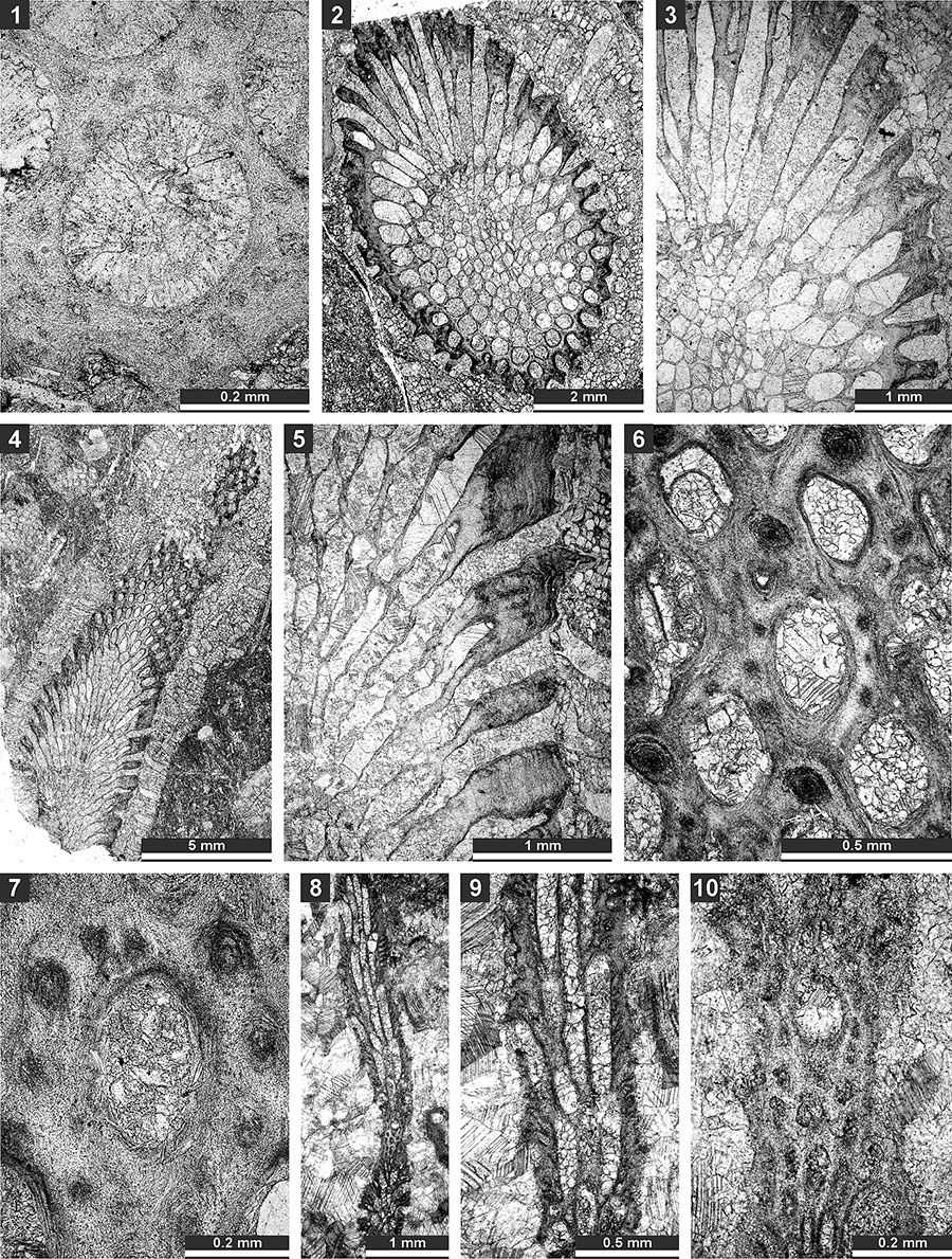
FIGURE 13. Thin section photographs of Streblotrypa ( Streblotrypa) parviformis n. sp., paratype SMF 23.260 (1) and paratype SMF 23.256 (2 and 3); Streblotrypa (Streblascopora) delicatula Sakagami, 1961, SMF 23.205 (4, 6, and 7), SMF 23.206 (5), and SMF 23.143 (8); and Streblotrypa (Streblascopora) marmionensis (Etheridge, 1926), SMF 23.145 (9). 1 and 8, branch transverse section; 2, 3, 5, and 9, longitudinal section; 4, branch oblique section; and 6 and 7, tangential section.
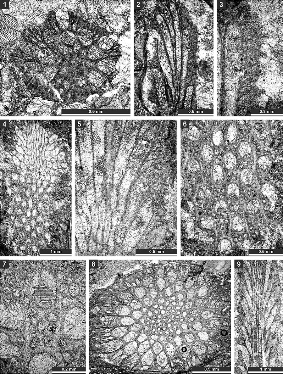
FIGURE 14. Thin section photographs of Streblotrypa ( Streblascopora) marmionensis (Etheridge, 1926), SMF 23.145 (1 and 2), and SMF 23.148 (3); and Rhabdomeson bretnalli Crockford, 1957, SMF 23.155 (4-6) and SMF 23.157 (7-9). 1, longitudinal section; 2 and 7, tangential section; 3 and 4, branch transverse section; and 5, 6, 8 and 9; branch oblique section.
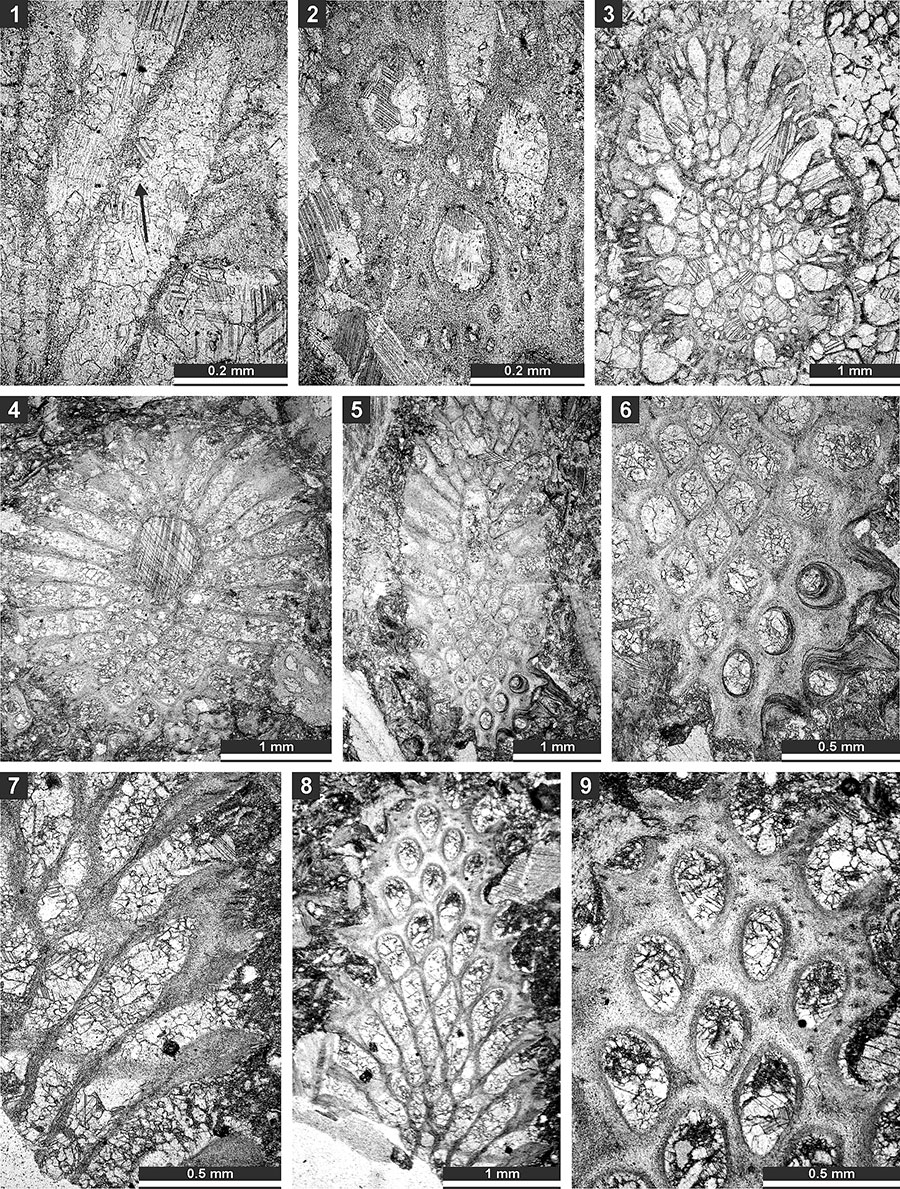
FIGURE 15. Thin section photographs of Rhabdomeson sp. SMF 23.160 (1-4) and SMF 23.158 (5); and Primorella rotunda Gorjunova, 1985, SMF 23.163 (6) and SMF 23.162 (7-9). 1-3, branch oblique section; 4 and 5, longitudinal section; 6, tangential section; and 7-9, oblique section.
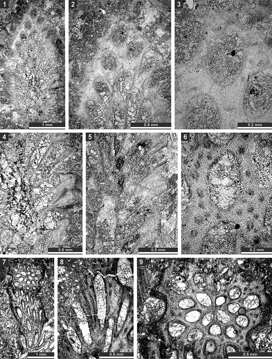
FIGURE 16. Thin section photographs of Timanotrypa australis n. sp., holotype SMF 23.165 (1), paratype SMF 23.166 (2 and 3), paratype SMF 23.169 (4), and paratype SMF 23.171 (5). 1, oblique section of the pinnate branch; 2 and 3, tangential section showing autozooecial apertures, nodes (arrows) and microstyles; and 4 and 5, branch transverse section.
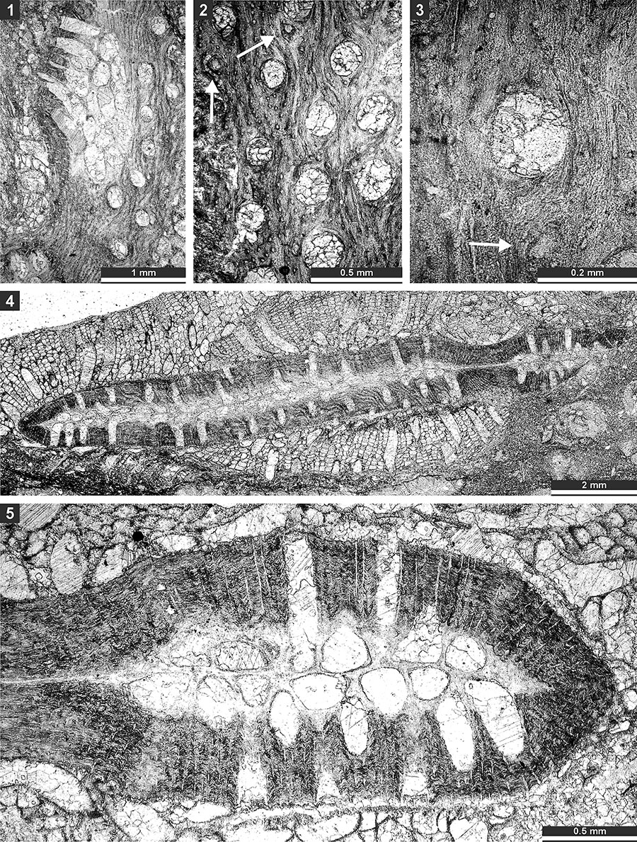
FIGURE 17. Thin section photographs of Spinofenestella sp., SMF 23.178 (1-5); and Spinofenestella subquadratopora (Schulga-Nesterenko, 1952), SMF 23. 179 (6-8). 1-3, tangential section of the reverse side; 4-7, tangential section of the obverse side; and 8, mid- tangential section.
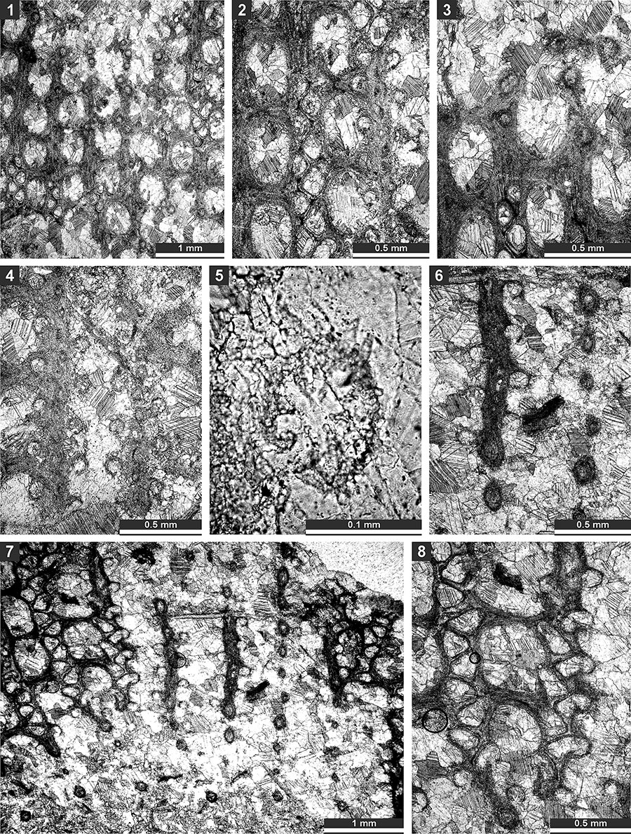
FIGURE 18. Thin section photographs of Polypora consanguinea Bassler, 1929, SMF 23.182 (1-4); and Polypora brouweri Bassler, 1929, SMF 23.183 (5-9). 1, oblique section; 2-5, tangential section showing autozooecial apertures, nodes and autozooecial chambers; 6, reverse side of the colony; and 7-9, tangential section showing autozooecial apertures and chambers.
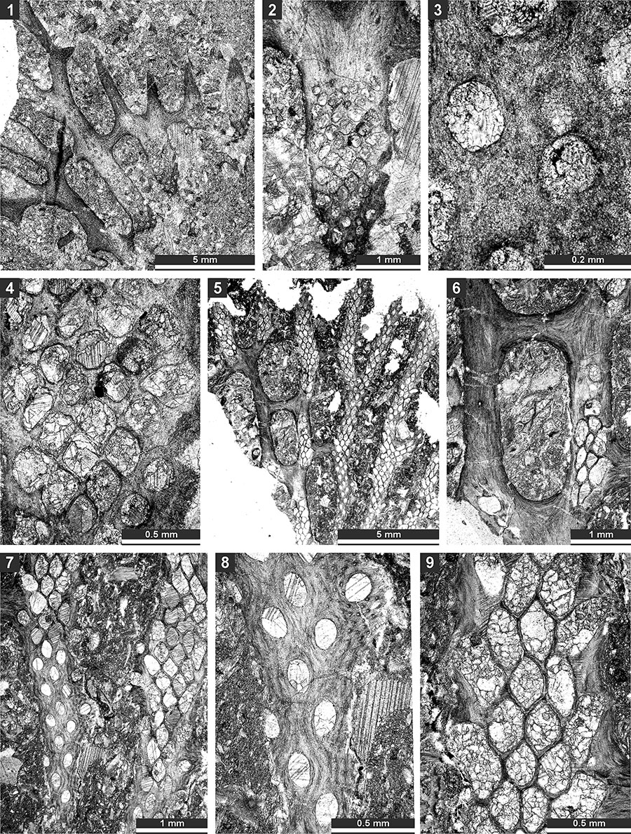
FIGURE 19. Thin section photographs of Polypora aff. voluminosa Trizna and Klautzan, 1961, SMF 23.187 (1-3, and 5) and SMF 23.185 (4 and 6); and Mackinneyella obesa (Crockford, 1957), SMF 23.191 (7) and SMF 23.192 (8). 1 and 2, tangential section branches and fenestrules; 3, tangential section of the reverse side showing nodes; 4, tangential section showing autozooecial apertures and chambers with hemisepta (arrow); 5, tangential section showing autozooecial apertures, chambers and heteromorph (arrow); 6, tangential section showing autozooecial apertures and nodes; and 7 and 8, tangential section showing autozooecial apertures and chambers.
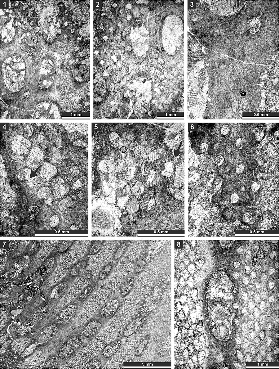
FIGURE 20. Thin section photographs of Mackinneyella obesa (Crockford, 1957), SMF 23.194 (1, 3), SMF 23.191 (2), and SMF 23.189 (4); and Protoretepora irregularis n. sp., paratype SMF 23.198 (5), SMF 23.268 (6), and SMF 23.195 (7). 1-3, and 7, tangential section showing autozooecial chambers; 4, branch transverse section; 5, branch longitudinal section; and 6, external view of the reverse side of the colony.
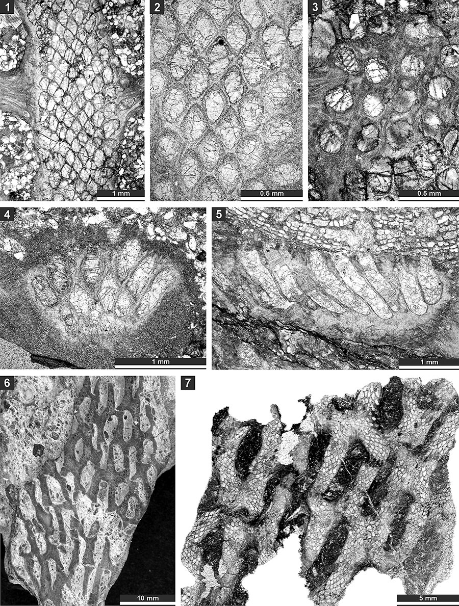
FIGURE 21. Thin section photographs of Protoretepora irregularis n. sp., paratype SMF 23.201 (1, 3, and 4), paratype SMF 23.202 (2), and holotype SMF 23.195 (5); and Tibetiporella ornata n. gen. n. sp., paratype SMF 23.224 (6 and 7) and holotype SMF 23.227 (8). 1, tangential section showing autozooecial apertures and chambers; 2, mid-tangential section showing autozooecial chambers; 3, 4, 7, and 8, tangential section showing autozooecial apertures, microstyles and nodes; 5, tangential section of obverse colony side with nodes; and 6, tangential section of the reverse side of the colony.

FIGURE 22. Thin section photographs of Tibetiporella ornata n. gen. n. sp., holotype SMF 23.227 (1 and 2), paratype SMF 23.211 (3 and 4), paratype SMF 23.210 (5 and 6), paratype SMF 23.213 (7 and 8), and paratype SMF 23.225 (9). 1 and 2, tangential section showing autozooecial apertures, nodes and heteromorphs; 3 and 4, mid-tangential section showing autozooecial chambers; 5 and 6, tangential section showing autozooecial apertures, nodes and heteromorphs; 7 and 8, longitudinal section showing autozooecial chambers and heteromorphs; and 9, transverse section showing autozooecial chambers.
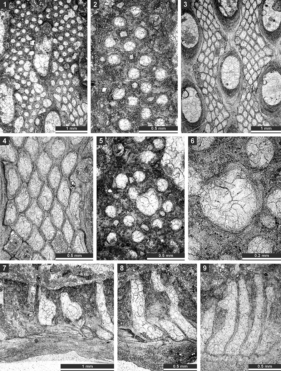
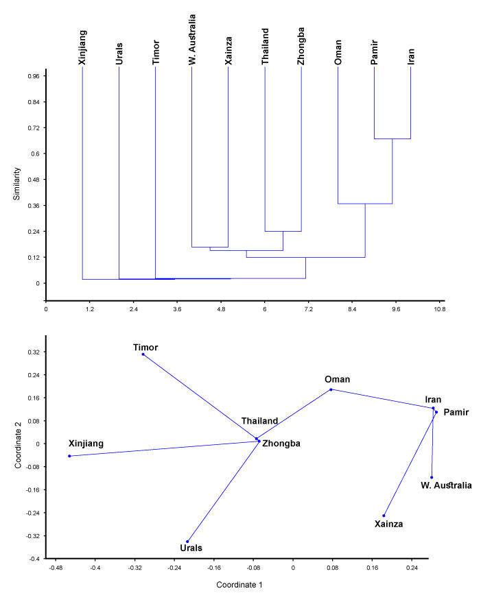
FIGURE 23. Palaeobiogeographical affinities of the bryozoan assemblage from the Zhongba Formation (Zhongba). Top: dendrogram of the cluster analysis using Jacquard's similarity index (unweighted pair-group average algorithm), and bottom: plot of non-metric MDS analysis made with Jacquard's similarity index. Areas: Pamir (Tajikistan), Oman (Batain Coast), Iran (central Iran, Chili Formation), Xainza (southwestern Tibet, Xiala Fm.), Thailand, Urals, Western Xinjiang (Baliqliq Group), and W. Australia (Noonkanbah Fm.). Japan, Malaysia, and Rutog (southwestern Tibet) were omitted as sharing only one species with Zhongba assemblage.