FIGURE 1. 1. Location map of the fossiliferous levels of the Uquía Formation exposed at San Roque, Humahuaca (Jujuy Province, Argentina). Relative location of the prospected area in: 2, Jujuy Province; and 3, Northwestern Argentina. Modified from Ortiz et al. (2012).
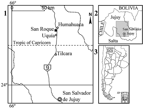
FIGURE 2. Line drawings (occlusal view) of Microtragulus bolivianus showing the anatomical nomenclature used in the text. 1. Right M3-4. 2. Right m1-4 (image reflected). Abbreviations: cf, central fossa; cfd, central fossid; ectf, ectoflexid; ectd, ectostylid; ent, entoconid; entd, entostylid?; entf, entoflexid; gr, groove; hyp, hypoconid; hypd, hypoconulid; mesl, mesiolabial lobe; met, metacone; metd, metaconid; metl, metaconule; prt, protocone; prtd, protoconid.
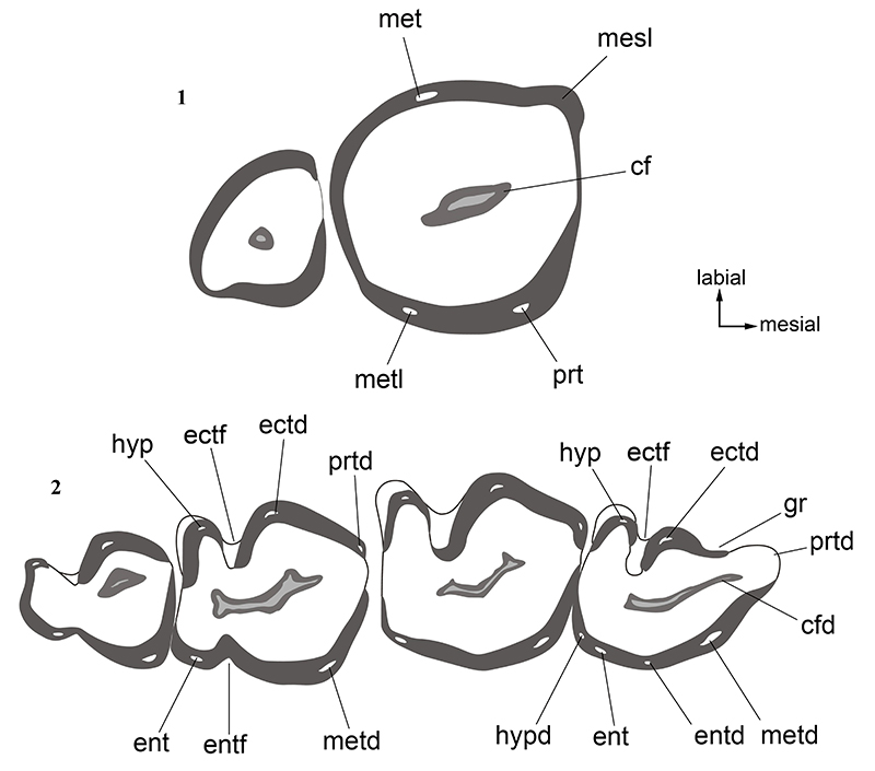
FIGURE 3. Microtragulus bolivianus. 1-3. JUY-P-0065, fragment of right maxilla with M3-M4 in: 1, lateral; 2, medial; and 3, occlusal views. 4-5. JUY-P-0066, right mandibular body with i1, alveoli of i2 and p3, and complete m1-4 in: 4, medial; and 5, lateral views. 6-7. JUY-P-0067, right mandibular body with complete i1 and i2, alveolus of p3, and complete m1-4 in: 6, medial; and 7, lateral views. 8-9. JUY-P-0068, anterior fragment of right mandibular body with i1 and alveoli of i2 and p3 in: 8, medial; and 9, lateral views. Scale bar equals 5 mm.
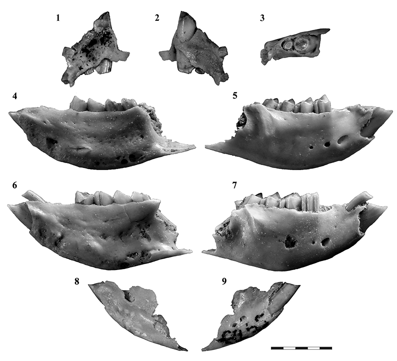
FIGURE 4. Microtragulus bolivianus, right lower dentition in occlusal view. 1. JUY-P-0066, detail of i1. 2. JUY-P-0066, m1-4. 3. JUY-P-0067, m1-4. Scale bar equals 5 mm.
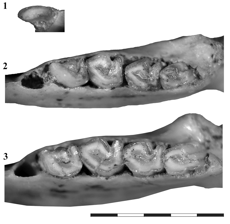
FIGURE 5. Microtragulus bolivianus, fore and hindlimb bones. 1-4. JUY-P-0069, right humerus almost complete except the proximal third in: 1, anterior; 2, posterior; 3, medial; and 4, lateral views. 5-8. JUY-P-0070, left humerus almost complete except the proximal third in: 5, anterior; 6, posterior; 7, medial; and 8, lateral views. 9-12. JUY-P-0071, distal half of left humerus in: 9, anterior; 10, posterior; 11, medial; and 12, lateral views. 13-16. JUY-P-0072, distal end of right humerus in 13, anterior; 14, posterior; 15, medial; and 16, lateral views. 17-19. JUY-P 59, proximal half of right ulna in: 17, anterior; 18, lateral; and 19, medial views. 20-22. JUY-P 60, proximal half of left ulna in: 20, anterior; 21, lateral; and 22, medial views. 23. JUY-P 61, proximal fragment of radius. 24-25. JUY-P 52, distal end of right tibiofibula in: 24, anterior; and 25, posterior views. Scale bar equals 10 mm.
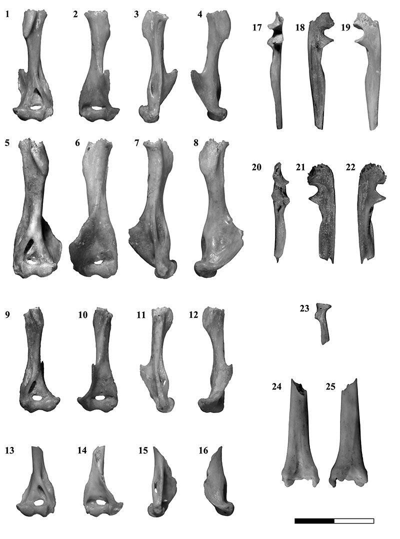
FIGURE 6. Microtragulus bolivianus, astragalus and calcaneus. 1-5. JUY-P-0075, complete right astragalus in: 1, dorsal; 2, plantar; 3, medial; 4, lateral; and 5, distal views. 6-10. JUY-P-0073, complete left calcaneus in: 6, dorsal; 7, plantar; 8, medial; 9, lateral; and 10, distal views. Scale bar equals 5 mm.
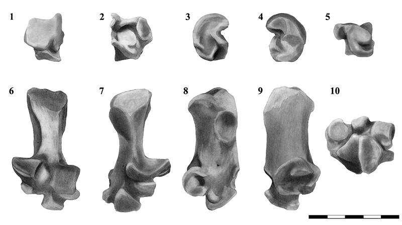
FIGURE 7. Microtragulus bolivianus, metatarsal III and IV and phalanges. JUY-P 55, complete right metatarsal III and JUY-P 56, distal portion of right metatarsal IV (not associated) in: 1, anterior; and 2, posterior views. Scale bar equals 10 mm. 3-5. JUY-P 58, ungual phalanges in lateral view. Scale bar equals 5 mm.
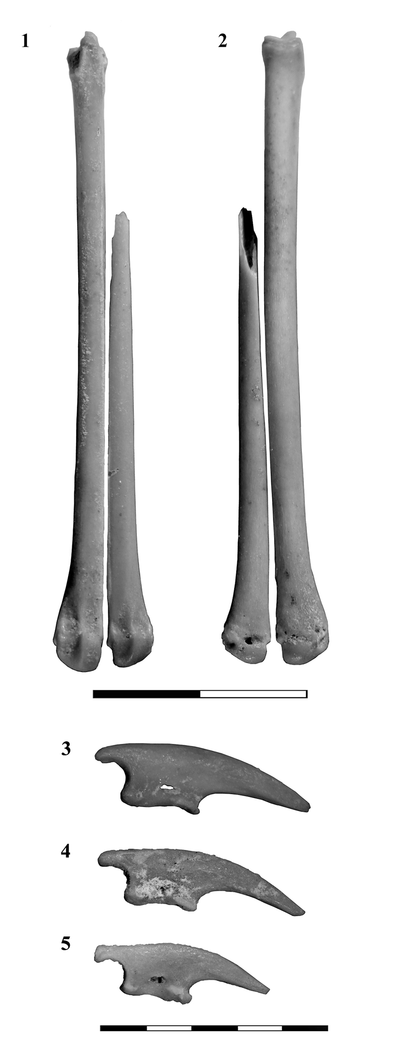
FIGURE 8. Microtragulus sp., upper and lower molars in occlusal views. 1. JUY-P 53, isolated left M3. 2. JUY-P 54, isolated left m2 or m3. Scale bar equals 2 mm.
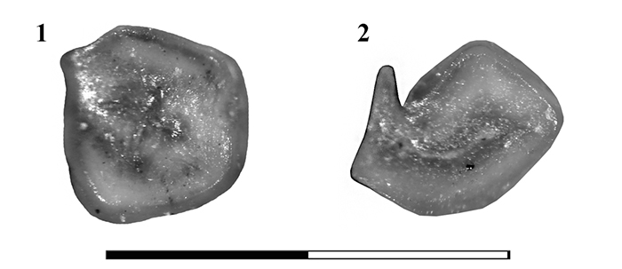
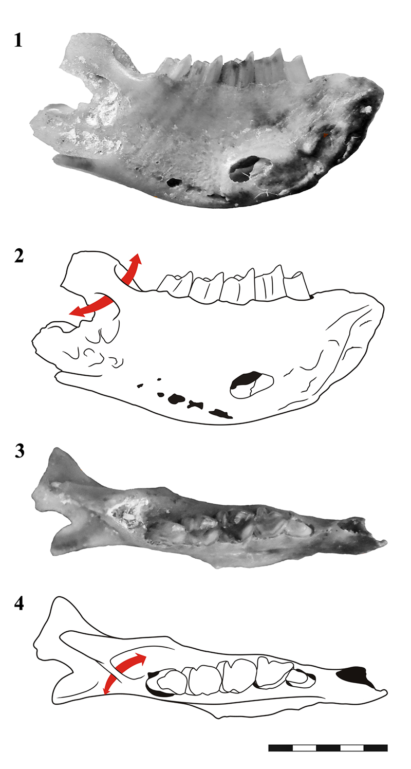
FIGURE 9. Argyrolagus sp. MACN 17590, left mandibular fragment with m1-4. 1-4. Photograph and line drawing in: 1-2, medial; and 3-4, dorsal views. The arrow points the maxillary canal. Scale bar equals 5 mm.