FIGURE 1. Simplified phylogenetic framework based on Scheyer et al. (2013) and Salas-Gismondi et al. (2015). The caimanine taxa present in the Urumaco Formation are marked in bold face.
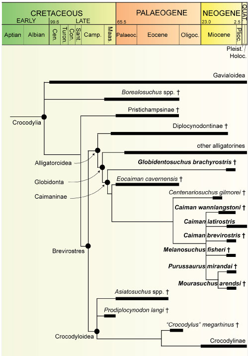
FIGURE 2. Photographs of Caiman brevirostris (MCNC-1829). 1, right dorsolateral view. 2, left ventrolateral view. 3, right lateral view. Note that the specimen is strongly crushed and folded in on itself.
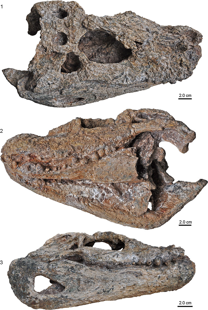
FIGURE 3. Photographs (1, 3, 5) and interpretative drawings (2, 4, 6) of Caiman brevirostris (MCNC-1829). 1,2, close-up of the fronto-nasal contact in right dorsolateral view. 3, 4, close-up of the skull roof in dorsal view. 5, 6, close-up of the craniomandibular articulation in slanted right dorsolateral view. Abbreviations: an, angular, ar, articular; d, dentary; f, frontal; j, jugal, itf, infratemporal fenestra; mf, mandibular fenestra; n, nasal; o, orbit; oc, occipital condyle; po, postorbital; prf, prefrontal, q, quadrate; qj, quadratojugal; sa, surangular; so, supraoccipital; sq, squamosal; stf, supratemporal fenestra.
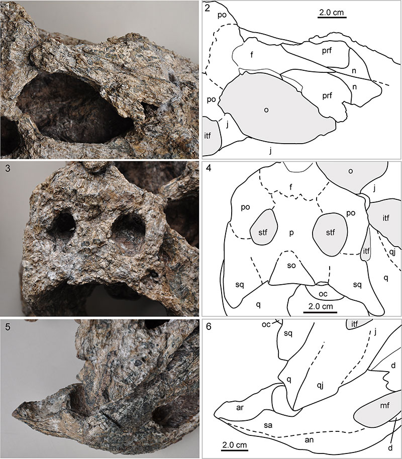
FIGURE 4. Interpretative drawings of skull bone configuration in the rostro-orbital region of three small sized caimanines from the Urumaco Formation, in comparison to three living caimanines (anterior is to the top). 1, Melanosuchus fisheri based on holotype skull MCNC-243. 2, Globidentosuchus brachyrostris based on holotype skull AMU-CURS-222. 3, Caiman brevirostris based on MCNC-1829 from the late Miocene Urumaco Formation, Urumaco, Venezuela. 4, Caiman brevirostris from the Pleistocene of Acre, Brazil (UFAC-196) originally described by Souza Filho (1987, also figured in Fortier et al., 2014). Note that the anterior projection of the frontal appears much broader in the Venezuelan specimen than in the Pleistocene specimen. 5, extant Melanosuchus niger based on ZSM 76/1911 (picture courtesy: Julia Desojo; see also Foth et al., 2015). 6, extant Caiman crocodilus based on Iordansky (1973, figure 7A). 7, extant Caiman latirostris based on Bona and Desojo (2011, figure 2). 8, extant Paleosuchus trigonatus and 9, extant Paleosuchus palpebrosus based on Medem (1958, figure 36). The bridge of the ‘spectacles’ between the orbits is indicated in 1, 5, 6, and 7. Drawings not to scale. Abbreviations: f, frontal; n, nasal; o, orbit; prf, prefrontal.
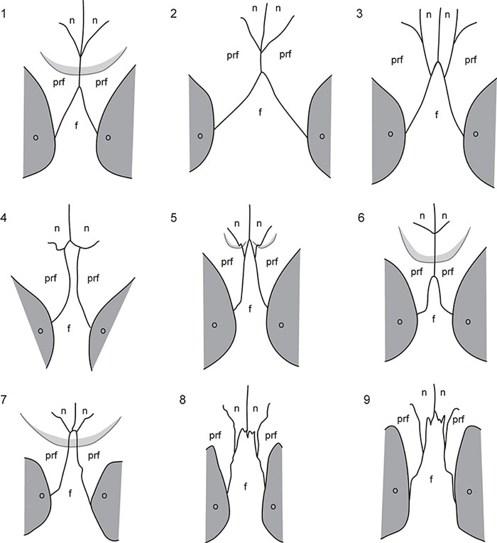
FIGURE 5. Photograph (1) and interpretative drawing (2) of Caiman brevirostris (MCNC-1829) in occipital view. Abbreviations: bo, basioccipital; eo, exoccipital; fm, foramen magnum; j, jugal; so, supraoccipital; sq, squamosal; stf, supratemporal fenestra; oc, occipital condyle; pt, pterygoid; q, quadrate; qj, quadratojugal.
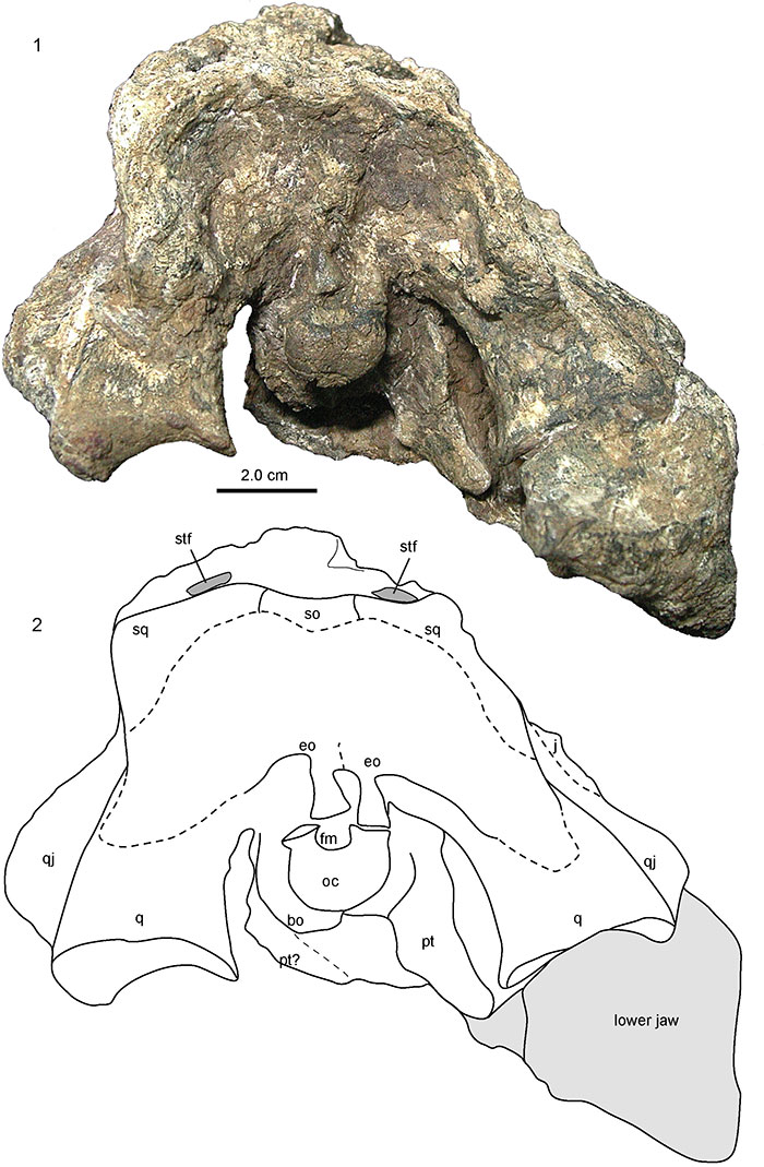
FIGURE 6. Photographs (1, 3, 5) and interpretative drawings (2, 4, 6) of Caiman wannlangstoni (AMU-CURS-49). 1,2, dorsal view. 3, 4, ventral view. 5, 6, right lateral view. Occlusion pits are marked in grey. The partially preserved teeth in the third premaxillary alveolus and the third and tenth maxillary alveoli are marked in dark grey. Note that the two occlusion pits anteromedial and posteromedial to the fourth premaxillary alveolus are preserved only as very faint impressions on the bone surface. Abbreviations: mx, maxilla; n, nasal; op, occlusion pit; pmx, premaxilla.
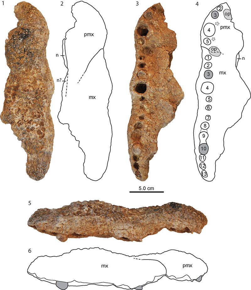
FIGURE 7. Photographs (1, 3) and interpretative drawings (2, 4) of Caiman latirostris (MCNC-URU-145-72V). 1, 2, dorsal view. 3, 4, ventral view. Alveoli are marked with numbers, occlusion pits in dark grey. The internal trabecular space within the maxilla is marked in light grey. Abbreviations: en, external naris; mx, maxilla; n, nasal; op, occlusion pit; pmx, premaxilla.
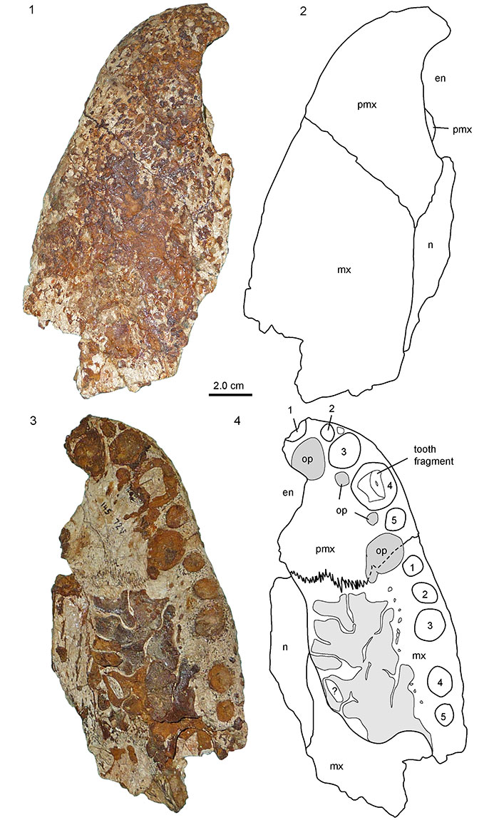
FIGURE 8. Photographs of Globidentosuchus brachyrostris (AMU-CURS-067). 1-3, left ramus of lower jaw in lateral (1), medial (2), and dorsal (3) view. 4-6, anterior, portion of right ramus in lateral (4), medial (5 ), and dorsal (6) view. 7, 8, fragment of a (right?) maxilla in ventral (7) and lateral (8) view. 9, 10, Fragment of a (left?) premaxilla in ventral (9 ) and dorsal (10) view. 11, indeterminate cranial fragment found associated with the other skeletal element and therefore referred to the same specimen.
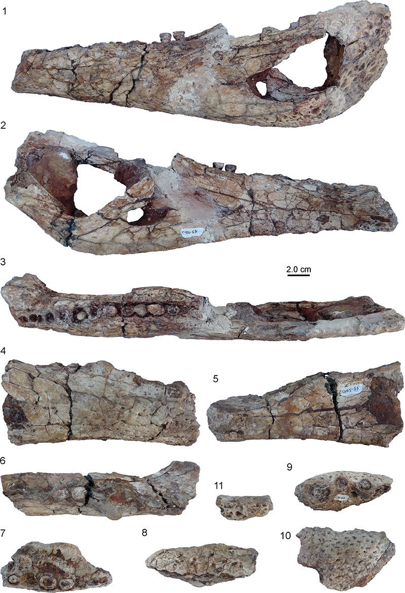
FIGURE 9. Photographs and interpretative drawings of Globidentosuchus brachyrostris lower jaw (AMU-CURS-067). 1, 2, assembled right and left ramus in dorsal view showing posterior crushing dentition and the splenials participating in symphysis. 3, 4, posterior part of left ramus in lateral view. 5, 6, posterior part of left ramus in medial view. Note that fragment of articular is still partially present, articulating with the angular ventrally. Abbreviations: a, angular; ar, articular; co, coronoid; d, dentary; fic, foramen intermandibularis caudalis; fme; external mandibular fenestra; path, area of pathology; sa, surangular; sed, sediment; sp, splenial.
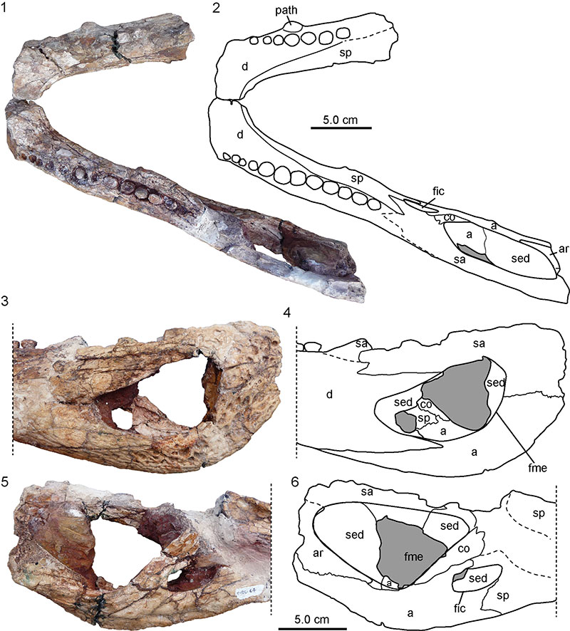
FIGURE 10. Photographs of Globidentosuchus cf. G. brachyrostris. 1-6, AMU-CURS-083. 7-18, AMU-CURS-084. 1, articulated left strongright associated dentary in dorsal view. 2,3, isolated right dentary in dorsal and angled medial view. 4-6, right dentary of (1) in lateral (4), dorsolateral (5) and angled medial view (6). 7, 8, right maxilla in angled lateral and angled medial view. 9, 10, right angular in medial and dorsal view. 11, 12, left articular and portion of surangular in angled medial and lateral view. 13, 14, right surangular sutured to part of dentary in angled medial and lateral view. 15, 16, left surangular in medial and lateral view. 17, 18, teeth. Abbreviations: d, dentary; ds, scar for dentary articulation; fic; foramen intermandibularis caudalis; fo, foramen; sa, surangular.
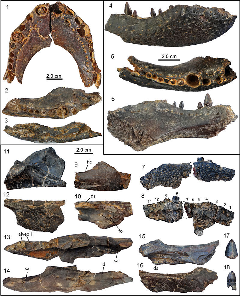
FIGURE 11. Images of Globidentosuchus cf. G. brachyrostris (UNEFM-VF-017) including three right dentaries (1-4), one left dentary (5, 6), and one left angular in angled dorsal (7) and medial (8) view. Images of dentaries in 3, 4, and 5 in dorsal view, 2 and 6 in ventral view. Note splenial associated with dentary and reaching the symphysis in (4).
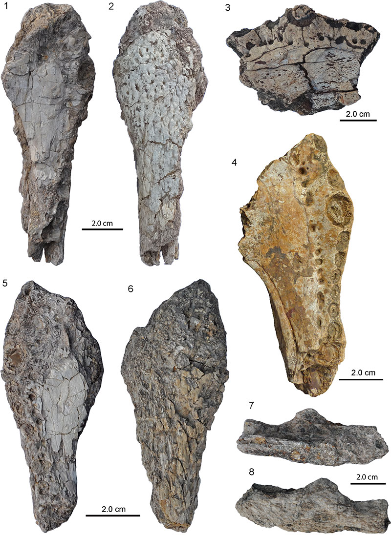
FIGURE 12. Photographs of the holotype skull of Melanosuchus fisheri (MCNC-243; 1-4) and the skull and associated lower jaw assignable to Caimaninae aff. Melanosuchus fisheri (AMU-CURS-234, 5-8). 1, skull in dorsal view. 2, close-up of the sutures between the frontal, prefrontals and posterior portion of nasals. Compare to interpretative drawing in Figure 4.1. 3, skull in left lateral view. 4, skull in occipital view. 5,6, skull in dorsal and ventral view. 7,8, lower jaw in dorsal and ventral view. Abbreviations: f, frontal; n, nasal; prf, prefrontal.
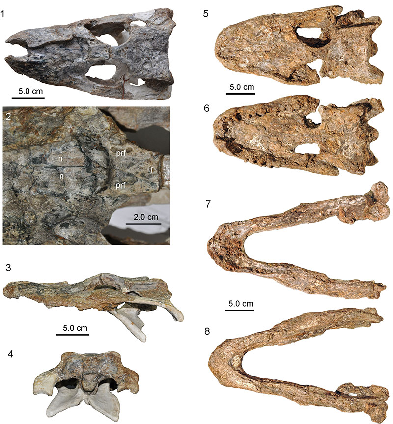
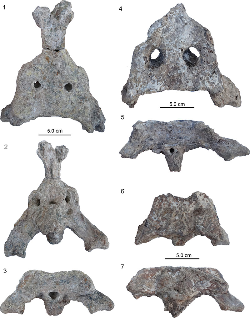
FIGURE 13. Photographs (1, 3, 4-6) and interpretative drawing (2) of the holotype skull and associated lower jaw remains (UNEFM-CIAAP-1297) (3) of Mourasuchus arendsi. 1, 2, skull in dorsal view. 3, right mandibular ramus in dorsal view. 4-6, Fragment of left mandibular ramus in dorsal (4), medial (5) and lateral (6) view. Abbreviations: f, frontal; j, jugal; la, lacrimal; mx, maxilla; n, nasal; p, parietal; pmx, premaxilla; po, postorbital; prf, prefrontal; pt, pterygoid; q, quadrate; qj, quadratojugal; so, supraoccipital; sq, squamosal.
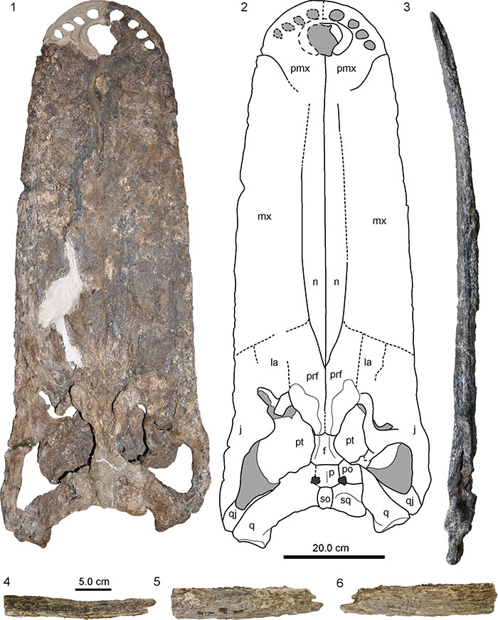
FIGURE 14. Photograph without (1) and with superimposed interpretative drawing (2) of the skull roof of holotype skull (UNEFM-CIAAP-1297) of Mourasuchus arendsi in left dorsolateral view. Abbreviations: f, frontal; mc, medial crest on parietal; p, parietal; po, postorbital; so, supraoccipital; sq, squamosal; stf, supratemporal fenestra.
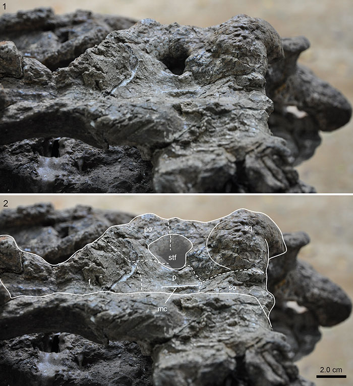
FIGURE 15. Photograph without (1) and with superimposed interpretative drawing (2) of the right side of the posterior portion of holotype skull (UNEFM-CIAAP-1297) of Mourasuchus arendsi in angled lateral view. Note clear notch of jugal and system of cracks (indicated by dotted white lines and arrows) leading to displacement of right posterolateral portion of skull. Abbreviations: j, jugal; po, postorbital; q, quadrate; qj, quadratojugal; sq, squamosal.
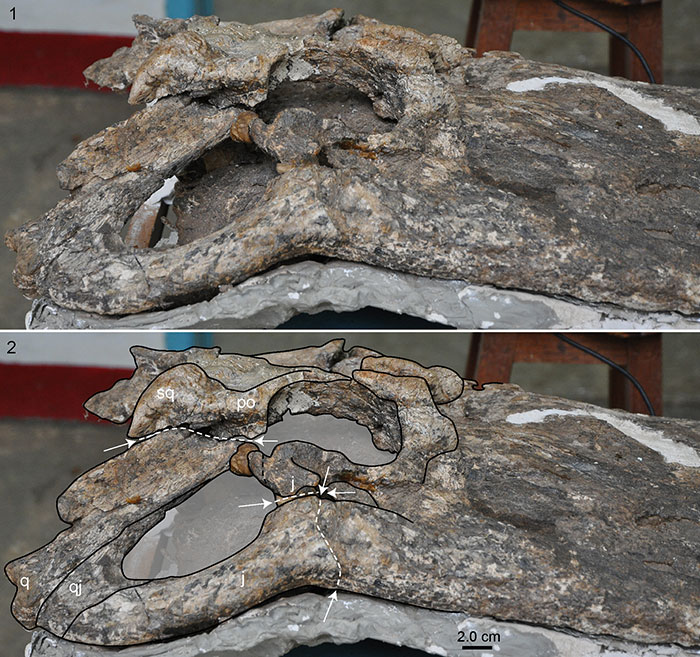
FIGURE 16. Photograph (1) and interpretative drawing (2 of the holotype skull (UNEFM-CIAAP-1297) of Mourasuchus arendsi in occipital view. Note dorsoventral compaction of specimen. Abbreviations: bo, basioccipital; eo, exoccipital; oc, occipital condyle; pt, pterygoid; q, quadrate; so, supraoccipital; sq, squamosal.
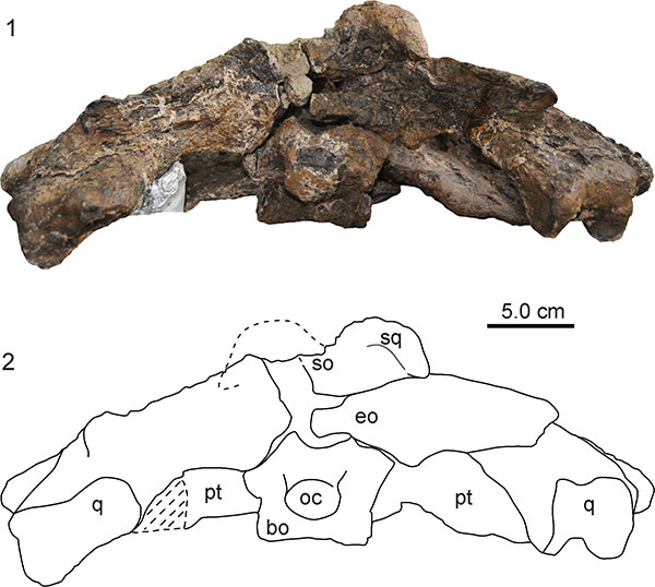
FIGURE 17. Photographs of skull and lower jaw (MCNC-URU-110-72V) assignable to Mourasuchus arendsi. 1, dorsal view. 2, ventral view. 3, dorsal view. 4, medial view. Note that large parts of the specimen have been heavily reconstructed and all tooth alveoli are covered by plaster-cast teeth.
FIGURE 18. Photographs and interpretative drawing of weathered posterior skull portion (UNEFM-VF-03) and large skull part (AMU-CURS-768) of Mourasuchus arendsi. 1, 2, incomplete skull in dorsal view. 3, 4, incomplete skull in occipital view. 5, 6, large porterior skull part in dorsal and ventral view. Note squamosal eminences and posterior median ridge on parietal in UNEFM-VF-03 and both, the squamosal eminences and the knobs in front of the orbits in AMU-CURS-768. Abbreviations: eo, exoccipital f, frontal; mx, maxilla; oc, occipital condyle; ok, ornamental knob on prefrontal; pa, parietal; po, postorbital; pt, pterygoid; q, quadrate; se, squamosal eminences; so, supraoccipital; sof, suborbital fenestra; sq, squamosal; stf, supratemporal fenestra.
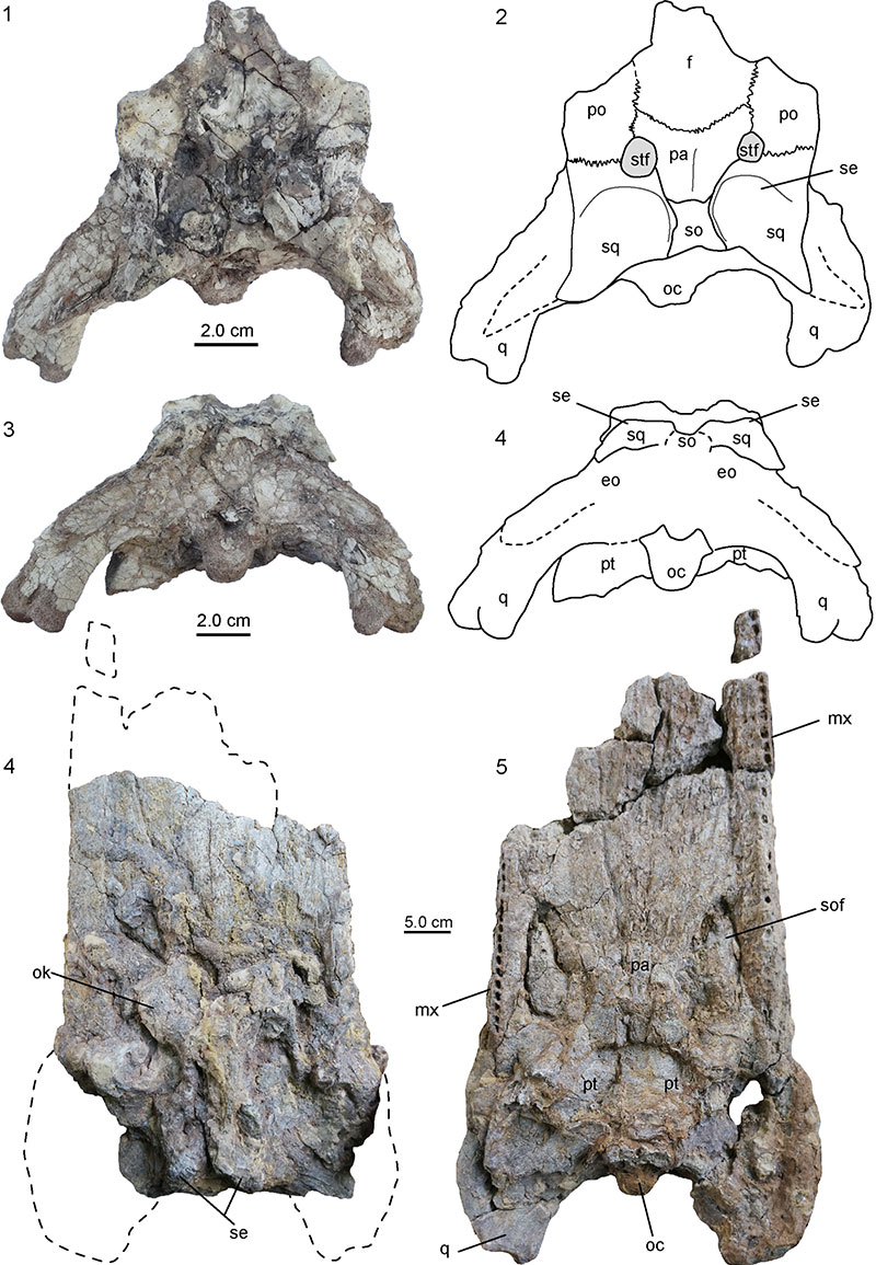
FIGURE 19. Photographs and interpretative drawings of the posterior skull portion and orbitofrontal region as well as additional fragments (possibly jugal and lower jaw fragments) of Mourasuchus arendsi (AMU-CURS-218). 1,2, skull fragment in dorsal view. Note squamosal eminences and orbital bony excrescences. 3, 4, skull fragment in occipital view. 5-9, skull fragments (partially from jugals?) found associated with the larger skull portions. Abbreviations: bo, basioccipital; eo, exoccipital; f, frontal; j, jugal; la, lacrimal; oc, occipital condyle; o, orbit, ok, orbital knob; p, parietal; po, postorbital; prf, prefrontal; pt, pterygoid; q, quadrate; qj, quadratojugal; so, supraoccipital; sq, squamosal; stf, supratemporal fenestra.
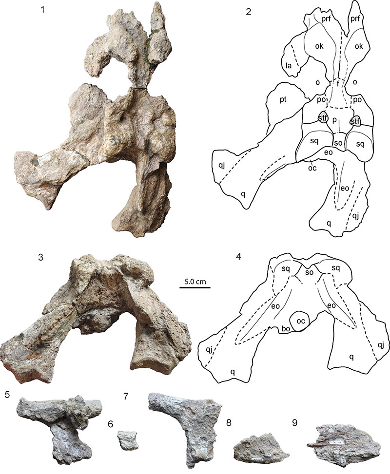
FIGURE 20. Photographs (1, 2, 5-12) and interpretative drawings (3, 4) of skull and lower jaw remains (AMU-CURS-073) of Mourasuchus sp. 1, 2, images of the largest fragment of the skull including the basioccipital-basisphenoid-pterygoid complex in dorsal (1) and occipital (2) view. 3, 4, interpretative drawings of the skull bones shown in 1 and 2. 5, 6, left quadrate in dorsal (5) and ventral (6) view. 7, fragment tentatively assigned to the postorbital bar of a right jugal in dorsal (?) view. 8, 9, large fragments of the maxillae in ventral view. 10, right lower jaw ramus in medial view. 11, small fragment of the left lower jaw ramus in dorsal view. 12, conical, slightly curved isolated tooth (the tip is set off by a break). Note different scale bar in 10 and 12. Abbreviations: bo, basioccipital; bs, basisphenoid; cf, carotid foramen; ec, ectopterygoid; eof, facet for exoccipital on basioccipital; ffm, floor of foramen magnum on basioccipital; oc, occipital condyle; pa, palatine; pt, pterygoid; vc, vertebral centra.
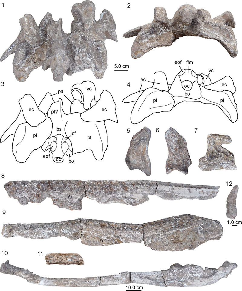
FIGURE 21. Photographs of weathered skull and lower jaw remains (AMU-CURS-396) of Mourasuchus sp. 1, posterior portion of skull and braincase in occipital view. Note squamosal eminences. 2, right premaxilla in dorsal view with perforations for anterior-most teeth of lower jaw. 3, fragment of maxilla with seven alveoli in ventral view. 4, portion of dentary in medial view. 5, unidentified bone fragment. 6, fragment of maxilla with about 26 alveoli in ventral view. 7, straight portion of lower jaw in dorsal view. The exact number of alveoli could not be elucidated.
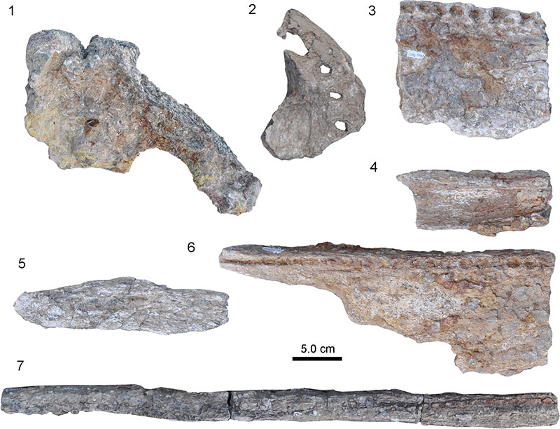
FIGURE 22. Photographs of skull and lower jaw remains of several individuals of Mourasuchus sp. 1, heavily encrusted posterior portion of skull (AMU-CURS-530) in occipital view. Note squamosal eminences. 2, 3, maxillary fragment (UNEFM-CIAPP-1378) in dorsal (2) and ventral (3) view. 4, two maxillary fragments (UNEFM-CIAPP-1447) preserved in overlapping position in ventral view. 5, 6, right premaxilla (AMU-CURS-395) in dorsal (5) and ventral (6) view. 7, small fragment of lower jaw in dorsal view 13 alveoli (AMU-CURS-537). 8, posterior portion of skull (AMU-CURS-695) in dorsal view. Note massive squamosal eminences. 9, twelve fragments of a lower jaw (AMU-CURS-430), with at least the larger fragments belonging to the right ramus. The largest pieces are in dorsal view, showing alveoli.
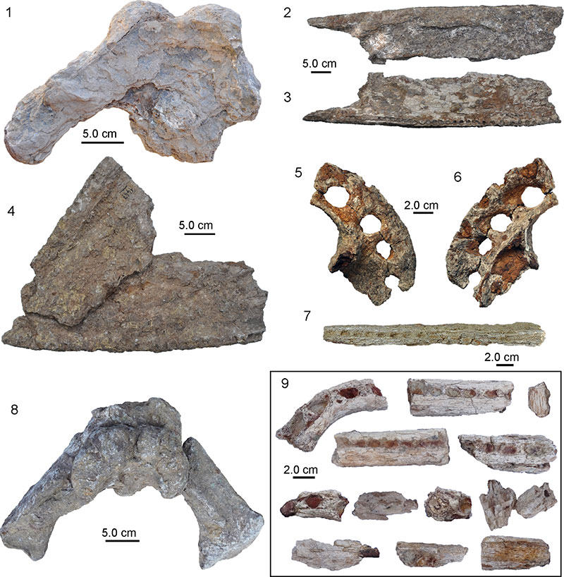
FIGURE 23. Photographs of lower jaw remains and associated left prefrontal of Mourasuchus sp. (AMU-CURS-748). 1, anterior and medial portion of lower jaw in dorsal view. Note lateromedial distortion of the right ramus and the break in the anterior portion of the left ramus. 2-6, posterior portions of the lower jaw in dorsal (2, 6), lateral (3, 5), and medial (4) view. 7-10, images and schematic interpretations of prefrontal in dorsal (7, 8) and lateral (9, 10) view. Abbreviations: a, angular; ar, articular; fme, external mandibular fenestra; ok, ornamental knob; prf.p, prefrontal pillar; prf.s, prefrontal shelf; ro, rim of orbit; rp, retroarticular process; sa, surangular.
FIGURE 24. Photographs of skull of holotype (1-5: UNEFM-CIAAP-1369; associated lower jaw not shown) and paratypes UNEFM-CIAAP 1445 (6) and AMU-CURS-135 (7) of Purussaurus mirandai. 1-3, holotype skull in dorsal (1), ventral (2) and angled rostral view (3); the latter is not to scale. 4, 5, skull fragment labelled as “quadrate” of UNEFM-CIAAP-1369, which possibly constitutes a proximal fragment of the left quadrate/quadratojugal complex of the holotype skull. 6, right lower jaw in dorsal view. 7, largely unprepared skull and attached left and right lower jaw rami in dorsal view (picture courtesy: Jorge Moreno, Colombia).
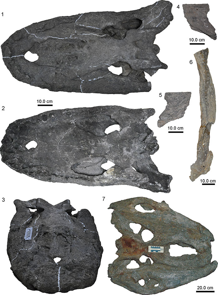
FIGURE 25. Photographs of skull and jaw material of Purussaurus. 1, right dentary fragment (MCNC-URU-115-72V) in dorsal view; 2-23, skull and lower jaw material belonging to several different individuals currently accessioned under a single collection number (MCNC-URU-158-72V). 2-3, anterior part of skull (electronically assembled to represent life position) in dorsal and ventral view. Only the right premaxilla and maxilla (2-4) were shown as part of the paratype series of P. mirandai by (Aguilera et al., 2006). 5, right part of skull table in dorsal view. 6, left part of skull table in lateral view. 7, isolated tooth. 8, 9, left quadrate in dorsal and ventral view. 10, right quadrate in dorsal view. 11, right ectopterygoid in posterolateral view. 12, lower jaw fragment in medial view with alveoli. 13, right jugal in lateral view. 14, 15, articulars with retroarticular processes in dorsal view. 16, 17, pterygoids? 18-21, lower jaw elements. 18, possible anterior fragment of left surangular in ventral view? 19, posterior fragment of surangular in lateral view? 20, fragment of right angular in medial view? 21, fragment of right surangular in medial view? 22, smaller skull table in the assemblage also assignable to Purussaurus in dorsal view. 23, anterior portion of lower jaw, initially referred to and figured as MCNC-URU-157-72V by (Aguilera, 2004).
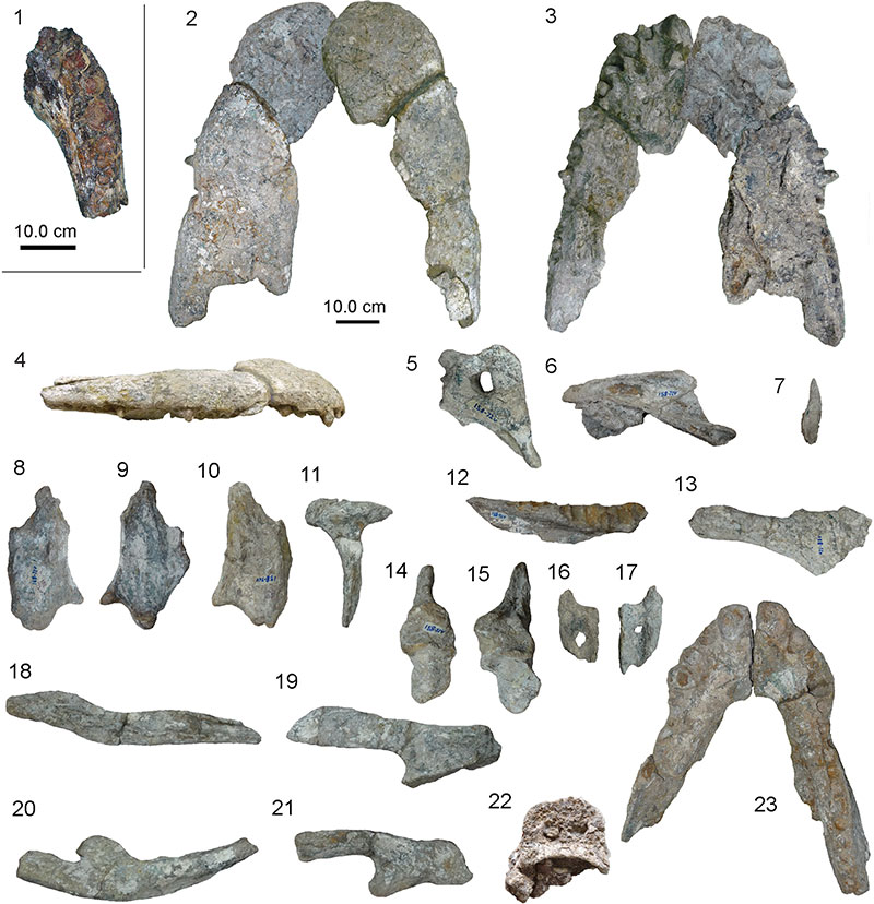
FIGURE 26. Photographs of specimens referable to Purussaurus cf. P. mirandai. 1, referred specimen AMU-CURS-057, right and left dentaries in dorsal view. 2, posterior skull fragment UNEFM-CIAAP-1368/1372 in dorsal view. 3, 4, premaxillae of UNEFM-CIAAP-1367 in dorsal and ventral view. 5, three isolated teeth of UNEFM-CIAAP-1367. 6, lower jaw of AMU-CURS-541 in dorsal view.
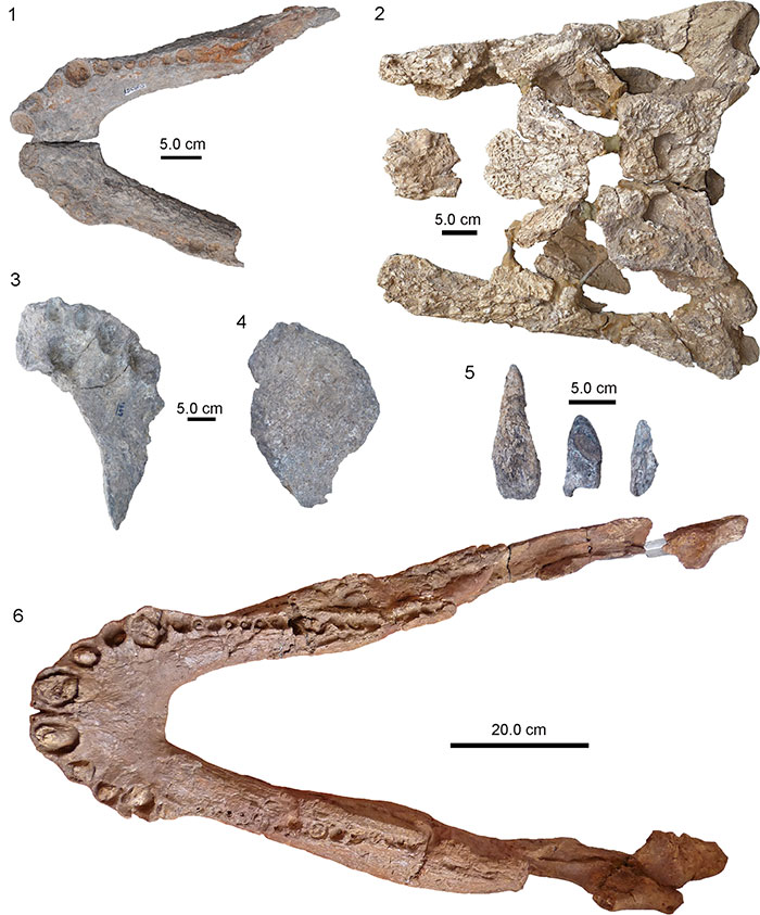
FIGURE 27. Photographs of lower jaw material of Purussaurus cf. P. mirandai in the show room of Museo Paleontológico Urumaco. 1, right mandibular ramus in lateral view (AMU-CURS-685). 2, lower jaw in dorsal view (MCN-USB no number).
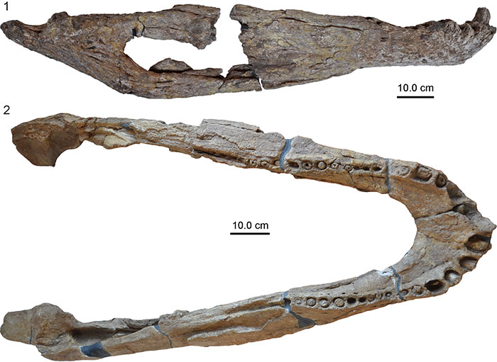
FIGURE 28. Photographs of isolated right premaxilla of Purussaurus cf. P. mirandai (AMU-CURS-602). 1, specimen prior to collection in the field (dorsal view). 2-4, surface scan model of specimen (taken with an Artec Spider Surface Scanner) in (2) dorsal, (3) ventral, and (4 lateral view.
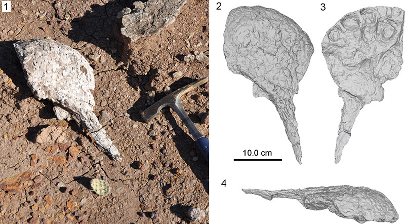
FIGURE 29. Photographs of associated cranial material (AMU-CURS-528) of Purussaurus cf. P. mirandai. 1-2, right premaxilla in dorsal (1) and ventral (2) view. 3-4, right jugal in lateral (3) and medial (4) view. 5-6, right ectopterygoid in ventral (5) and dorsal (6) view. 7-8, left ectopterygoid in ventral (7) and dorsal (8) view. 9-10, dentary fragment in external (9) and internal (10) view. 11, isolated teeth (note different scale bar).
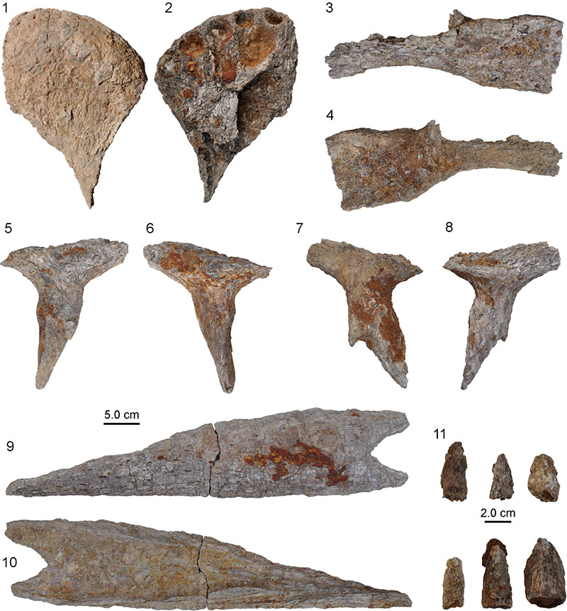
FIGURE 30. Photographs of cranial bones of several individuals of Purussaurus. 1, Strongly weathered lower jaw ramus (UNEFM-CIAAP-1434) in medial view. 2-7, associated lower jaw fragments (MCNC-76-72V). 2, 3, dentary symphyseal region showing the symphyseal region in ventral (2) and dorsal (3) view. 4-6, posterior portions of the left (4) and right dentary, the latter in medial (5) and lateral (6) view. Note that part of splenial is still attached to dentary in (5). 7, left articular, surangular and portion of angular in medial view. 8, right articular and surangular in medial view. 9-16, associated skull and lower jaw fragments (MCNC-URU-90-72V). 9,10, left ramus in ventral and dorsal view. 11-14, two premaxillary fragments in dorsal and ventral view each. 15, 16, two dentary fragments in dorsal view. 17-25, skull and lower jaw fragments (MCNC-URU-111-72V), which were found in an assemblage of at least two individuals of different size. 17, partial skull in dorsal view. 18, smaller partial skull in dorsal view. 19, strongly weathered maxillary fragment (?) in ventral view. 20, left dentary fragment in dorsal view. 21, 22, right articular and surangular in dorsolateral (21) and dorsal (22) view. 23,24, left angular, surangular, and articular in medial (23) and lateral (24) view. 25, dentary symphyseal region and portion of right dentary in dorsal view.
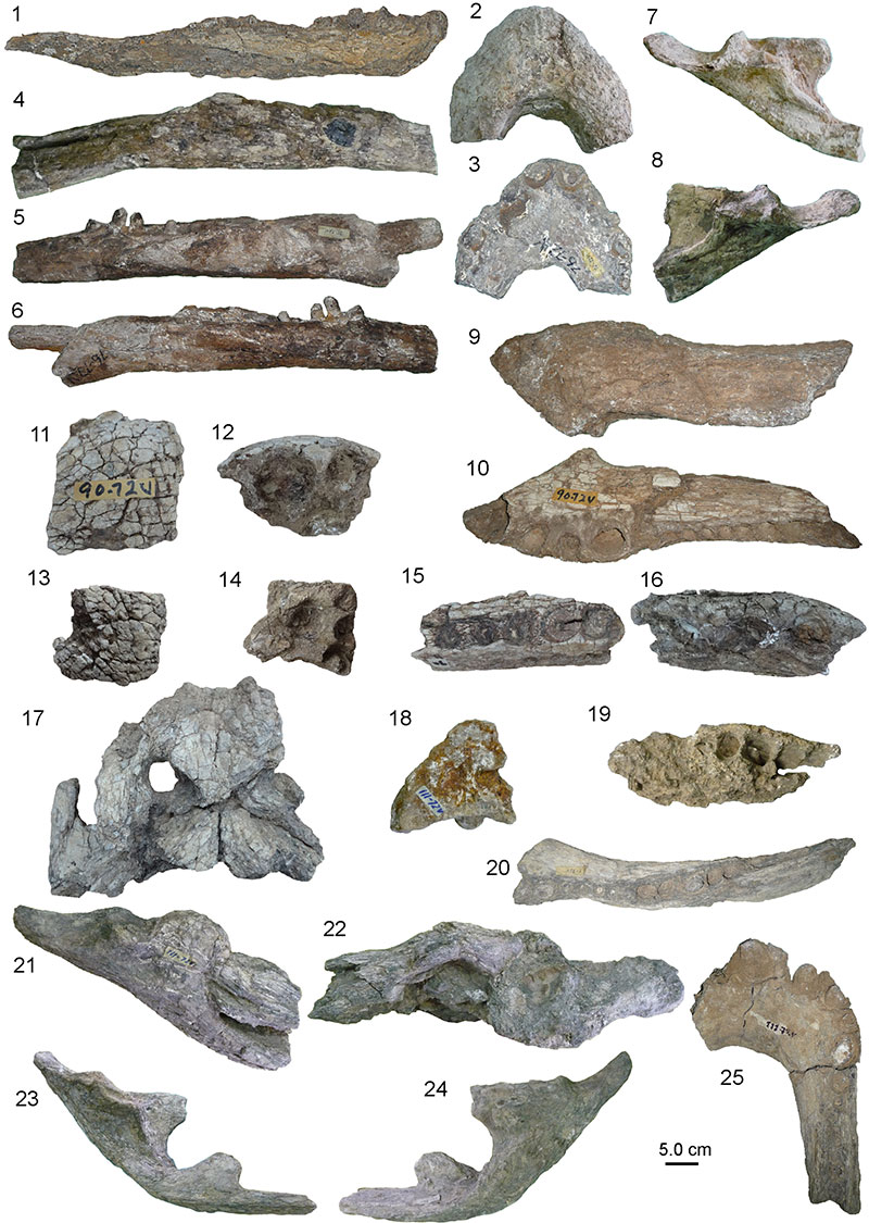
FIGURE 31. Cranial and lower jaw specimens of Purussaurus sp. 1-4, lower jaw fragments (AMU-CURS-384). 1, 2, left dentary in dorsal and medial view. 3, 4, left articular and angular in dorsal and lateral view. 5, 6, quadrate and skull table (AMU-CURS-606) in dorsal view. 7, dentaries of lower jaw (AMU-CURS-394) in dorsal view. Note the evident deformation of the left dentary.
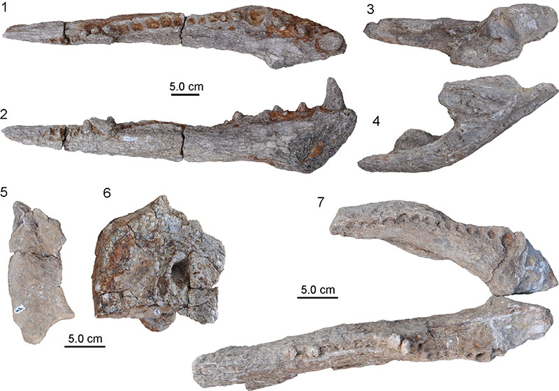
FIGURE 32. Photographs (1, 2) of the anterior portion of the lower jaw of Purussaurus sp. (AMU-CURS-671) and interpretative drawing of the natural cross-section (3) of the right dentary. Abbreviations: ab, alveolar bone; rf; functional tooth; rt1, first replacement tooth; rt2, second replacement tooth.
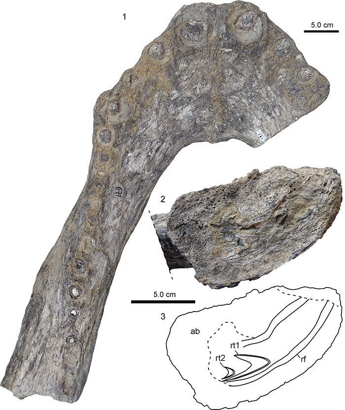
FIGURE 33. Photographs of isolated giant crocodylian teeth assignable to Purussaurus sp. All but the smallest specimen (19 mm) show maximal cross-sectional diameters between 27 and 39 mm, which lie in the alveolar diameters of the largest Purussaurus specimens from Urumaco. 1, 2, tooth AMU-CURS-006 in rostral (1) and medial (2) view, carrying a wear facet. 3, 4, strongly weathered tooth AMU-CURS-046 in rostral (3) and medial (4) view. 5,6, two teeth labelled AMU-CURS-033. 7, tooth AMU-CURS-101. 8, small tooth AMU-CURS-022. 9, 10, tooth UNEFM-CIAAP-1432 in medial view.
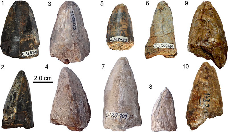
FIGURE 34. Photographs of caimanine lower jaw fragment (AMU-CURS-090) in lateral (1) and medial (2) view.
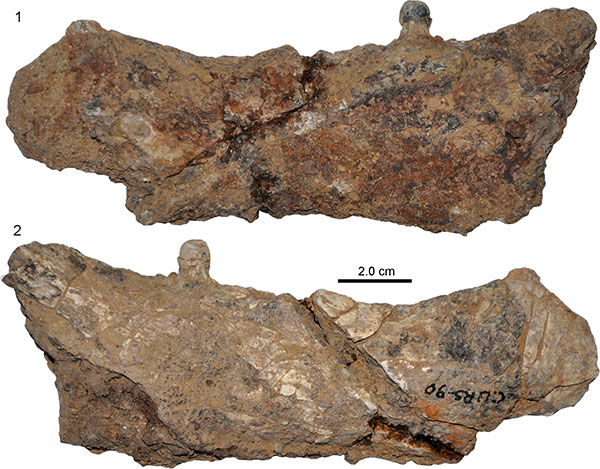
FIGURE 35. Photographs of heavily encrusted anterior portion of a partial caimanine skull and lower jaw (AMU-CURS-100) in dorsal (1) and ventral (2) view.
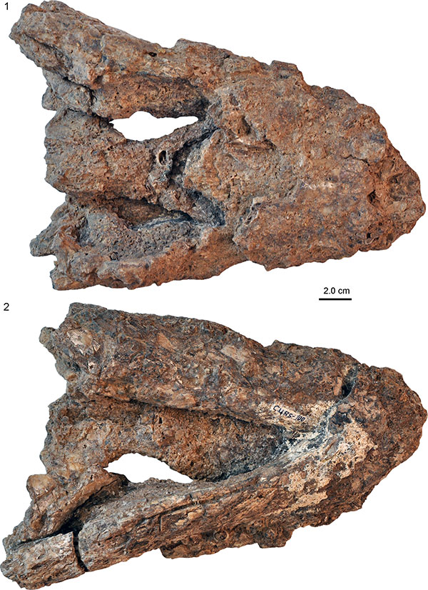
FIGURE 36. Photographs of skull (1-3, 5) and associated lower jaw fragment (4, 5) of small Caimaninae indet. (AMU-CURS105/106). 1, dorsal view. 2, ventral view. 3, occipital view. 4, left dentary in dorsal view. 5, Close-up of mounted specimen (not to scale) showing a preserved knob-like crushing tooth in the posterior part of the maxilla (marked by white arrow).
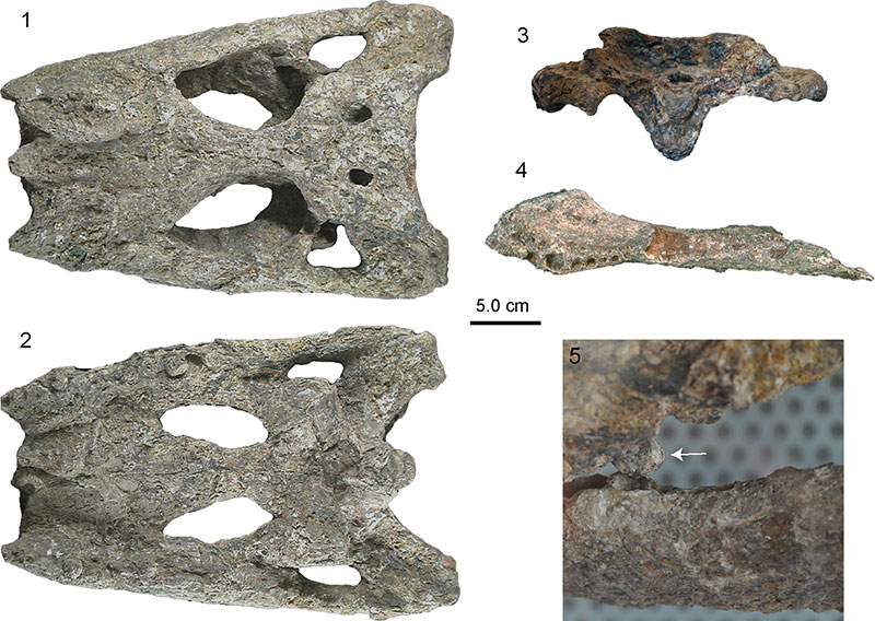
FIGURE 37. Photographs of heavily weathered and altered caimanine lower jaw fragment (AMU-CURS-110) in lateral (1), medial (2), and dorsal (3) view.
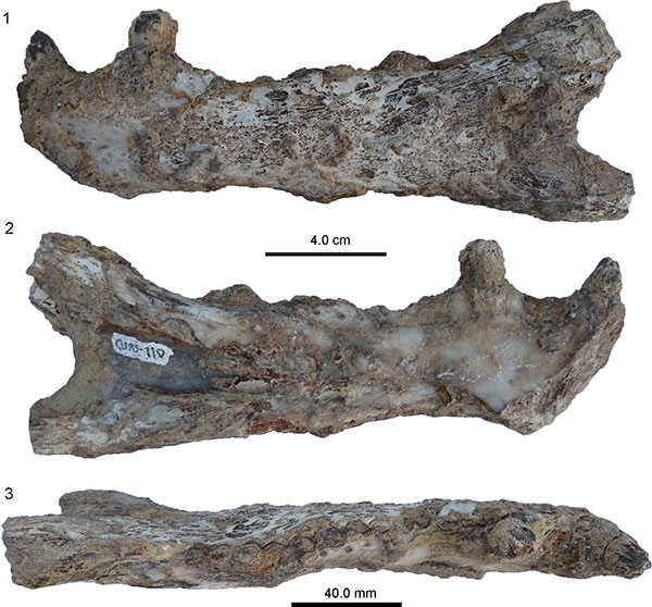
FIGURE 38. Photographs of strongly deformed partial caimanine skull (AMU-CURS-113) in dorsal (1) and ventral (2) view.
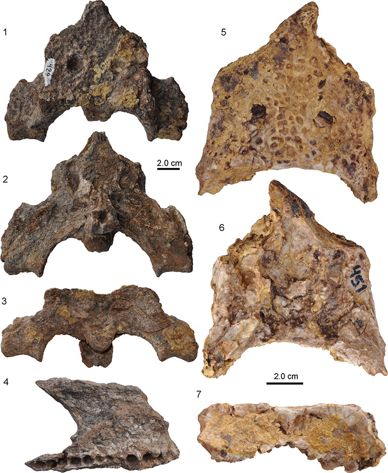
FIGURE 39. Photographs of remains including an associated posterior skull and maxilla fragments (AMU-CURS-429: 1-4) and a weathered and partly encrusted, isolated skull table of a small caimanine (AMU-CURS-451: 5-7). Posterior skull table and braincase in 1, dorsal, 2, ventral, and 3, occipital view. Maxillary fragment (4) in ventral view. Isolated skull table in dorsal (5), ventral (6), and occipital (7) view showing small exposure of supraoccipital on the skull roof.
FIGURE 40. Photographs of small caimanine skull part (AMU-CURS-601) consisting of an articulated left jugal and partial quadratojugal in lateral (1) and medial (2) view.
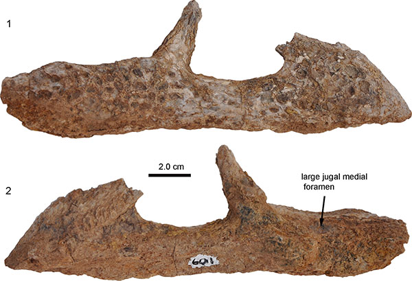
FIGURE 41. Photographs of small caimanine skull roofs. A-C, UNEFM-VF-06 in dorsal (1), ventral (2), and occipital (3) view. 4, 5, UNEFM-VF-019 in dorsal and occipital view. 6, 7, UNEFM-VF-022 in dorsal and occipital view.